6YPT
 
 | |
3M6Q
 | |
3M6R
 | |
3M6O
 | |
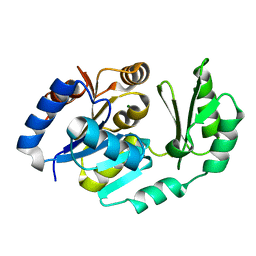
1JB1
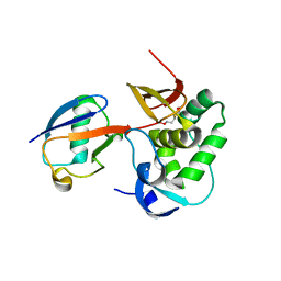
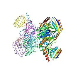
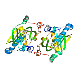
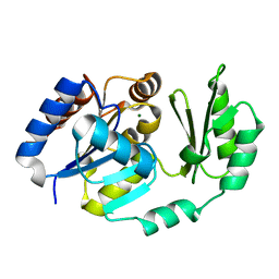
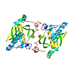
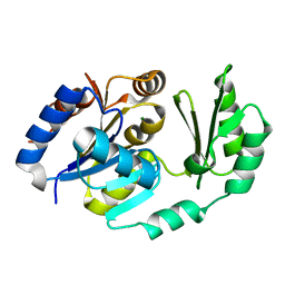
 | | Lactobacillus casei HprK/P Bound to Phosphate | | 分子名称: | HPRK PROTEIN, PHOSPHATE ION | | 著者 | Fieulaine, S, Morera, S, Poncet, S, Monedero, V, Gueguen-Chaignon, V, Galinier, A, Janin, J, Deutscher, J, Nessler, S. | | 登録日 | 2001-06-01 | | 公開日 | 2001-08-08 | | 最終更新日 | 2017-10-04 | | 実験手法 | X-RAY DIFFRACTION (2.8 Å) | | 主引用文献 | X-ray structure of HPr kinase: a bacterial protein kinase with a P-loop nucleotide-binding domain.
EMBO J., 20, 2001
|
|
1KKL
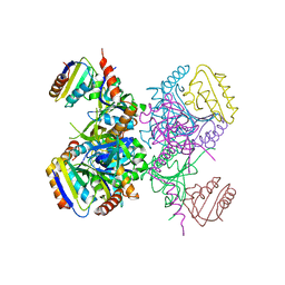 | | L.casei HprK/P in complex with B.subtilis HPr | | 分子名称: | CALCIUM ION, HprK protein, PHOSPHOCARRIER PROTEIN HPR | | 著者 | Fieulaine, S, Morera, S, Poncet, S, Galinier, A, Janin, J, Deutscher, J, Nessler, S. | | 登録日 | 2001-12-10 | | 公開日 | 2002-08-28 | | 最終更新日 | 2023-08-16 | | 実験手法 | X-RAY DIFFRACTION (2.8 Å) | | 主引用文献 | X-ray structure of a bifunctional protein kinase in complex with its protein substrate HPr.
Proc.Natl.Acad.Sci.USA, 99, 2002
|
|
1KKM
 | | L.casei HprK/P in complex with B.subtilis P-Ser-HPr | | 分子名称: | CALCIUM ION, HprK protein, PHOSPHATE ION, ... | | 著者 | Fieulaine, S, Morera, S, Poncet, S, Galinier, A, Janin, J, Deutscher, J, Nessler, S. | | 登録日 | 2001-12-10 | | 公開日 | 2002-08-28 | | 最終更新日 | 2023-08-16 | | 実験手法 | X-RAY DIFFRACTION (2.8 Å) | | 主引用文献 | X-ray structure of a bifunctional protein kinase in complex with its protein substrate HPr.
Proc.Natl.Acad.Sci.USA, 99, 2002
|
|
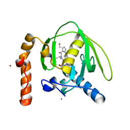
4JE6
 | | Crystal structure of a human-like mitochondrial peptide deformylase | | 分子名称: | Peptide deformylase 1A, chloroplastic/mitochondrial, ZINC ION | | 著者 | Fieulaine, S, Meinnel, T, Giglione, C. | | 登録日 | 2013-02-26 | | 公開日 | 2014-02-26 | | 最終更新日 | 2023-11-08 | | 実験手法 | X-RAY DIFFRACTION (2 Å) | | 主引用文献 | Understanding the highly efficient catalysis of prokaryotic peptide deformylases by shedding light on the determinants specifying the low activity of the human counterpart.
Acta Crystallogr.,Sect.D, 70, 2014
|
|
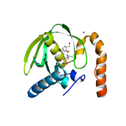
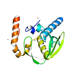
4JE8
 | | Crystal structure of a human-like mitochondrial peptide deformylase in complex with Met-Ala-Ser | | 分子名称: | Peptide deformylase 1A, chloroplastic/mitochondrial, ZINC ION, ... | | 著者 | Fieulaine, S, Meinnel, T, Giglione, C. | | 登録日 | 2013-02-26 | | 公開日 | 2014-02-26 | | 最終更新日 | 2023-11-08 | | 実験手法 | X-RAY DIFFRACTION (2.4 Å) | | 主引用文献 | Understanding the highly efficient catalysis of prokaryotic peptide deformylases by shedding light on the determinants specifying the low activity of the human counterpart.
Acta Crystallogr.,Sect.D, 70, 2014
|
|
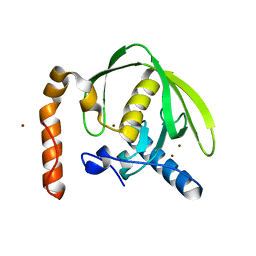
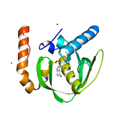
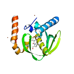
4JE7
 | | Crystal structure of a human-like mitochondrial peptide deformylase in complex with actinonin | | 分子名称: | ACTINONIN, Peptide deformylase 1A, chloroplastic/mitochondrial, ... | | 著者 | Fieulaine, S, Meinnel, T, Giglione, C. | | 登録日 | 2013-02-26 | | 公開日 | 2014-02-26 | | 最終更新日 | 2023-11-08 | | 実験手法 | X-RAY DIFFRACTION (2.1 Å) | | 主引用文献 | Understanding the highly efficient catalysis of prokaryotic peptide deformylases by shedding light on the determinants specifying the low activity of the human counterpart.
Acta Crystallogr.,Sect.D, 70, 2014
|
|
1ZY1
 | | X-ray structure of peptide deformylase from Arabidopsis thaliana (AtPDF1A) in complex with Met-Ala-Ser | | 分子名称: | Peptide deformylase, mitochondrial, ZINC ION, ... | | 著者 | Fieulaine, S, Juillan-Binard, C, Serero, A, Dardel, F, Giglione, C, Meinnel, T, Ferrer, J.-L. | | 登録日 | 2005-06-09 | | 公開日 | 2005-09-27 | | 最終更新日 | 2023-08-23 | | 実験手法 | X-RAY DIFFRACTION (3 Å) | | 主引用文献 | The crystal structure of mitochondrial (Type 1A) peptide deformylase provides clear guidelines for the design of inhibitors specific for the bacterial forms
J.Biol.Chem., 280, 2005
|
|
2B1R
 | |
2B1Q
 | |
2D2V
 | |
1ZXZ
 | | X-ray structure of peptide deformylase from Arabidopsis thaliana (AtPDF1A); crystals grown in PEG-5000 MME as precipitant | | 分子名称: | Peptide deformylase, mitochondrial, ZINC ION | | 著者 | Fieulaine, S, Juillan-Binard, C, Serero, A, Dardel, F, Giglione, C, Meinnel, T, Ferrer, J.-L. | | 登録日 | 2005-06-09 | | 公開日 | 2005-09-27 | | 最終更新日 | 2023-08-23 | | 実験手法 | X-RAY DIFFRACTION (2.8 Å) | | 主引用文献 | The crystal structure of mitochondrial (Type 1A) peptide deformylase provides clear guidelines for the design of inhibitors specific for the bacterial forms
J.Biol.Chem., 280, 2005
|
|
1ZY0
 | | X-ray structure of peptide deformylase from Arabidopsis thaliana (AtPDF1A); crystals grown in PEG-6000 | | 分子名称: | Peptide deformylase, mitochondrial, ZINC ION | | 著者 | Fieulaine, S, Juillan-Binard, C, Serero, A, Dardel, F, Giglione, C, Meinnel, T, Ferrer, J.-L. | | 登録日 | 2005-06-09 | | 公開日 | 2005-09-27 | | 最終更新日 | 2023-08-23 | | 実験手法 | X-RAY DIFFRACTION (2.9 Å) | | 主引用文献 | The crystal structure of mitochondrial (Type 1A) peptide deformylase provides clear guidelines for the design of inhibitors specific for the bacterial forms
J.Biol.Chem., 280, 2005
|
|
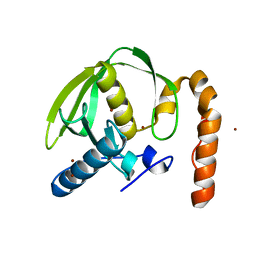
1U2T
 | | X-Ray structure of the sucrose-phosphatase (SPP) from Synechocystis sp. PCC6803 in complex with sucrose6P | | 分子名称: | 6-O-phosphono-beta-D-fructofuranose-(2-1)-alpha-D-glucopyranose, sucrose-phosphatase (SPP) | | 著者 | Fieulaine, S, Lunn, J.E, Borel, F, Ferrer, J.-L. | | 登録日 | 2004-07-20 | | 公開日 | 2005-06-14 | | 最終更新日 | 2023-08-23 | | 実験手法 | X-RAY DIFFRACTION (2.9 Å) | | 主引用文献 | The structure of a cyanobacterial sucrose-phosphatase reveals the sugar tongs that release free sucrose in the cell
PLANT CELL, 17, 2005
|
|
3O3J
 | |
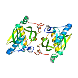
1S2O
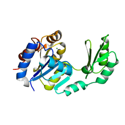 | | X-Ray structure of the sucrose-phosphatase (SPP) from Synechocystis sp. PCC6803 at 1.40 A resolution | | 分子名称: | MAGNESIUM ION, sucrose-phosphatase | | 著者 | Fieulaine, S, Lunn, J.E, Borel, F, Ferrer, J.L. | | 登録日 | 2004-01-09 | | 公開日 | 2005-02-22 | | 最終更新日 | 2024-02-14 | | 実験手法 | X-RAY DIFFRACTION (1.4 Å) | | 主引用文献 | The structure of a cyanobacterial sucrose-phosphatase reveals the sugar tongs that release free sucrose in the cell.
Plant Cell, 17, 2005
|
|
3M6P
 | |
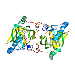
1U2S
 | | X-Ray structure of the sucrose-phosphatase (SPP) from Synechocystis sp. PCC6803 in complex with glucose | | 分子名称: | MAGNESIUM ION, alpha-D-glucopyranose, sucrose-phosphatase | | 著者 | Fieulaine, S, Lunn, J.E, Borel, F, Ferrer, J.-L. | | 登録日 | 2004-07-20 | | 公開日 | 2005-06-14 | | 最終更新日 | 2023-08-23 | | 実験手法 | X-RAY DIFFRACTION (2.5 Å) | | 主引用文献 | The structure of a cyanobacterial sucrose-phosphatase reveals the sugar tongs that release free sucrose in the cell
PLANT CELL, 17, 2005
|
|
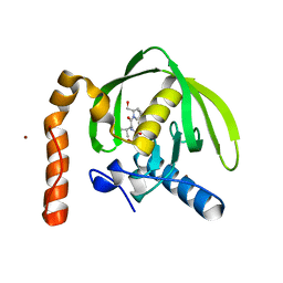
3PN4
 | | Crystal structure of Arabidopsis thaliana petide deformylase 1B (AtPDF1B) in complex with actinonin (crystallized in PEG-550-MME) | | 分子名称: | ACTINONIN, Peptide deformylase 1B, chloroplastic, ... | | 著者 | Fieulaine, S, Meinnel, T, Giglione, C. | | 登録日 | 2010-11-18 | | 公開日 | 2011-06-08 | | 最終更新日 | 2023-09-06 | | 実験手法 | X-RAY DIFFRACTION (1.9 Å) | | 主引用文献 | Trapping conformational States along ligand-binding dynamics of Peptide deformylase: the impact of induced fit on enzyme catalysis.
Plos Biol., 9, 2011
|
|
3PN2
 | |
3PN6
 | |
1TJ3
 | | X-Ray structure of the Sucrose-Phosphatase (SPP) from Synechocystis sp. PCC6803 in a closed conformation | | 分子名称: | MAGNESIUM ION, Sucrose-Phosphatase | | 著者 | Fieulaine, S, Lunn, J.E, Borel, F, Ferrer, J.-L. | | 登録日 | 2004-06-03 | | 公開日 | 2005-06-14 | | 最終更新日 | 2023-08-23 | | 実験手法 | X-RAY DIFFRACTION (2.8 Å) | | 主引用文献 | The structure of a cyanobacterial sucrose-phosphatase reveals the sugar tongs that release free sucrose in the cell.
Plant Cell, 17, 2005
|
|


