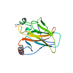
1BRG
 
 | |
1BSB
 | |
1BSC
 | |
2BIO
 | |
2BIP
 | |
2BIN
 | |
2BIM
 | |
2BIQ
 | |
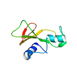
1CIR
 | | COMPLEX OF TWO FRAGMENTS OF CI2 [(1-40)(DOT)(41-64)] | | 分子名称: | CHYMOTRYPSIN INHIBITOR 2 | | 著者 | Davis, B.J, Fersht, A.R. | | 登録日 | 1995-10-02 | | 公開日 | 1996-01-29 | | 最終更新日 | 2024-05-22 | | 実験手法 | SOLUTION NMR | | 主引用文献 | Towards the complete structural characterization of a protein folding pathway: the structures of the denatured, transition and native states for the association/folding of two complementary fragments of cleaved chymotrypsin inhibitor 2. Direct evidence for a nucleation-condensation mechanism
Structure Fold.Des., 1, 1996
|
|
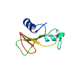
1CIQ
 | | COMPLEX OF TWO FRAGMENTS OF CI2, RESIDUES 1-40 AND 41-64 | | 分子名称: | CHYMOTRYPSIN INHIBITOR 2 | | 著者 | Buckle, A.M, Fersht, A.R. | | 登録日 | 1995-10-02 | | 公開日 | 1996-03-08 | | 最終更新日 | 2024-02-07 | | 実験手法 | X-RAY DIFFRACTION (2.2 Å) | | 主引用文献 | Towards the complete structural characterization of a protein folding pathway: the structures of the denatured, transition and native states for the association/folding of two complementary fragments of cleaved chymotrypsin inhibitor 2. Direct evidence for a nucleation-condensation mechanism
Structure Fold.Des., 1, 1996
|
|
5LAP
 | |
1FY9
 | |
1FYA
 | |
1BNG
 | |
1BTA
 | |
1BRN
 | |
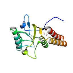
2C6A
 | | Solution structure of the C4 zinc-finger domain of HDM2 | | 分子名称: | UBIQUITIN-PROTEIN LIGASE E3 MDM2, ZINC ION | | 著者 | Yu, G.W, Allen, M.D, Andreeva, A, Fersht, A.R, Bycroft, M. | | 登録日 | 2005-11-08 | | 公開日 | 2006-01-04 | | 最終更新日 | 2024-05-15 | | 実験手法 | SOLUTION NMR | | 主引用文献 | Solution Structure of the C4 Zinc Finger Domain of Hdm2.
Protein Sci., 15, 2006
|
|
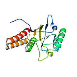
2YBG
 | | Structure of Lys120-acetylated p53 core domain | | 分子名称: | CELLULAR TUMOR ANTIGEN P53, ZINC ION | | 著者 | Arbely, E, Allen, M.D, Joerger, A.C, Fersht, A.R. | | 登録日 | 2011-03-08 | | 公開日 | 2011-05-04 | | 最終更新日 | 2011-07-13 | | 実験手法 | X-RAY DIFFRACTION (1.9 Å) | | 主引用文献 | Acetylation of Lysine 120 of P53 Endows DNA- Binding Specificity at Effective Physiological Salt Concentration.
Proc.Natl.Acad.Sci.USA, 108, 2011
|
|
1UOL
 | |
2FEJ
 | | Solution structure of human p53 DNA binding domain. | | 分子名称: | Cellular tumor antigen p53, ZINC ION | | 著者 | Perez-Canadillas, J.M, Tidow, H, Freund, S.M, Rutherford, T.J, Ang, H.C, Fersht, A.R. | | 登録日 | 2005-12-16 | | 公開日 | 2006-01-31 | | 最終更新日 | 2024-05-29 | | 実験手法 | SOLUTION NMR | | 主引用文献 | Solution structure of p53 core domain: Structural basis for its instability
Proc.Natl.Acad.Sci.Usa, 103, 2006
|
|
1HL2
 | |
1BTB
 | |
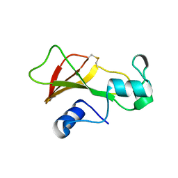
1BNI
 | | BARNASE WILDTYPE STRUCTURE AT PH 6.0 | | 分子名称: | BARNASE | | 著者 | Cameron, A, Henrick, K, Fersht, A.R, Dodson, G, Buckle, A.M. | | 登録日 | 1995-05-17 | | 公開日 | 1995-09-15 | | 最終更新日 | 2024-02-07 | | 実験手法 | X-RAY DIFFRACTION (2.1 Å) | | 主引用文献 | Crystal structural analysis of mutations in the hydrophobic cores of barnase.
J.Mol.Biol., 234, 1993
|
|
1JON
 | |
1COA
 | | THE EFFECT OF CAVITY CREATING MUTATIONS IN THE HYDROPHOBIC CORE OF CHYMOTRYPSIN INHIBITOR 2 | | 分子名称: | CHYMOTRYPSIN INHIBITOR 2 | | 著者 | Jackson, S.E, Moracci, M, Elmasry, N, Johnson, C.M, Fersht, A.R. | | 登録日 | 1993-05-14 | | 公開日 | 1994-01-31 | | 最終更新日 | 2024-02-07 | | 実験手法 | X-RAY DIFFRACTION (2.2 Å) | | 主引用文献 | Effect of cavity-creating mutations in the hydrophobic core of chymotrypsin inhibitor 2.
Biochemistry, 32, 1993
|
|


