2LMA
 
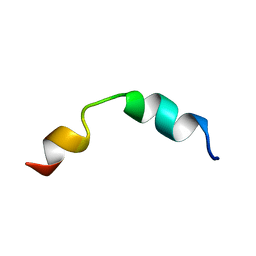 | | Solution structure of CD4+ T cell derived peptide Thp5 | | 分子名称: | Thp5 peptide | | 著者 | Pandey, N.K, Khan, M.M, Chatterjee, S, Dwivedi, V.P, Tousif, S, Das, J, Van Kaer, L. | | 登録日 | 2011-11-29 | | 公開日 | 2011-12-28 | | 最終更新日 | 2024-05-15 | | 実験手法 | SOLUTION NMR | | 主引用文献 | CD4+ T cell derived novel peptide Thp5 induces IL-4 production in CD4+ T cells to direct T helper 2 cell differentiation
J.Biol.Chem., 2011
|
|
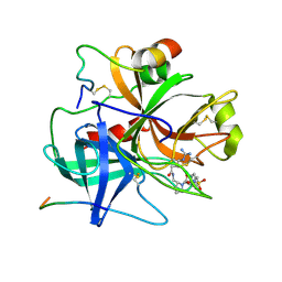
4FKD
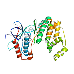
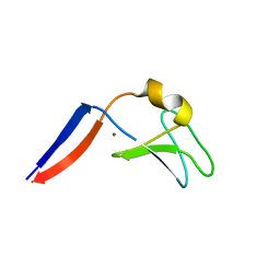 | | Identification of the Activator Binding Residues in the Second Cysteine-Rich Regulatory Domain of Protein Kinase C Theta | | 分子名称: | Protein kinase C theta type, ZINC ION | | 著者 | Rahman, G.M, Shanker, S, Lewin, N.E, Prasad, B.V.V, Blumberg, P.M, Das, J. | | 登録日 | 2012-06-13 | | 公開日 | 2013-01-23 | | 最終更新日 | 2024-02-28 | | 実験手法 | X-RAY DIFFRACTION (1.633 Å) | | 主引用文献 | Identification of the Activator Binding Residues in the Second Cysteine-Rich Regulatory Domain of Protein Kinase C Theta.
Biochem.J., 451, 2013
|
|
7W61
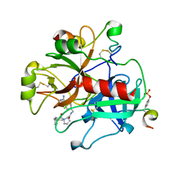
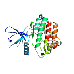
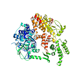
 | | Crystal structure of farnesol dehydrogenase from Helicoverpa armigera | | 分子名称: | 1,2-ETHANEDIOL, ACETATE ION, Farnesol dehydrogenase, ... | | 著者 | Kumar, R, Das, J, Mahto, J.K, Sharma, M, Kumar, P, Sharma, A.K. | | 登録日 | 2021-12-01 | | 公開日 | 2022-07-27 | | 最終更新日 | 2023-11-29 | | 実験手法 | X-RAY DIFFRACTION (1.6 Å) | | 主引用文献 | Crystal structure and molecular characterization of NADP + -farnesol dehydrogenase from cotton bollworm, Helicoverpaarmigera.
Insect Biochem.Mol.Biol., 147, 2022
|
|
3TU7
 | |
3L8X
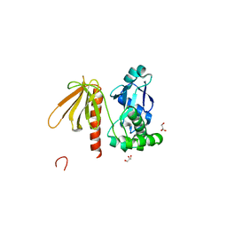
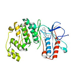 | | P38 alpha kinase complexed with a pyrazolo-pyrimidine based inhibitor | | 分子名称: | Mitogen-activated protein kinase 14, N,4-dimethyl-3-[(1-phenyl-1H-pyrazolo[3,4-d]pyrimidin-4-yl)amino]benzamide | | 著者 | Sack, J.S. | | 登録日 | 2010-01-04 | | 公開日 | 2010-03-02 | | 最終更新日 | 2024-02-21 | | 実験手法 | X-RAY DIFFRACTION (2.4 Å) | | 主引用文献 | Pyrazolo-Pyrimidines: A Novel Heterocyclic Scaffold for Potent and Selective P38 Alpha Inhibitors.
Bioorg.Med.Chem.Lett., 18, 2008
|
|
3OCG
 | |
5WFJ
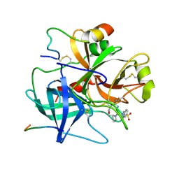
 | | THE JAK3 KINASE DOMAIN IN COMPLEX WITH A COVALENT INHIBITOR | | 分子名称: | 4-({[3-(propanoylamino)phenyl]methyl}amino)pyrrolo[1,2-b]pyridazine-3-carboxamide, Tyrosine-protein kinase JAK3 | | 著者 | Sack, J. | | 登録日 | 2017-07-12 | | 公開日 | 2017-10-04 | | 最終更新日 | 2017-10-11 | | 実験手法 | X-RAY DIFFRACTION (2.48 Å) | | 主引用文献 | Discovery of highly potent, selective, covalent inhibitors of JAK3.
Bioorg. Med. Chem. Lett., 27, 2017
|
|
7CT1
 | |
1BMN
 | |
1BMM
 | |
2BYV
 | |
3CF6
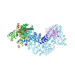 | | Structure of Epac2 in complex with cyclic-AMP and Rap | | 分子名称: | 6-(6-AMINO-PURIN-9-YL)-2-THIOXO-TETRAHYDRO-2-FURO[3,2-D][1,3,2]DIOXAPHOSPHININE-2,7-DIOL, Rap guanine nucleotide exchange factor (GEF) 4, Ras-related protein Rap-1b, ... | | 著者 | Rehmann, H, Arias-Palomo, E, Hadders, M.A, Schwede, F, Llorca, O, Bos, J.L. | | 登録日 | 2008-03-02 | | 公開日 | 2008-07-29 | | 最終更新日 | 2023-11-01 | | 実験手法 | X-RAY DIFFRACTION (2.2 Å) | | 主引用文献 | Structure of Epac2 in complex with a cyclic AMP analogue and RAP1B
Nature, 455, 2008
|
|
3UEJ
 | |
3UGL
 | | Structural and functional characterization of an anesthetic binding site in the second cysteine-rich domain of protein kinase C delta | | 分子名称: | PHOSPHATE ION, Proteine kinase C delta type, ZINC ION, ... | | 著者 | Shanmugasundararaj, S, Stehle, T, Miller, K.W. | | 登録日 | 2011-11-02 | | 公開日 | 2012-12-12 | | 最終更新日 | 2023-09-13 | | 実験手法 | X-RAY DIFFRACTION (1.357 Å) | | 主引用文献 | Structural and Functional Characterization of an Anesthetic Binding Site in the Second Cysteine-Rich Domain of Protein Kinase Cdelta
Biophys.J., 103, 2012
|
|
3UFF
 | |
3UGI
 | | Structural and functional characterization of an anesthetic binding site in the second cysteine-rich domain of protein kinase C delta | | 分子名称: | (methoxymethyl)cyclopropane, PHOSPHATE ION, Protein kinase C delta type, ... | | 著者 | Shanmugasundararaj, S, Stehle, T, Miller, K.W. | | 登録日 | 2011-11-02 | | 公開日 | 2012-12-12 | | 最終更新日 | 2023-09-13 | | 実験手法 | X-RAY DIFFRACTION (1.361 Å) | | 主引用文献 | Structural and Functional Characterization of an Anesthetic Binding Site in the Second Cysteine-Rich Domain of Protein Kinase Cdelta
Biophys.J., 103, 2012
|
|
3UEY
 | |
3UGD
 | | Structural and functional characterization of an anesthetic binding site in the second cysteine-rich domain of protein kinase C delta | | 分子名称: | 1,2-ETHANEDIOL, PHOSPHATE ION, Protein kinase C delta type, ... | | 著者 | Shanmugasundararaj, S, Stehle, T, Miller, K.W. | | 登録日 | 2011-11-02 | | 公開日 | 2012-12-12 | | 最終更新日 | 2023-09-13 | | 実験手法 | X-RAY DIFFRACTION (1.45 Å) | | 主引用文献 | Structural and functional characterization of an anesthetic binding site in the second cysteine-rich domain of protein kinase C delta
Biophys.J., 103, 2012
|
|


