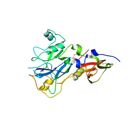
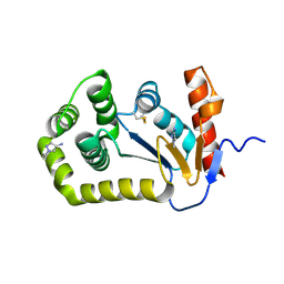
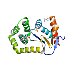
2JM2
 
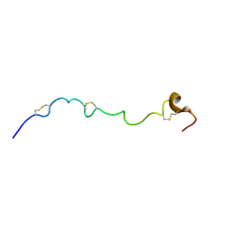 | | Structure of the N-terminal subdomain of insulin-like growth factor (IGF) binding protein-6 and its interactions with IGFs | | 分子名称: | Insulin-like growth factor-binding protein 6 | | 著者 | Chandrashekaran, I.R, Yao, S, Wang, C.C, Bansal, P.S, Alewood, P.F, Forbes, B.E, Wallace, J.C, Bach, L.A, Norton, R.S. | | 登録日 | 2006-09-18 | | 公開日 | 2007-03-27 | | 最終更新日 | 2023-12-20 | | 実験手法 | SOLUTION NMR | | 主引用文献 | The N-Terminal Subdomain of Insulin-like Growth Factor (IGF) Binding Protein 6. Structure and Interaction with IGFs
Biochemistry, 46, 2007
|
|
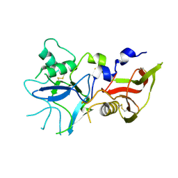
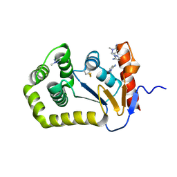
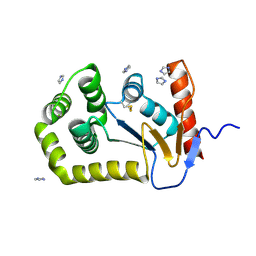
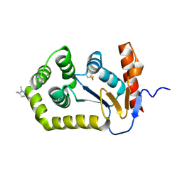
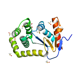
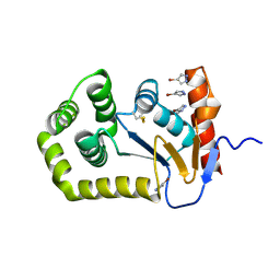
2N34
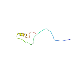 | | NMR assignments and solution structure of the JAK interaction region of SOCS5 | | 分子名称: | Suppressor of cytokine signaling 5 | | 著者 | Chandrashekaran, I.R, Mohanty, B, Linossi, E.M, Nicholson, S.E, Babon, J, Norton, R.S, Dagley, L.F, Leung, E.W.W, Murphy, J.M. | | 登録日 | 2015-05-21 | | 公開日 | 2015-07-29 | | 最終更新日 | 2024-05-15 | | 実験手法 | SOLUTION NMR | | 主引用文献 | Structure and Functional Characterization of the Conserved JAK Interaction Region in the Intrinsically Disordered N-Terminus of SOCS5.
Biochemistry, 54, 2015
|
|
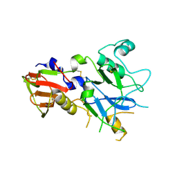
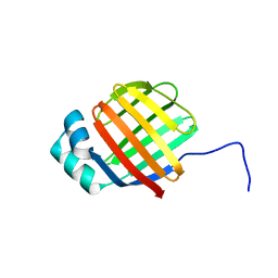
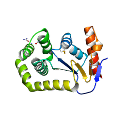
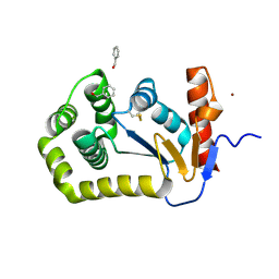
7N3K
 | | Oridonin-bound SARS-CoV-2 Nsp9 | | 分子名称: | (1beta,6beta,7beta,8alpha,9beta,10alpha,13alpha,14R,16beta)-1,6,7,14-tetrahydroxy-7,20-epoxykauran-15-one, Non-structural protein 9, SULFATE ION | | 著者 | Littler, D.R, Gully, B.S, Rossjohn, J. | | 登録日 | 2021-06-01 | | 公開日 | 2021-10-27 | | 最終更新日 | 2023-10-18 | | 実験手法 | X-RAY DIFFRACTION (3 Å) | | 主引用文献 | A natural product compound inhibits coronaviral replication in vitro by binding to the conserved Nsp9 SARS-CoV-2 protein.
J.Biol.Chem., 297, 2021
|
|
4R1C
 | |
4R19
 | |
4R1A
 | |
4R1B
 | |
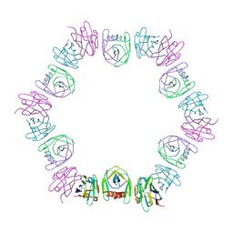
8DG1
 | |
8D11
 | |
8CXE
 | |
8D10
 | |
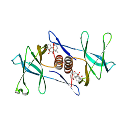
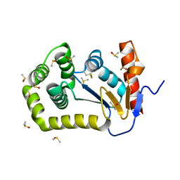
8DG2
 | | Crystal Structure of EcDsbA in a complex with DMSO | | 分子名称: | COPPER (II) ION, DIMETHYL SULFOXIDE, GLYCEROL, ... | | 著者 | Whitehouse, R.L, Ilyichova, O.V, Taylor, A.J. | | 登録日 | 2022-06-23 | | 公開日 | 2022-12-07 | | 最終更新日 | 2023-10-25 | | 実験手法 | X-RAY DIFFRACTION (1.95 Å) | | 主引用文献 | Fragment screening libraries for the identification of protein hot spots and their minimal binding pharmacophores.
Rsc Med Chem, 14, 2023
|
|
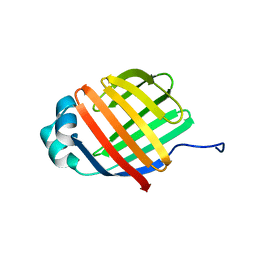
8DQU
 | | Nanobody bound SARS-CoV-2 Nsp9 | | 分子名称: | Nanobody, Non-structural protein 9 | | 著者 | Littler, D.R, Gully, B.S, Rossjohn, J. | | 登録日 | 2022-07-20 | | 公開日 | 2023-02-08 | | 最終更新日 | 2023-10-25 | | 実験手法 | X-RAY DIFFRACTION (2.45003176 Å) | | 主引用文献 | Inside-out: Antibody-binding reveals potential folding hinge-points within the SARS-CoV-2 replication co-factor nsp9.
Plos One, 18, 2023
|
|
6DO6
 | | NMR solution structure of wild type apo hFABP1 at 308 K | | 分子名称: | Fatty acid-binding protein, liver | | 著者 | Scanlon, M.J, Mohanty, B, Doak, B.C, Patil, R. | | 登録日 | 2018-06-09 | | 公開日 | 2018-12-26 | | 最終更新日 | 2024-05-01 | | 実験手法 | SOLUTION NMR | | 主引用文献 | A ligand-induced structural change in fatty acid-binding protein 1 is associated with potentiation of peroxisome proliferator-activated receptor alpha agonists.
J. Biol. Chem., 294, 2019
|
|
6DO7
 | | NMR solution structure of wild type hFABP1 with GW7647 | | 分子名称: | Fatty acid-binding protein, liver | | 著者 | Scanlon, M.J, Mohanty, B, Doak, B.C, Patil, R. | | 登録日 | 2018-06-09 | | 公開日 | 2019-01-02 | | 最終更新日 | 2024-05-01 | | 実験手法 | SOLUTION NMR | | 主引用文献 | A ligand-induced structural change in fatty acid-binding protein 1 is associated with potentiation of peroxisome proliferator-activated receptor alpha agonists.
J. Biol. Chem., 294, 2019
|
|
6DRG
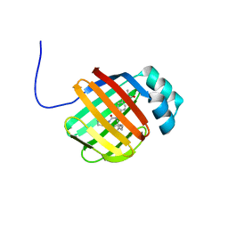 | | NMR solution structure of wild type hFABP1 with GW7647 | | 分子名称: | 2-[(4-{2-[(4-cyclohexylbutyl)(cyclohexylcarbamoyl)amino]ethyl}phenyl)sulfanyl]-2-methylpropanoic acid, Fatty acid-binding protein, liver | | 著者 | Scanlon, M.J, Mohanty, B, Doak, B.C, Patil, R. | | 登録日 | 2018-06-11 | | 公開日 | 2018-12-26 | | 最終更新日 | 2024-05-01 | | 実験手法 | SOLUTION NMR | | 主引用文献 | A ligand-induced structural change in fatty acid-binding protein 1 is associated with potentiation of peroxisome proliferator-activated receptor alpha agonists.
J. Biol. Chem., 294, 2019
|
|
8D12
 | |
8CZM
 | |
8DG0
 | |
8CXD
 | |
8CZN
 | |


