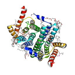
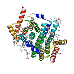
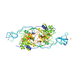
1IHC
 
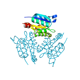 | | X-ray Structure of Gephyrin N-terminal Domain | | 分子名称: | Gephyrin | | 著者 | Sola, M, Kneussel, M, Heck, I.S, Betz, H, Weissenhorn, W. | | 登録日 | 2001-04-21 | | 公開日 | 2001-05-16 | | 最終更新日 | 2024-02-07 | | 実験手法 | X-RAY DIFFRACTION (1.9 Å) | | 主引用文献 | X-ray crystal structure of the trimeric N-terminal domain of gephyrin.
J.Biol.Chem., 276, 2001
|
|
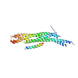
1EPU
 | | X-RAY crystal structure of neuronal SEC1 from squid | | 分子名称: | S-SEC1 | | 著者 | Bracher, A, Perrakis, A, Dresbach, T, Betz, H, Weissenhorn, W. | | 登録日 | 2000-03-29 | | 公開日 | 2000-08-09 | | 最終更新日 | 2017-10-04 | | 実験手法 | X-RAY DIFFRACTION (2.4 Å) | | 主引用文献 | The X-ray crystal structure of neuronal Sec1 from squid sheds new light on the role of this protein in exocytosis.
Structure Fold.Des., 8, 2000
|
|
1L4A
 | | X-RAY STRUCTURE OF THE NEURONAL COMPLEXIN/SNARE COMPLEX FROM THE SQUID LOLIGO PEALEI | | 分子名称: | S-SNAP25 fusion protein, S-SYNTAXIN, SYNAPHIN A, ... | | 著者 | Bracher, A, Kadlec, J, Betz, H, Weissenhorn, W. | | 登録日 | 2002-03-04 | | 公開日 | 2002-07-31 | | 最終更新日 | 2023-08-16 | | 実験手法 | X-RAY DIFFRACTION (2.95 Å) | | 主引用文献 | X-ray structure of a neuronal complexin-SNARE complex from squid.
J.Biol.Chem., 277, 2002
|
|
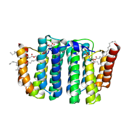
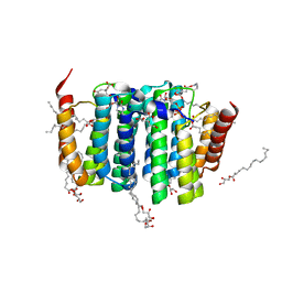
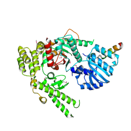
1KJT
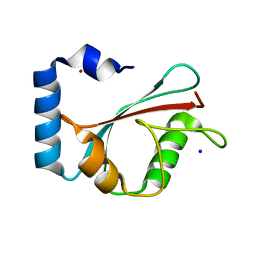 | | Crystal Structure of the GABA(A) Receptor Associated Protein, GABARAP | | 分子名称: | GABARAP, NICKEL (II) ION, SODIUM ION | | 著者 | Bavro, V.N, Sola, M, Bracher, A, Kneussel, M, Betz, H, Weissenhorn, W. | | 登録日 | 2001-12-05 | | 公開日 | 2002-04-24 | | 最終更新日 | 2024-02-14 | | 実験手法 | X-RAY DIFFRACTION (2 Å) | | 主引用文献 | Crystal structure of the GABA(A)-receptor-associated protein, GABARAP.
EMBO Rep., 3, 2002
|
|
1FVF
 | |
1FVH
 | |
7POW
 | | Crystal structure of phosphatidyl serine synthase (PSS) in transition state. | | 分子名称: | (2R)-2,3-dihydroxypropyl (9Z)-octadec-9-enoate, (2S)-2-amino-3-[[[(2R,3S,4R,5R)-5-(4-amino-2-oxo-pyrimidin-1-yl)-3,4-dihydroxy-tetrahydrofuran-2-yl]methoxy-hydroxy-phosphoryl]oxy-[(2R)-2,3-bis[[(Z)-octadec-9-enoyl]oxy]propoxy]-dihydroxy-lambda^5-phosphanyl]oxy-propanoic acid, 5'-O-[(R)-{[(S)-{(2R)-2,3-bis[(9E)-octadec-9-enoyloxy]propoxy}(hydroxy)phosphoryl]oxy}(hydroxy)phosphoryl]cytidine, ... | | 著者 | Yildiz, O, Centola, M. | | 登録日 | 2021-09-10 | | 公開日 | 2021-12-08 | | 最終更新日 | 2023-03-01 | | 実験手法 | X-RAY DIFFRACTION (2.51 Å) | | 主引用文献 | Crystal structures of phosphatidyl serine synthase PSS reveal the catalytic mechanism of CDP-DAG alcohol O-phosphatidyl transferases
Nat Commun, 12, 2021
|
|
7B1N
 | |
7B1K
 | | Crystal structure of phosphatidyl serine synthase (PSS) in the closed conformation with bound citrate. | | 分子名称: | (2R)-2,3-dihydroxypropyl (9Z)-octadec-9-enoate, 5'-O-[(R)-{[(S)-{(2R)-2,3-bis[(9E)-octadec-9-enoyloxy]propoxy}(hydroxy)phosphoryl]oxy}(hydroxy)phosphoryl]cytidine, CALCIUM ION, ... | | 著者 | Yildiz, O, Centola, M. | | 登録日 | 2020-11-25 | | 公開日 | 2021-12-08 | | 実験手法 | X-RAY DIFFRACTION (2.2 Å) | | 主引用文献 | Crystal structures of phosphatidyl serine synthase PSS reveal the catalytic mechanism of CDP-DAG alcohol O-phosphatidyl transferases
Nat Commun, 12, 2021
|
|
7B1L
 | | Crystal structure of phosphatidyl serine synthase (PSS) in the closed conformation with bound citrate. | | 分子名称: | (2R)-2,3-dihydroxypropyl (9Z)-octadec-9-enoate, 5'-O-[(R)-{[(S)-{(2R)-2,3-bis[(9E)-octadec-9-enoyloxy]propoxy}(hydroxy)phosphoryl]oxy}(hydroxy)phosphoryl]cytidine, CALCIUM ION, ... | | 著者 | Yildiz, O, Centola, M. | | 登録日 | 2020-11-25 | | 公開日 | 2021-12-08 | | 最終更新日 | 2024-10-09 | | 実験手法 | X-RAY DIFFRACTION (1.85 Å) | | 主引用文献 | Crystal structures of phosphatidyl serine synthase PSS reveal the catalytic mechanism of CDP-DAG alcohol O-phosphatidyl transferases
Nat Commun, 12, 2021
|
|
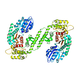
1T3E
 | | Structural basis of dynamic glycine receptor clustering | | 分子名称: | 49-mer fragment of Glycine receptor beta chain, Gephyrin, SULFATE ION | | 著者 | Sola, M, Bavro, V.N, Timmins, J, Franz, T, Ricard-Blum, S, Schoehn, G, Ruigrok, R.W.H, Paarmann, I, Saiyed, T, O'Sullivan, G.A. | | 登録日 | 2004-04-26 | | 公開日 | 2004-07-27 | | 最終更新日 | 2024-04-03 | | 実験手法 | X-RAY DIFFRACTION (3.25 Å) | | 主引用文献 | Structural basis of dynamic glycine receptor clustering by gephyrin
Embo J., 23, 2004
|
|
1MQS
 | |


