Loading
PDBj
メニューPDBj@FacebookPDBj@TwitterPDBj@YouTubewwPDB FoundationwwPDB
RCSB PDBPDBeBMRBAdv. SearchSearch help
Search by PDB author
5V3W
DownloadVisualize
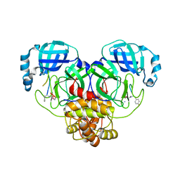
BU of 5v3w by Molmil
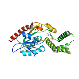
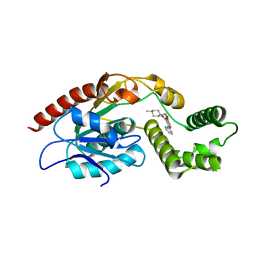
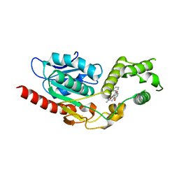
Crystal Structure of the Apo form of Thioesterase domain of Mtb Pks13
分子名称: (2R)-2-{[(2R)-2-{[(2S)-2-{[(2R)-2-hydroxypropyl]oxy}propyl]oxy}propyl]oxy}propan-1-ol, Polyketide synthase Pks13 (Termination polyketide synthase)
著者Aggarwal, A, Sacchettini, J.C.
登録日2017-03-08
公開日2017-07-05
最終更新日2023-10-04
実験手法X-RAY DIFFRACTION (1.723 Å)
主引用文献Development of a Novel Lead that Targets M. tuberculosis Polyketide Synthase 13.
Cell, 170, 2017
5V40
DownloadVisualize
BU of 5v40 by Molmil
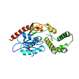
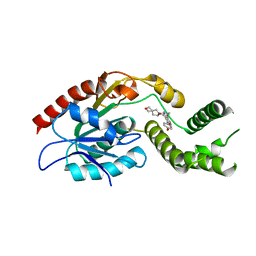
Crystal Structure of Mtb Pks13 Thioesterase domain in complex with inhibitor TAM6
分子名称: Polyketide synthase Pks13 (Termination polyketide synthase), ethyl 4-[(dimethylamino)methyl]-5-hydroxy-2-phenyl-1-benzofuran-3-carboxylate
著者Aggarwal, A, Sacchettini, J.C.
登録日2017-03-08
公開日2017-07-05
最終更新日2023-10-04
実験手法X-RAY DIFFRACTION (1.991 Å)
主引用文献Development of a Novel Lead that Targets M. tuberculosis Polyketide Synthase 13.
Cell, 170, 2017
5V42
DownloadVisualize
BU of 5v42 by Molmil
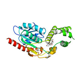
Crystal Structure of Mtb Pks13 Thioesterase domain in complex with inhibitor TAM3
分子名称: Polyketide synthase Pks13 (Termination polyketide synthase), ethyl 5-hydroxy-2-phenyl-4-(pyrrolidin-1-ylmethyl)-1-benzofuran-3-carboxylate
著者Aggarwal, A, Sacchettini, J.C.
登録日2017-03-08
公開日2017-07-05
最終更新日2023-10-04
実験手法X-RAY DIFFRACTION (1.987 Å)
主引用文献Development of a Novel Lead that Targets M. tuberculosis Polyketide Synthase 13.
Cell, 170, 2017
5V3Y
DownloadVisualize
BU of 5v3y by Molmil
Crystal Structure of Mtb Pks13 Thioesterase domain in complex with inhibitor TAM16
分子名称: 2-(4-hydroxyphenyl)-~{N}-methyl-5-oxidanyl-4-(piperidin-1-ylmethyl)-1-benzofuran-3-carboxamide, Polyketide synthase Pks13 (Termination polyketide synthase), SULFATE ION
著者Aggarwal, A, Sacchettini, J.C.
登録日2017-03-08
公開日2017-07-05
最終更新日2023-10-04
実験手法X-RAY DIFFRACTION (1.98 Å)
主引用文献Development of a Novel Lead that Targets M. tuberculosis Polyketide Synthase 13.
Cell, 170, 2017
5V41
DownloadVisualize
BU of 5v41 by Molmil
Crystal Structure of Mtb Pks13 Thioesterase domain in complex with inhibitor TAM5
分子名称: Polyketide synthase Pks13 (Termination polyketide synthase), ethyl 5-hydroxy-4-(morpholin-4-ylmethyl)-2-phenyl-1-benzofuran-3-carboxylate
著者Aggarwal, A, Sacchettini, J.C.
登録日2017-03-08
公開日2017-07-05
最終更新日2023-10-04
実験手法X-RAY DIFFRACTION (2.051 Å)
主引用文献Development of a Novel Lead that Targets M. tuberculosis Polyketide Synthase 13.
Cell, 170, 2017
5V3Z
DownloadVisualize
BU of 5v3z by Molmil
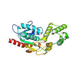
Crystal Structure of the D1607N mutant form of Thioesterase domain of Mtb Pks13
分子名称: PENTAETHYLENE GLYCOL, Polyketide synthase Pks13 (Termination polyketide synthase), SULFATE ION
著者Aggarwal, A, Sacchettini, J.C.
登録日2017-03-08
公開日2017-07-05
最終更新日2023-10-04
実験手法X-RAY DIFFRACTION (1.881 Å)
主引用文献Development of a Novel Lead that Targets M. tuberculosis Polyketide Synthase 13.
Cell, 170, 2017
5V3X
DownloadVisualize
BU of 5v3x by Molmil
Crystal Structure of Mtb Pks13 Thioesterase domain in complex with inhibitor TAM1
分子名称: Polyketide synthase Pks13 (Termination polyketide synthase), ethyl 5-hydroxy-4-[(4-methylpiperidin-1-yl)methyl]-2-phenyl-1-benzofuran-3-carboxylate
著者Aggarwal, A, Sacchettini, J.C.
登録日2017-03-08
公開日2017-07-05
最終更新日2023-10-04
実験手法X-RAY DIFFRACTION (1.936 Å)
主引用文献Development of a Novel Lead that Targets M. tuberculosis Polyketide Synthase 13.
Cell, 170, 2017
7M7V
DownloadVisualize
BU of 7m7v by Molmil
Crystal Structure of Mtb Pks13 Thioesterase domain in complex with Compound 6
分子名称: 5-hydroxy-2-(4-hydroxyphenyl)-N-methyl-4-[(2-oxa-6-azaspiro[3.4]octan-6-yl)methyl]-1-benzofuran-3-carboxamide, Polyketide synthase Pks13
著者Aggarwal, A, Sacchettini, J.C.
登録日2021-03-29
公開日2022-02-16
最終更新日2023-10-18
実験手法X-RAY DIFFRACTION (2.29 Å)
主引用文献Optimization of TAM16, a Benzofuran That Inhibits the Thioesterase Activity of Pks13; Evaluation toward a Preclinical Candidate for a Novel Antituberculosis Clinical Target.
J.Med.Chem., 65, 2022
2VU5
DownloadVisualize
BU of 2vu5 by Molmil
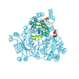
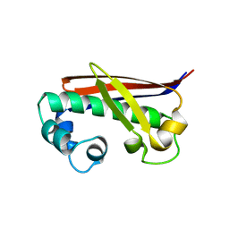
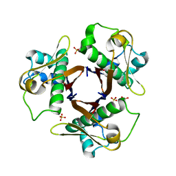
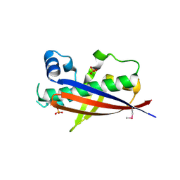
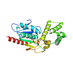
Crystal structure of Pndk from Bacillus anthracis
分子名称: NUCLEOSIDE DIPHOSPHATE KINASE
著者Misra, G, Aggarwal, A, Dube, D, Zaman, M.S, Singh, Y, Ramachandran, R.
登録日2008-05-21
公開日2009-03-10
最終更新日2024-05-08
実験手法X-RAY DIFFRACTION (2 Å)
主引用文献Crystal Structure of the Bacillus Anthracis Nucleoside Diphosphate Kinase and its Characterization Reveals an Enzyme Adapted to Perform Under Stress Conditions.
Proteins, 76, 2009
7M5A
DownloadVisualize
BU of 7m5a by Molmil
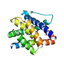
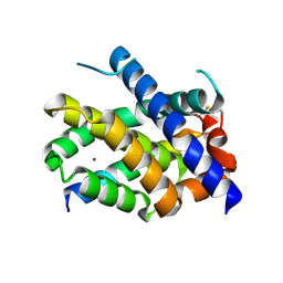
Crystal Structure of human BAK in complex with W3W5_BID
分子名称: BH3-interacting domain death agonist p15, Bcl-2 homologous antagonist/killer
著者Singh, G, Aggarwal, A, Moldoveanu, T.
登録日2021-03-23
公開日2022-01-12
最終更新日2024-10-09
実験手法X-RAY DIFFRACTION (1.5 Å)
主引用文献Structural basis of BAK activation in mitochondrial apoptosis initiation.
Nat Commun, 13, 2022
7M5B
DownloadVisualize
BU of 7m5b by Molmil
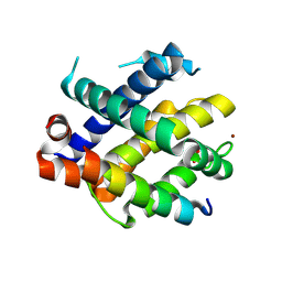
Crystal Structure of human BAK in complex with M3W5_BID
分子名称: BH3-interacting domain death agonist p15, Bcl-2 homologous antagonist/killer, COPPER (II) ION
著者Singh, G, Aggarwal, A, Moldoveanu, T.
登録日2021-03-23
公開日2022-01-12
最終更新日2024-10-23
実験手法X-RAY DIFFRACTION (1.85 Å)
主引用文献Structural basis of BAK activation in mitochondrial apoptosis initiation.
Nat Commun, 13, 2022
7M5C
DownloadVisualize
BU of 7m5c by Molmil
Crystal Structure of human BAK in complex with WT BAK BH3 peptide
分子名称: Bcl-2 homologous antagonist/killer, COPPER (II) ION, SULFATE ION
著者Singh, G, Aggarwal, A, Moldoveanu, T.
登録日2021-03-23
公開日2022-01-12
最終更新日2024-10-09
実験手法X-RAY DIFFRACTION (3.06 Å)
主引用文献Structural basis of BAK activation in mitochondrial apoptosis initiation.
Nat Commun, 13, 2022
6W32
DownloadVisualize
BU of 6w32 by Molmil
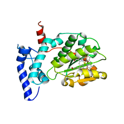
Crystal structure of Sfh5
分子名称: PROTOPORPHYRIN IX CONTAINING FE, Phosphatidylinositol transfer protein SFH5
著者Gulten, G, Khan, D, Aggarwal, A, Krieger, I, Sacchettini, J.C, Bankaitis, V.A.
登録日2020-03-08
公開日2020-11-25
最終更新日2024-03-06
実験手法X-RAY DIFFRACTION (2.9 Å)
主引用文献A Sec14-like phosphatidylinositol transfer protein paralog defines a novel class of heme-binding proteins.
Elife, 9, 2020
6UWA
DownloadVisualize
BU of 6uwa by Molmil
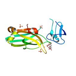
Mouse PKC C1B and C2 domains
分子名称: CITRIC ACID, LEAD (II) ION, Protein kinase C, ...
著者Aggarwal, A, Mire, J, Sacchettini, J.C, Igumenova, T.
登録日2019-11-04
公開日2020-12-09
最終更新日2024-03-06
実験手法X-RAY DIFFRACTION (1.2 Å)
主引用文献Mouse PKC C1B and C2 domains
To Be Published
2LWE
DownloadVisualize
BU of 2lwe by Molmil
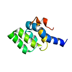
Solution structure of mutant (T170E) second CARD of human RIG-I
分子名称: Probable ATP-dependent RNA helicase DDX58
著者Dutta, K, Ferrage, F, Aggarwal, A.
登録日2012-07-27
公開日2012-10-31
最終更新日2024-05-01
実験手法SOLUTION NMR
主引用文献Structure and Dynamics of the Second CARD of Human RIG-I Provide Mechanistic Insights into Regulation of RIG-I Activation.
Structure, 20, 2012
2LWD
DownloadVisualize
BU of 2lwd by Molmil
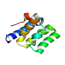
Solution structure of second CARD of human RIG-I
分子名称: Probable ATP-dependent RNA helicase DDX58
著者Dutta, K, Ferrage, F, Aggarwal, A.
登録日2012-07-27
公開日2012-10-31
最終更新日2024-05-01
実験手法SOLUTION NMR
主引用文献Structure and Dynamics of the Second CARD of Human RIG-I Provide Mechanistic Insights into Regulation of RIG-I Activation.
Structure, 20, 2012
1IF1
DownloadVisualize
BU of 1if1 by Molmil
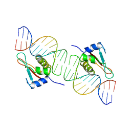
INTERFERON REGULATORY FACTOR 1 (IRF-1) COMPLEX WITH DNA
分子名称: DNA (26-MER), PROTEIN (INTERFERON REGULATORY FACTOR 1)
著者Escalante, C.R, Yie, J, Thanos, D, Aggarwal, A.
登録日1997-09-12
公開日1998-02-25
最終更新日2024-02-07
実験手法X-RAY DIFFRACTION (3 Å)
主引用文献Structure of IRF-1 with bound DNA reveals determinants of interferon regulation.
Nature, 391, 1998
8BON
DownloadVisualize
BU of 8bon by Molmil
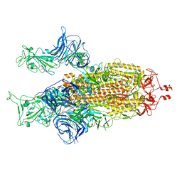
Structure of the SARS-CoV-2 spike glycoprotein in complex with the macrocyclic peptide S1B3inL1
分子名称: 2-acetamido-2-deoxy-beta-D-glucopyranose, 2-acetamido-2-deoxy-beta-D-glucopyranose-(1-4)-2-acetamido-2-deoxy-beta-D-glucopyranose, Macrocyclic peptide S1B3inL1, ...
著者Hurdiss, D.L.
登録日2022-11-15
公開日2023-06-28
最終更新日2023-07-05
実験手法ELECTRON MICROSCOPY (3.2 Å)
主引用文献A broad-spectrum macrocyclic peptide inhibitor of the SARS-CoV-2 spike protein.
Proc.Natl.Acad.Sci.USA, 120, 2023
3NE3
DownloadVisualize
BU of 3ne3 by Molmil
Mycobacterium tuberculosis Acyl Carrier Protein Synthase Apo structure
分子名称: Holo-[acyl-carrier-protein] synthase
著者Gokulan, K, Mycobacterium Tuberculosis Structural Proteomics Project (XMTB)
登録日2010-06-08
公開日2010-07-28
最終更新日2024-02-21
実験手法X-RAY DIFFRACTION (1.9 Å)
主引用文献Mycobacterium tuberculosis acyl carrier protein synthase adopts two different pH-dependent structural conformations.
Acta Crystallogr.,Sect.D, 67, 2011
3NE1
DownloadVisualize
BU of 3ne1 by Molmil
Mycobacterium tuberculosis Acyl Carrier Protein Synthase in complex with sulfate ion
分子名称: Holo-[acyl-carrier-protein] synthase, SULFATE ION
著者Gokulan, K.
登録日2010-06-08
公開日2010-07-21
最終更新日2024-02-21
実験手法X-RAY DIFFRACTION (2.51 Å)
主引用文献Mycobacterium tuberculosis acyl carrier protein synthase adopts two different pH-dependent structural conformations.
Acta Crystallogr.,Sect.D, 67, 2011
3NE9
DownloadVisualize
BU of 3ne9 by Molmil
Chronobacterium ammoiniagenes apo-ACPS structure
分子名称: Phosphopantetheine protein transferase, Ppt1p, SULFATE ION
著者Gokulan, K, Mycobacterium Tuberculosis Structural Proteomics Project (XMTB)
登録日2010-06-08
公開日2010-08-25
最終更新日2020-09-09
実験手法X-RAY DIFFRACTION (2.5 Å)
主引用文献Mycobacterium tuberculosis acyl carrier protein synthase adopts two different pH-dependent structural conformations.
Acta Crystallogr.,Sect.D, 67, 2011
7E9Y
DownloadVisualize
BU of 7e9y by Molmil
Crystal structure of eLACCO1
分子名称: (2S)-2-HYDROXYPROPANOIC ACID, CALCIUM ION, Lactate-binding periplasmic protein TTHA0766,Lactate-binding periplasmic protein TTHA0766
著者Wen, Y, Campbell, R.E, Lemieux, M.J, Nasu, Y.
登録日2021-03-05
公開日2021-12-22
最終更新日2023-11-29
実験手法X-RAY DIFFRACTION (2.25 Å)
主引用文献A genetically encoded fluorescent biosensor for extracellular L-lactate.
Nat Commun, 12, 2021
7RNW
DownloadVisualize
BU of 7rnw by Molmil
SARS-CoV-2 Main Protease in complex with a cyclic peptide inhibitor
分子名称: 3C-like proteinase, ACE-DTY-LEU-GLN-TYR-ALA-VAL-LEU-ARG-HIS-LYS-ARG-ARG-GLU-SEC
著者Frkic, R.L, Jackson, C.J.
登録日2021-07-30
公開日2022-03-16
最終更新日2023-10-18
実験手法X-RAY DIFFRACTION (2.35 Å)
主引用文献Antiviral cyclic peptides targeting the main protease of SARS-CoV-2.
Chem Sci, 13, 2022
7VCM
DownloadVisualize
BU of 7vcm by Molmil
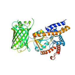
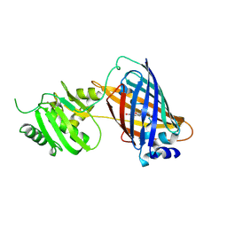
crystal structure of GINKO1
分子名称: Green fluorescent protein,Potassium binding protein Kbp,Green fluorescent protein, POTASSIUM ION
著者Wen, Y, Campbell, R.E, Lemieux, M.J.
登録日2021-09-03
公開日2022-07-27
最終更新日2024-11-06
実験手法X-RAY DIFFRACTION (1.85 Å)
主引用文献A sensitive and specific genetically-encoded potassium ion biosensor for in vivo applications across the tree of life.
Plos Biol., 20, 2022
8TOQ
DownloadVisualize
BU of 8toq by Molmil
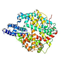
ACE2-peptide 1 complex
分子名称: 2-acetamido-2-deoxy-beta-D-glucopyranose, Angiotensin-converting enzyme 2, CHLORIDE ION, ...
著者Christie, M, Payne, R.J.
登録日2023-08-03
公開日2024-02-07
実験手法X-RAY DIFFRACTION (2.3 Å)
主引用文献Discovery of High Affinity Cyclic Peptide Ligands for Human ACE2 with SARS-CoV-2 Entry Inhibitory Activity.
Acs Chem.Biol., 19, 2024


12>

227111

件を2024-11-06に公開中

PDB statisticsPDBj update infoContact PDBjnumon