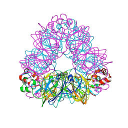
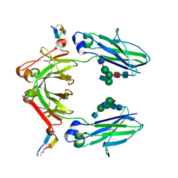
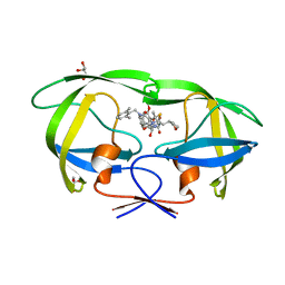
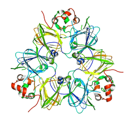
6A28
 
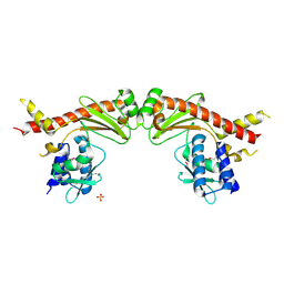 | | Crystal structure of PprA W183R mutant form 2 | | 分子名称: | DNA repair protein PprA, SULFATE ION | | 著者 | Adachi, M, Shibazaki, C, Shimizu, R, Arai, S, Satoh, K, Narumi, I, Kuroki, R. | | 登録日 | 2018-06-09 | | 公開日 | 2018-12-26 | | 最終更新日 | 2023-11-22 | | 実験手法 | X-RAY DIFFRACTION (2.193 Å) | | 主引用文献 | Extended structure of pleiotropic DNA repair-promoting protein PprA from Deinococcus radiodurans.
FASEB J., 33, 2019
|
|
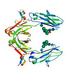
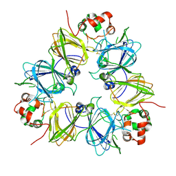
6A27
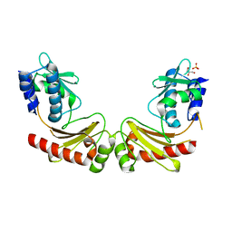 | | Crystal structure of PprA W183R mutant form 1 | | 分子名称: | DNA repair protein PprA, GLYCEROL, SULFATE ION | | 著者 | Adachi, M, Shibazaki, C, Shimizu, R, Arai, S, Satoh, K, Narumi, I, Kuroki, R. | | 登録日 | 2018-06-09 | | 公開日 | 2018-12-26 | | 最終更新日 | 2023-11-22 | | 実験手法 | X-RAY DIFFRACTION (1.353 Å) | | 主引用文献 | Extended structure of pleiotropic DNA repair-promoting protein PprA from Deinococcus radiodurans.
FASEB J., 33, 2019
|
|
6A29
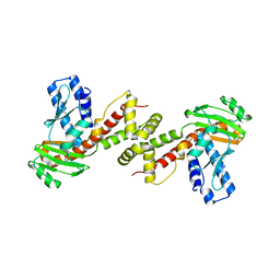 | | Crystal structure of PprA A139R mutant | | 分子名称: | DNA repair protein PprA | | 著者 | Adachi, M, Shibazaki, C, Shimizu, R, Arai, S, Satoh, K, Narumi, I, Kuroki, R. | | 登録日 | 2018-06-09 | | 公開日 | 2018-12-26 | | 最終更新日 | 2024-03-27 | | 実験手法 | X-RAY DIFFRACTION (2.399 Å) | | 主引用文献 | Extended structure of pleiotropic DNA repair-promoting protein PprA from Deinococcus radiodurans.
FASEB J., 33, 2019
|
|
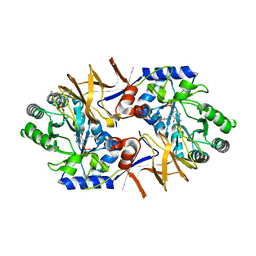
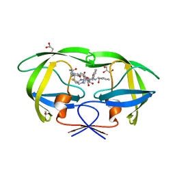
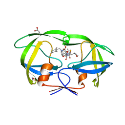
1BFN
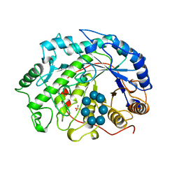 | | BETA-AMYLASE/BETA-CYCLODEXTRIN COMPLEX | | 分子名称: | BETA-AMYLASE, Cycloheptakis-(1-4)-(alpha-D-glucopyranose), SULFATE ION | | 著者 | Adachi, M, Mikami, B, Katsube, T, Utsumi, S. | | 登録日 | 1998-05-22 | | 公開日 | 1998-10-28 | | 最終更新日 | 2024-02-07 | | 実験手法 | X-RAY DIFFRACTION (2.07 Å) | | 主引用文献 | Crystal structure of recombinant soybean beta-amylase complexed with beta-cyclodextrin.
J.Biol.Chem., 273, 1998
|
|
3FX5
 | | Structure of HIV-1 Protease in Complex with Potent Inhibitor KNI-272 Determined by High Resolution X-ray Crystallography | | 分子名称: | (4R)-N-tert-butyl-3-[(2S,3S)-2-hydroxy-3-({N-[(isoquinolin-5-yloxy)acetyl]-S-methyl-L-cysteinyl}amino)-4-phenylbutanoyl]-1,3-thiazolidine-4-carboxamide, GLYCEROL, protease | | 著者 | Adachi, M, Ohhara, T, Tamada, T, Okazaki, N, Kuroki, R. | | 登録日 | 2009-01-20 | | 公開日 | 2009-03-24 | | 最終更新日 | 2023-11-01 | | 実験手法 | X-RAY DIFFRACTION (0.93 Å) | | 主引用文献 | Structure of HIV-1 protease in complex with potent inhibitor KNI-272 determined by high-resolution X-ray and neutron crystallography.
Proc.Natl.Acad.Sci.USA, 2009
|
|
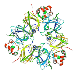
1OD5
 | | Crystal structure of glycinin A3B4 subunit homohexamer | | 分子名称: | CARBONATE ION, GLYCININ, MAGNESIUM ION | | 著者 | Adachi, M, Kanamori, J, Masuda, T, Yagasaki, K, Kitamura, K, Mikami, B, Utsumi, S. | | 登録日 | 2003-02-13 | | 公開日 | 2003-06-02 | | 最終更新日 | 2023-12-13 | | 実験手法 | X-RAY DIFFRACTION (2.1 Å) | | 主引用文献 | Crystal Structure of Soybean 11S Globulin: Glycinin A3B4 Homohexamer
Proc.Natl.Acad.Sci.USA, 100, 2003
|
|
2ZYE
 | | Structure of HIV-1 Protease in Complex with Potent Inhibitor KNI-272 Determined by Neutron Crystallography | | 分子名称: | (4R)-N-tert-butyl-3-[(2S,3S)-2-hydroxy-3-({N-[(isoquinolin-5-yloxy)acetyl]-S-methyl-L-cysteinyl}amino)-4-phenylbutanoyl]-1,3-thiazolidine-4-carboxamide, protease | | 著者 | Adachi, M, Ohhara, T, Tamada, T, Okazaki, N, Kuroki, R. | | 登録日 | 2009-01-20 | | 公開日 | 2009-03-24 | | 最終更新日 | 2024-05-29 | | 実験手法 | NEUTRON DIFFRACTION (1.9 Å) | | 主引用文献 | Structure of HIV-1 protease in complex with potent inhibitor KNI-272 determined by high-resolution X-ray and neutron crystallography.
Proc.Natl.Acad.Sci.USA, 2009
|
|
1FXZ
 | | CRYSTAL STRUCTURE OF SOYBEAN PROGLYCININ A1AB1B HOMOTRIMER | | 分子名称: | GLYCININ G1 | | 著者 | Adachi, M, Takenaka, Y, Gidamis, A.B, Mikami, B, Utsumi, S. | | 登録日 | 2000-09-28 | | 公開日 | 2001-10-03 | | 最終更新日 | 2011-07-13 | | 実験手法 | X-RAY DIFFRACTION (2.8 Å) | | 主引用文献 | Crystal structure of soybean proglycinin A1aB1b homotrimer.
J.Mol.Biol., 305, 2001
|
|
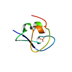
5ZL6
 | | Histidine Racemase from Leuconostoc mesenteroides subsp. sake NBRC 102480 | | 分子名称: | Histidine racemase, PYRIDOXAL-5'-PHOSPHATE | | 著者 | Adachi, M, Shimizu, R, Oikawa, T. | | 登録日 | 2018-03-27 | | 公開日 | 2018-11-28 | | 最終更新日 | 2023-11-22 | | 実験手法 | X-RAY DIFFRACTION (2.1 Å) | | 主引用文献 | The first identification and characterization of a histidine-specific amino acid racemase, histidine racemase from a lactic acid bacterium, Leuconostoc mesenteroides subsp. sake NBRC 102480.
Amino Acids, 51, 2019
|
|
8GOS
 | |
6IQG
 | | X-ray crystal structure of Fc and peptide complex | | 分子名称: | 18-mer peptide G(HCS)DCAYHRGELVWCT(HCS)H(NH2), 2-acetamido-2-deoxy-beta-D-glucopyranose-(1-2)-alpha-D-mannopyranose-(1-3)-[2-acetamido-2-deoxy-beta-D-glucopyranose-(1-2)-alpha-D-mannopyranose-(1-6)]beta-D-mannopyranose-(1-4)-2-acetamido-2-deoxy-beta-D-glucopyranose-(1-4)-[alpha-L-fucopyranose-(1-6)]2-acetamido-2-deoxy-beta-D-glucopyranose, CHLORIDE ION, ... | | 著者 | Adachi, M, Ito, Y. | | 登録日 | 2018-11-08 | | 公開日 | 2019-02-13 | | 最終更新日 | 2024-03-13 | | 実験手法 | X-RAY DIFFRACTION (2.998 Å) | | 主引用文献 | Site-Specific Chemical Conjugation of Antibodies by Using Affinity Peptide for the Development of Therapeutic Antibody Format.
Bioconjug. Chem., 30, 2019
|
|
6IQH
 | | X-ray crystal structure of covalent-bonded complex of Fc and peptide | | 分子名称: | 17-mer peptide (GPDCAYHKGELVWCTFH), 2-acetamido-2-deoxy-beta-D-glucopyranose-(1-2)-alpha-D-mannopyranose-(1-3)-[2-acetamido-2-deoxy-beta-D-glucopyranose-(1-2)-alpha-D-mannopyranose-(1-6)]beta-D-mannopyranose-(1-4)-2-acetamido-2-deoxy-beta-D-glucopyranose-(1-4)-[alpha-L-fucopyranose-(1-6)]2-acetamido-2-deoxy-beta-D-glucopyranose, Immunoglobulin gamma-1 heavy chain | | 著者 | Adachi, M, Ito, Y. | | 登録日 | 2018-11-08 | | 公開日 | 2019-02-13 | | 最終更新日 | 2024-01-24 | | 実験手法 | X-RAY DIFFRACTION (2.999 Å) | | 主引用文献 | Site-Specific Chemical Conjugation of Antibodies by Using Affinity Peptide for the Development of Therapeutic Antibody Format.
Bioconjug. Chem., 30, 2019
|
|
6IXD
 | | X-ray crystal structure of bPI-11 hiv-1 protease complex | | 分子名称: | (4R)-3-[(2S,3S)-3-[2-[4-[5-[(3aS,4S,6aR)-2-oxidanylidene-1,3,3a,4,6,6a-hexahydrothieno[3,4-d]imidazol-4-yl]pentanoylamino]-2,6-dimethyl-phenoxy]ethanoylamino]-2-oxidanyl-4-phenyl-butanoyl]-5,5-dimethyl-N-[(1S,2R)-2-oxidanyl-2,3-dihydro-1H-inden-1-yl]-1,3-thiazolidine-4-carboxamide, CHLORIDE ION, GLYCEROL, ... | | 著者 | Adachi, M, Hidaka, K. | | 登録日 | 2018-12-10 | | 公開日 | 2019-05-22 | | 最終更新日 | 2023-11-22 | | 実験手法 | X-RAY DIFFRACTION (1 Å) | | 主引用文献 | Acquired Removability of Aspartic Protease Inhibitors by Direct Biotinylation.
Bioconjug.Chem., 30, 2019
|
|
6L27
 | | X-ray crystal structure of the mutant green fluorescent protein | | 分子名称: | Green fluorescent protein | | 著者 | Adachi, M, Shimizu, R, Shibazaki, C, Kagotani, Y, Ostermann, A, Schrader, T.E. | | 登録日 | 2019-10-02 | | 公開日 | 2020-04-01 | | 最終更新日 | 2023-11-15 | | 実験手法 | X-RAY DIFFRACTION (0.77 Å) | | 主引用文献 | Direct Observation of the Protonation States in the Mutant Green Fluorescent Protein.
J Phys Chem Lett, 11, 2020
|
|
6L26
 | | Neutron crystal structure of the mutant green fluorescent protein (EGFP) | | 分子名称: | Green fluorescent protein, trideuteriooxidanium | | 著者 | Adachi, M, Shimizu, R, Shibazaki, C, Kagotani, Y, Ostermann, A, Schrader, T.E. | | 登録日 | 2019-10-02 | | 公開日 | 2020-04-08 | | 最終更新日 | 2023-11-22 | | 実験手法 | NEUTRON DIFFRACTION (1.444 Å) | | 主引用文献 | Direct Observation of the Protonation States in the Mutant Green Fluorescent Protein.
J Phys Chem Lett, 11, 2020
|
|
5YOJ
 | | Structure of A17 HIV-1 Protease in Complex with Inhibitor KNI-1657 | | 分子名称: | (4R)-N-[(2,6-dimethylphenyl)methyl]-3-[(2S,3S)-3-[[(2S)-2-[(7-methoxy-1-benzofuran-2-yl)carbonylamino]-2-[(3R)-oxolan-3 -yl]ethanoyl]amino]-2-oxidanyl-4-phenyl-butanoyl]-5,5-dimethyl-1,3-thiazolidine-4-carboxamide, A17 HIV-1 protease, GLYCEROL | | 著者 | Adachi, M, Hidaka, K, Kuroki, R, Kiso, Y. | | 登録日 | 2017-10-29 | | 公開日 | 2018-07-11 | | 最終更新日 | 2024-03-27 | | 実験手法 | X-RAY DIFFRACTION (1.5 Å) | | 主引用文献 | Identification of Highly Potent Human Immunodeficiency Virus Type-1 Protease Inhibitors against Lopinavir and Darunavir Resistant Viruses from Allophenylnorstatine-Based Peptidomimetics with P2 Tetrahydrofuranylglycine.
J. Med. Chem., 61, 2018
|
|
5YOK
 | | Structure of HIV-1 Protease in Complex with Inhibitor KNI-1657 | | 分子名称: | (4R)-N-[(2,6-dimethylphenyl)methyl]-3-[(2S,3S)-3-[[(2S)-2-[(7-methoxy-1-benzofuran-2-yl)carbonylamino]-2-[(3R)-oxolan-3 -yl]ethanoyl]amino]-2-oxidanyl-4-phenyl-butanoyl]-5,5-dimethyl-1,3-thiazolidine-4-carboxamide, GLYCEROL, HIV-1 PROTEASE | | 著者 | Adachi, M, Hidaka, K, Kuroki, R, Kiso, Y. | | 登録日 | 2017-10-29 | | 公開日 | 2018-07-18 | | 最終更新日 | 2024-03-27 | | 実験手法 | X-RAY DIFFRACTION (0.85 Å) | | 主引用文献 | Identification of Highly Potent Human Immunodeficiency Virus Type-1 Protease Inhibitors against Lopinavir and Darunavir Resistant Viruses from Allophenylnorstatine-Based Peptidomimetics with P2 Tetrahydrofuranylglycine.
J. Med. Chem., 61, 2018
|
|
5XQN
 | | Crystal structure of Notched-fin eelpout type III antifreeze protein (NFE6, AFP), C2221 form. | | 分子名称: | Ice-structuring protein, SULFATE ION | | 著者 | Adachi, M, Kondo, H, Tsuda, S. | | 登録日 | 2017-06-07 | | 公開日 | 2018-05-16 | | 最終更新日 | 2024-03-27 | | 実験手法 | X-RAY DIFFRACTION (1.19 Å) | | 主引用文献 | Polypentagonal ice-like water networks emerge solely in an activity-improved variant of ice-binding protein
Proc. Natl. Acad. Sci. U.S.A., 115, 2018
|
|
5XQR
 | | Crystal structure of Notched-fin eelpout type III antifreeze protein A20V mutant (NFE6, AFP), C2221 form | | 分子名称: | ACETATE ION, Ice-structuring protein | | 著者 | Adachi, M, Shimizu, R, Shibazaki, C, Kondo, H, Tsuda, S. | | 登録日 | 2017-06-07 | | 公開日 | 2018-05-16 | | 最終更新日 | 2023-11-22 | | 実験手法 | X-RAY DIFFRACTION (1.3 Å) | | 主引用文献 | Polypentagonal ice-like water networks emerge solely in an activity-improved variant of ice-binding protein
Proc. Natl. Acad. Sci. U.S.A., 115, 2018
|
|
5XQU
 | | Crystal structure of Notched-fin eelpout type III antifreeze protein A20I mutant (NFE6, AFP), P212121 form | | 分子名称: | Ice-structuring protein | | 著者 | Adachi, M, Shimizu, R, Shibazaki, C, Kondo, H, Tsuda, S. | | 登録日 | 2017-06-07 | | 公開日 | 2018-05-16 | | 最終更新日 | 2023-11-22 | | 実験手法 | X-RAY DIFFRACTION (1 Å) | | 主引用文献 | Polypentagonal ice-like water networks emerge solely in an activity-improved variant of ice-binding protein
Proc. Natl. Acad. Sci. U.S.A., 115, 2018
|
|
5XQV
 | | Crystal structure of Notched-fin eelpout type III antifreeze protein A20L mutant (NFE6, AFP), P21 form | | 分子名称: | Ice-structuring protein | | 著者 | Adachi, M, Shimizu, R, Shibazaki, C, Kondo, H, Tsuda, S. | | 登録日 | 2017-06-07 | | 公開日 | 2018-05-16 | | 最終更新日 | 2023-11-22 | | 実験手法 | X-RAY DIFFRACTION (0.97 Å) | | 主引用文献 | Polypentagonal ice-like water networks emerge solely in an activity-improved variant of ice-binding protein
Proc. Natl. Acad. Sci. U.S.A., 115, 2018
|
|
5XR0
 | | Crystal structure of Notched-fin eelpout type III antifreeze protein A20T mutant (NFE6, AFP), P21 form | | 分子名称: | Ice-structuring protein | | 著者 | Adachi, M, Shimizu, R, Shibazaki, C, Kondo, H, Tsuda, S. | | 登録日 | 2017-06-07 | | 公開日 | 2018-05-16 | | 最終更新日 | 2023-11-22 | | 実験手法 | X-RAY DIFFRACTION (0.98 Å) | | 主引用文献 | Polypentagonal ice-like water networks emerge solely in an activity-improved variant of ice-binding protein
Proc. Natl. Acad. Sci. U.S.A., 115, 2018
|
|
5XQP
 | | Crystal structure of Notched-fin eelpout type III antifreeze protein (NFE6, AFP), P212121 form | | 分子名称: | Ice-structuring protein, SULFATE ION | | 著者 | Adachi, M, Shimizu, R, Shibazaki, C, Kondo, H, Tsuda, S. | | 登録日 | 2017-06-07 | | 公開日 | 2018-05-16 | | 最終更新日 | 2023-11-22 | | 実験手法 | X-RAY DIFFRACTION (1 Å) | | 主引用文献 | Polypentagonal ice-like water networks emerge solely in an activity-improved variant of ice-binding protein
Proc. Natl. Acad. Sci. U.S.A., 115, 2018
|
|
1UD1
 | |
1UCX
 | |


