1AUA
 
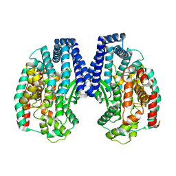
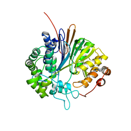
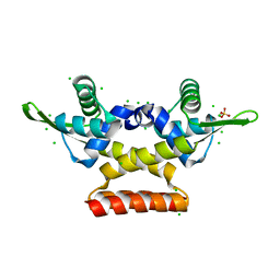
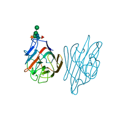
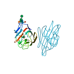
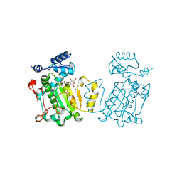 | | PHOSPHATIDYLINOSITOL TRANSFER PROTEIN SEC14P FROM SACCHAROMYCES CEREVISIAE | | 分子名称: | PHOSPHATIDYLINOSITOL TRANSFER PROTEIN SEC14P, octyl beta-D-glucopyranoside | | 著者 | Sha, B, Phillips, S.E, Bankaitis, V.A, Luo, M. | | 登録日 | 1997-08-20 | | 公開日 | 1997-12-24 | | 最終更新日 | 2024-02-07 | | 実験手法 | X-RAY DIFFRACTION (2.5 Å) | | 主引用文献 | Crystal structure of the Saccharomyces cerevisiae phosphatidylinositol-transfer protein.
Nature, 391, 1998
|
|
1C3G
 | |
1AA7
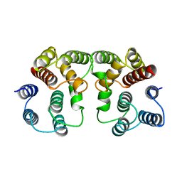 | |
1HHO
 | |
8BEO
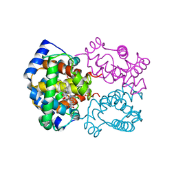
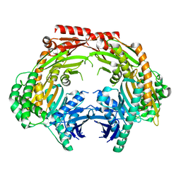
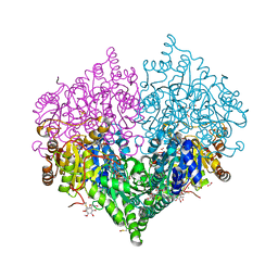 | | Crystal structure of E. coli glyoxylate carboligase mutant I393A with MAP | | 分子名称: | (2R,3S)-1,4-DIMERCAPTOBUTANE-2,3-DIOL, 2,3-DIHYDROXY-1,4-DITHIOBUTANE, 2,3-DIMETHOXY-5-METHYL-1,4-BENZOQUINONE, ... | | 著者 | Shaanan, B, Binshtein, E. | | 登録日 | 2022-10-21 | | 公開日 | 2023-11-08 | | 最終更新日 | 2023-11-15 | | 実験手法 | X-RAY DIFFRACTION (1.96 Å) | | 主引用文献 | Crystal structure of E. coli glyoxylate carboligase mutant I393A with MAP
To Be Published
|
|
5GIV
 | | Crystal structure of M32 carboxypeptidase from Deinococcus radiodurans R1 | | 分子名称: | ACETATE ION, Carboxypeptidase 1, ZINC ION | | 著者 | Sharma, B, Singh, R, Yadav, P, Ghosh, B, Kumar, A, Jamdar, S.N, Makde, R.D. | | 登録日 | 2016-06-25 | | 公開日 | 2017-07-12 | | 最終更新日 | 2023-11-08 | | 実験手法 | X-RAY DIFFRACTION (2.4 Å) | | 主引用文献 | Active site gate of M32 carboxypeptidases illuminated by crystal structure and molecular dynamics simulations
Biochim. Biophys. Acta, 1865, 2017
|
|
1IOB
 | |
1LTE
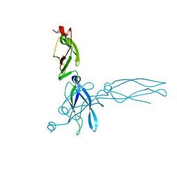
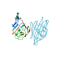
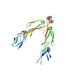
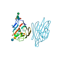 | | STRUCTURE OF A LEGUME LECTIN WITH AN ORDERED N-LINKED CARBOHYDRATE IN COMPLEX WITH LACTOSE | | 分子名称: | CALCIUM ION, CORAL TREE LECTIN, MANGANESE (II) ION, ... | | 著者 | Shaanan, B, Lis, H, Sharon, N. | | 登録日 | 1991-06-25 | | 公開日 | 1994-01-31 | | 最終更新日 | 2020-07-29 | | 実験手法 | X-RAY DIFFRACTION (2 Å) | | 主引用文献 | Structure of a legume lectin with an ordered N-linked carbohydrate in complex with lactose.
Science, 254, 1991
|
|
2V36
 | | Crystal structure of gamma-glutamyl transferase from Bacillus subtilis | | 分子名称: | GAMMA-GLUTAMYLTRANSPEPTIDASE LARGE CHAIN, GAMMA-GLUTAMYLTRANSPEPTIDASE SMALL CHAIN | | 著者 | Sharath, B, Prabhune, A.A, Suresh, C.G, Wilkinson, A.J, Brannigan, J.A. | | 登録日 | 2007-06-13 | | 公開日 | 2008-07-01 | | 最終更新日 | 2023-12-13 | | 実験手法 | X-RAY DIFFRACTION (1.85 Å) | | 主引用文献 | Crystal Structure of Gamma-Glutamyl Transferase
To be Published
|
|
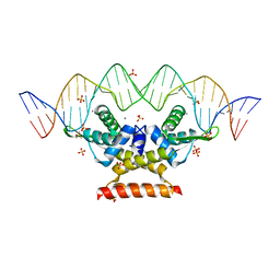
6QH0
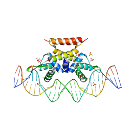 | | The complex structure of hsRosR-S5 (VNG0258H/RosR-S5) | | 分子名称: | DNA (28-MER), MANGANESE (II) ION, SULFATE ION, ... | | 著者 | Shaanan, B, Kutnowski, N. | | 登録日 | 2019-01-14 | | 公開日 | 2019-07-10 | | 最終更新日 | 2024-01-24 | | 実験手法 | X-RAY DIFFRACTION (2.436 Å) | | 主引用文献 | Specificity of protein-DNA interactions in hypersaline environment: structural studies on complexes of Halobacterium salinarum oxidative stress-dependent protein hsRosR.
Nucleic Acids Res., 47, 2019
|
|
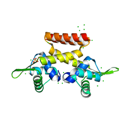
6QIL
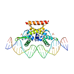 | | The complex structure of hsRosR-S1 (VNG0258H/RosR-S1) | | 分子名称: | 2-(N-MORPHOLINO)-ETHANESULFONIC ACID, DNA (28-MER), DNA binding protein, ... | | 著者 | Shaanan, B, Kutnowski, N. | | 登録日 | 2019-01-21 | | 公開日 | 2019-07-10 | | 最終更新日 | 2024-01-24 | | 実験手法 | X-RAY DIFFRACTION (2 Å) | | 主引用文献 | Specificity of protein-DNA interactions in hypersaline environment: structural studies on complexes of Halobacterium salinarum oxidative stress-dependent protein hsRosR.
Nucleic Acids Res., 47, 2019
|
|
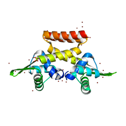
6QUA
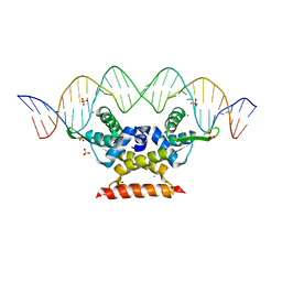 | | The complex structure of hsRosR-SG (vng0258/RosR-SG) | | 分子名称: | DNA (28-MER), MANGANESE (II) ION, SULFATE ION, ... | | 著者 | Shaanan, B, Kutnowski, N. | | 登録日 | 2019-02-27 | | 公開日 | 2019-07-10 | | 最終更新日 | 2024-01-24 | | 実験手法 | X-RAY DIFFRACTION (2.681 Å) | | 主引用文献 | Specificity of protein-DNA interactions in hypersaline environment: structural studies on complexes of Halobacterium salinarum oxidative stress-dependent protein hsRosR.
Nucleic Acids Res., 47, 2019
|
|
6QFD
 | | The complex structure of hsRosR-S4 (vng0258/RosR-S4) | | 分子名称: | DNA (28-MER), DNA-binding protein, MANGANESE (II) ION, ... | | 著者 | Shaanan, B, Kutnowski, N. | | 登録日 | 2019-01-10 | | 公開日 | 2019-07-10 | | 最終更新日 | 2024-01-24 | | 実験手法 | X-RAY DIFFRACTION (2.133 Å) | | 主引用文献 | Specificity of protein-DNA interactions in hypersaline environment: structural studies on complexes of Halobacterium salinarum oxidative stress-dependent protein hsRosR.
Nucleic Acids Res., 47, 2019
|
|
5ZWT
 | |
6F5C
 | |
6EZ1
 | |
6FDH
 | |
6FAQ
 | |
1AX0
 | |
1AXZ
 | |
1AX2
 | |
1AXY
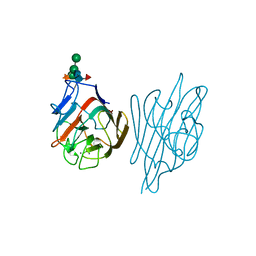
 | | ERYTHRINA CORALLODENDRON LECTIN | | 分子名称: | CALCIUM ION, LECTIN, MANGANESE (II) ION, ... | | 著者 | Shaanan, B, Elgavish, S. | | 登録日 | 1997-10-24 | | 公開日 | 1998-05-06 | | 最終更新日 | 2023-08-02 | | 実験手法 | X-RAY DIFFRACTION (1.95 Å) | | 主引用文献 | Structures of the Erythrina corallodendron lectin and of its complexes with mono- and disaccharides.
J.Mol.Biol., 277, 1998
|
|
1AX1
 | |
5ZF6
 | | Crystal structure of the dimeric human PNPase | | 分子名称: | Polyribonucleotide nucleotidyltransferase 1, mitochondrial | | 著者 | Yuan, H.S, Golzarroshan, B. | | 登録日 | 2018-03-02 | | 公開日 | 2018-08-22 | | 最終更新日 | 2023-11-22 | | 実験手法 | X-RAY DIFFRACTION (2.796 Å) | | 主引用文献 | Crystal structure of dimeric human PNPase reveals why disease-linked mutants suffer from low RNA import and degradation activities.
Nucleic Acids Res., 46, 2018
|
|
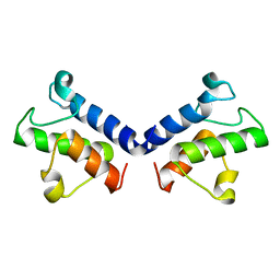
2B26
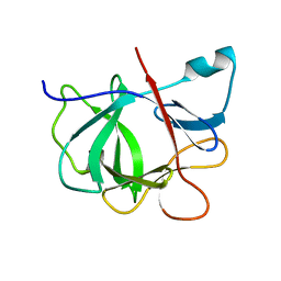
 | | The crystal structure of the protein complex of yeast Hsp40 Sis1 and Hsp70 Ssa1 | | 分子名称: | Heat shock 70 kDa protein cognate 2, SIS1 protein | | 著者 | Li, J, Wu, Y, Qian, X, Sha, B. | | 登録日 | 2005-09-16 | | 公開日 | 2006-09-19 | | 最終更新日 | 2024-02-14 | | 実験手法 | X-RAY DIFFRACTION (3.2 Å) | | 主引用文献 | Crystal structure of yeast Sis1 peptide-binding fragment and Hsp70 Ssa1 C-terminal complex.
Biochem.J., 398, 2006
|
|


