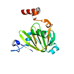
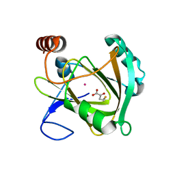
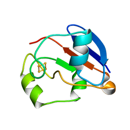
1PDX
 
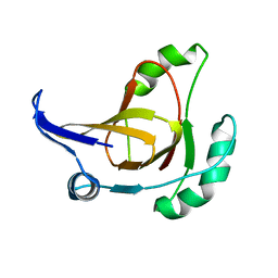
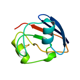 | | PUTIDAREDOXIN | | 分子名称: | FE2/S2 (INORGANIC) CLUSTER, PROTEIN (PUTIDAREDOXIN) | | 著者 | Pochapsky, T.C, Jain, N.U, Kuti, M, Lyons, T.A, Heymont, J. | | 登録日 | 1999-02-15 | | 公開日 | 1999-05-12 | | 最終更新日 | 2023-12-27 | | 実験手法 | SOLUTION NMR | | 主引用文献 | A refined model for the solution structure of oxidized putidaredoxin.
Biochemistry, 38, 1999
|
|
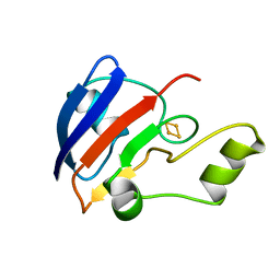
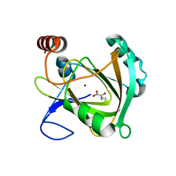
1PUT
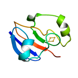 | | AN NMR-DERIVED MODEL FOR THE SOLUTION STRUCTURE OF OXIDIZED PUTIDAREDOXIN, A 2FE, 2-S FERREDOXIN FROM PSEUDOMONAS | | 分子名称: | FE2/S2 (INORGANIC) CLUSTER, PUTIDAREDOXIN | | 著者 | Pochapsky, T.C, Ye, X.M, Ratnaswamy, G, Lyons, T.A. | | 登録日 | 1994-07-09 | | 公開日 | 1994-09-30 | | 最終更新日 | 2024-05-01 | | 実験手法 | SOLUTION NMR | | 主引用文献 | An NMR-derived model for the solution structure of oxidized putidaredoxin, a 2-Fe, 2-S ferredoxin from Pseudomonas.
Biochemistry, 33, 1994
|
|
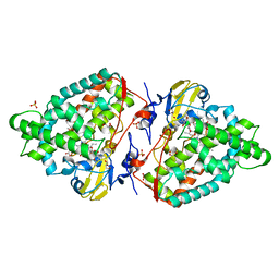
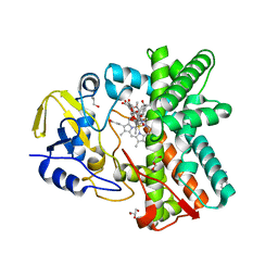
1ZRR
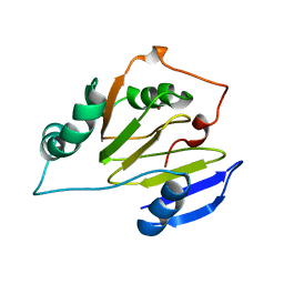 | | Residual Dipolar Coupling Refinement of Acireductone Dioxygenase from Klebsiella | | 分子名称: | E-2/E-2' protein, NICKEL (II) ION | | 著者 | Pochapsky, T.C, Pochapsky, S.S, Ju, T, Hoefler, C, Liang, J. | | 登録日 | 2005-05-19 | | 公開日 | 2005-12-06 | | 最終更新日 | 2024-05-22 | | 実験手法 | SOLUTION NMR | | 主引用文献 | A refined model for the structure of acireductone dioxygenase from Klebsiella ATCC 8724 incorporating residual dipolar couplings
J.Biomol.NMR, 34, 2006
|
|
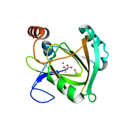
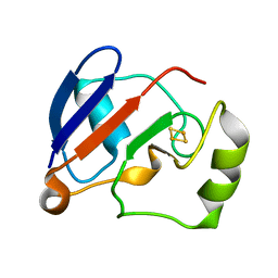
1GPX
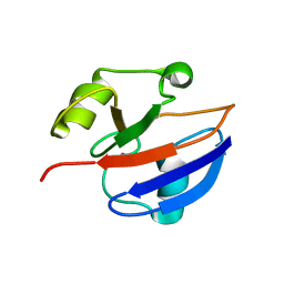 | | C85S GAPDX, NMR, 20 STRUCTURES | | 分子名称: | GALLIUM (III) ION, PUTIDAREDOXIN | | 著者 | Pochapsky, T.C, Kuti, M, Kazanis, S. | | 登録日 | 1998-06-10 | | 公開日 | 1999-01-27 | | 最終更新日 | 2024-05-01 | | 実験手法 | SOLUTION NMR | | 主引用文献 | The solution structure of a gallium-substituted putidaredoxin mutant: GaPdx C85S.
J.Biomol.NMR, 12, 1998
|
|
2LQD
 | | Reduced and CO-bound cytochrome P450cam (CYP101A1) | | 分子名称: | CARBON MONOXIDE, Camphor 5-monooxygenase, POTASSIUM ION, ... | | 著者 | Pochapsky, T.C, Pochapsky, S.S, Asciutto, E, Madura, J, Young, M.J. | | 登録日 | 2012-03-02 | | 公開日 | 2012-05-09 | | 最終更新日 | 2024-05-01 | | 実験手法 | SOLUTION NMR | | 主引用文献 | Solution Structural Ensembles of Substrate-Free Cytochrome P450(cam).
Biochemistry, 51, 2012
|
|
2HJI
 | | Structural model for the Fe-containing isoform of acireductone dioxygenase | | 分子名称: | E-2/E-2' protein, FE (II) ION | | 著者 | Pochapsky, T.C, Ju, T, Maroney, M.J, Chai, S.C. | | 登録日 | 2006-06-30 | | 公開日 | 2006-10-24 | | 最終更新日 | 2024-05-29 | | 実験手法 | SOLUTION NMR | | 主引用文献 | One Protein, Two Enzymes Revisited: A Structural Entropy Switch Interconverts the Two Isoforms of Acireductone Dioxygenase
J.Mol.Biol., 363, 2006
|
|
2L8M
 | | Reduced and CO-bound cytochrome P450cam (CYP101A1) | | 分子名称: | CAMPHOR, CARBON MONOXIDE, CHLORIDE ION, ... | | 著者 | Pochapsky, T.C, Pochapsky, S.S, Dang, M, Asciutto, E, Madura, J. | | 登録日 | 2011-01-19 | | 公開日 | 2011-02-16 | | 最終更新日 | 2024-05-15 | | 実験手法 | SOLUTION NMR | | 主引用文献 | Experimentally Restrained Molecular Dynamics Simulations for Characterizing the Open States of Cytochrome P450(cam).
Biochemistry, 50, 2011
|
|
5UHU
 | |
7JXG
 | | Structural model for Fe-containing human acireductone dioxygenase | | 分子名称: | 1,2-dihydroxy-3-keto-5-methylthiopentene dioxygenase, FE (II) ION | | 著者 | Pochapsky, T.C, Liu, X, Deshpande, A, Ringe, D, Garber, A, Ryan, J. | | 登録日 | 2020-08-27 | | 公開日 | 2020-11-18 | | 最終更新日 | 2024-05-01 | | 実験手法 | SOLUTION NMR | | 主引用文献 | A Model for the Solution Structure of Human Fe(II)-Bound Acireductone Dioxygenase and Interactions with the Regulatory Domain of Matrix Metalloproteinase I (MMP-I).
Biochemistry, 59, 2020
|
|
1B9R
 | |
4AW3
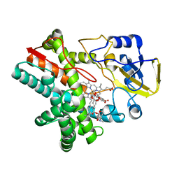
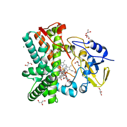
 | | Structure of the mixed-function P450 MycG F286V mutant in complex with mycinamicin V in P1 space group | | 分子名称: | GLYCEROL, MYCINAMICIN V, P-450-LIKE PROTEIN, ... | | 著者 | Li, S, Tietz, D.R, Rutaganira, F.U, Kells, P.M, Anzai, Y, Kato, F, Pochapsky, T.C, Sherman, D.H, Podust, L.M. | | 登録日 | 2012-05-30 | | 公開日 | 2012-09-05 | | 最終更新日 | 2023-12-20 | | 実験手法 | X-RAY DIFFRACTION (2.05 Å) | | 主引用文献 | Substrate Recognition by the Multifunctional Cytochrome P450 Mycg in Mycinamicin Hydroxylation and Epoxidation Reactions.
J.Biol.Chem., 287, 2012
|
|
5I91
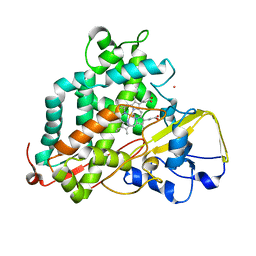
 | | Structure of Mouse Acirecutone dioxygenase with to Ni2+ and 2-keto-4-(methylthio)-butyric acid in the active site | | 分子名称: | 1,2-dihydroxy-3-keto-5-methylthiopentene dioxygenase, 4-(METHYLSULFANYL)-2-OXOBUTANOIC ACID, NICKEL (II) ION | | 著者 | Deshpande, A.R, Robinson, H, Wagenpfeil, K, Pochapsky, T.C, Petsko, G.A, Ringe, D. | | 登録日 | 2016-02-19 | | 公開日 | 2016-03-09 | | 最終更新日 | 2023-09-27 | | 実験手法 | X-RAY DIFFRACTION (1.76 Å) | | 主引用文献 | Metal-Dependent Function of a Mammalian Acireductone Dioxygenase.
Biochemistry, 55, 2016
|
|
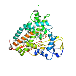
5I8S
 | | Structure of Mouse Acireductone dioxygenase with Ni2+ ion and pentanoic acid in the active site | | 分子名称: | 1,2-dihydroxy-3-keto-5-methylthiopentene dioxygenase, NICKEL (II) ION, PENTANOIC ACID | | 著者 | Deshpande, A.R, Wagenpfeil, K, Pochapsky, T.C, Petsko, G.A, Ringe, D. | | 登録日 | 2016-02-19 | | 公開日 | 2016-03-09 | | 最終更新日 | 2023-09-27 | | 実験手法 | X-RAY DIFFRACTION (1.89 Å) | | 主引用文献 | Metal-Dependent Function of a Mammalian Acireductone Dioxygenase.
Biochemistry, 55, 2016
|
|
5I8T
 | | Structure of Mouse Acireductone dioxygenase with Ni2+ ion and D-lactic acid in the active site | | 分子名称: | 1,2-dihydroxy-3-keto-5-methylthiopentene dioxygenase, ISOPROPYL ALCOHOL, LACTIC ACID, ... | | 著者 | Deshpande, A.R, Wagenpfeil, K, Pochapsky, T.C, Petsko, G.A, Ringe, D. | | 登録日 | 2016-02-19 | | 公開日 | 2016-03-09 | | 最終更新日 | 2023-11-15 | | 実験手法 | X-RAY DIFFRACTION (1.751 Å) | | 主引用文献 | Metal-Dependent Function of a Mammalian Acireductone Dioxygenase.
Biochemistry, 55, 2016
|
|
5I8Y
 | | Structure of Mouse Acireductone Dioxygenase bound to Co2+ and 2-keto-4-(methylthio)-butyric acid | | 分子名称: | 1,2-dihydroxy-3-keto-5-methylthiopentene dioxygenase, 4-(METHYLSULFANYL)-2-OXOBUTANOIC ACID, COBALT (II) ION | | 著者 | Deshpande, A.R, Wagenpfeil, K, Pochapsky, T.C, Petsko, G.A, Ringe, D. | | 登録日 | 2016-02-19 | | 公開日 | 2016-03-09 | | 最終更新日 | 2023-09-27 | | 実験手法 | X-RAY DIFFRACTION (1.942 Å) | | 主引用文献 | Metal-Dependent Function of a Mammalian Acireductone Dioxygenase.
Biochemistry, 55, 2016
|
|
5I93
 | | Structure of Mouse Acireductone dioxygenase with Ni2+ and 2-ketopentanoic acid in the active site | | 分子名称: | 1,2-dihydroxy-3-keto-5-methylthiopentene dioxygenase, 2-oxopentanoic acid, NICKEL (II) ION | | 著者 | Deshpande, A.R, Wagenpfeil, K, Pochapsky, T.C, Petsko, G.A, Ringe, D. | | 登録日 | 2016-02-19 | | 公開日 | 2016-03-09 | | 最終更新日 | 2023-09-27 | | 実験手法 | X-RAY DIFFRACTION (2.236 Å) | | 主引用文献 | Metal-Dependent Function of a Mammalian Acireductone Dioxygenase.
Biochemistry, 55, 2016
|
|
3ZSN
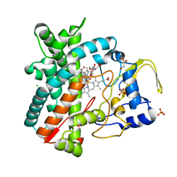
 | | Structure of the mixed-function P450 MycG F286A mutant in complex with mycinamicin IV | | 分子名称: | BENZAMIDINE, GLYCEROL, MYCINAMICIN IV, ... | | 著者 | Li, S, Kells, P.M, Rutaganira, F.U, Anzai, Y, Kato, F, Sherman, D.H, Podust, L.M. | | 登録日 | 2011-06-29 | | 公開日 | 2012-05-09 | | 最終更新日 | 2023-12-20 | | 実験手法 | X-RAY DIFFRACTION (1.9 Å) | | 主引用文献 | Substrate Recognition by the Multifunctional Cytochrome P450 Mycg in Mycinamicin Hydroxylation and Epoxidation Reactions.
J.Biol.Chem., 287, 2012
|
|
1YJI
 | | RDC-refined Solution NMR structure of reduced putidaredoxin | | 分子名称: | FE2/S2 (INORGANIC) CLUSTER, Putidaredoxin | | 著者 | Jain, N.U, Tjioe, E, Savidor, A, Boulie, J. | | 登録日 | 2005-01-14 | | 公開日 | 2005-06-28 | | 最終更新日 | 2024-05-22 | | 実験手法 | SOLUTION NMR | | 主引用文献 | Redox-dependent structural differences in putidaredoxin derived from homologous structure refinement via residual dipolar couplings.
Biochemistry, 44, 2005
|
|
8FDA
 | | Human Cytochrome P450 17A1 in complex with steroidal isonitrile inhibitor | | 分子名称: | PROTOPORPHYRIN IX CONTAINING FE, Steroid 17-alpha-hydroxylase/17,20 lyase, [(1~{R})-1-[(3~{S},5~{S},8~{R},9~{S},10~{S},13~{S},17~{R})-3-methanoyloxy-10,13-dimethyl-1,2,3,4,5,6,7,8,9,11,12,13,14,15,16,17-hexadecahydrocyclopenta[a]phenanthren-17-yl]ethyl]-methylidyne-azanium | | 著者 | Richard, A.M, Scott, E.E. | | 登録日 | 2022-12-02 | | 公開日 | 2023-08-23 | | 最終更新日 | 2023-09-13 | | 実験手法 | X-RAY DIFFRACTION (2.2 Å) | | 主引用文献 | Selective steroidogenic cytochrome P450 haem iron ligation by steroid-derived isonitriles.
Commun Chem, 6, 2023
|
|
1YJJ
 | | RDC-refined Solution NMR structure of oxidized putidaredoxin | | 分子名称: | FE2/S2 (INORGANIC) CLUSTER, Putidaredoxin | | 著者 | Jain, N.U, Tjioe, E, Savidor, A, Boulie, J. | | 登録日 | 2005-01-14 | | 公開日 | 2005-06-28 | | 最終更新日 | 2024-05-22 | | 実験手法 | SOLUTION NMR | | 主引用文献 | Redox-dependent structural differences in putidaredoxin derived from homologous structure refinement via residual dipolar couplings.
Biochemistry, 44, 2005
|
|
2YGX
 | | Structure of the mixed-function P450 MycG in P21 space group | | 分子名称: | GLYCEROL, P-450-LIKE PROTEIN, PROTOPORPHYRIN IX CONTAINING FE, ... | | 著者 | Li, S, Kells, P.M, Rutaganira, F.U, Sherman, D.H, Podust, L.M. | | 登録日 | 2011-04-22 | | 公開日 | 2012-02-29 | | 最終更新日 | 2023-12-20 | | 実験手法 | X-RAY DIFFRACTION (2.39 Å) | | 主引用文献 | Substrate Recognition by the Multifunctional Cytochrome P450 Mycg in Mycinamicin Hydroxylation and Epoxidation Reactions.
J.Biol.Chem., 287, 2012
|
|
2Y5Z
 | | Mixed-function P450 MycG in complex with mycinamicin III in C2221 space group | | 分子名称: | BENZAMIDINE, GLYCEROL, MYCINAMICIN III, ... | | 著者 | Li, S, Kells, P.M, Sherman, D.H, Podust, L.M. | | 登録日 | 2011-01-19 | | 公開日 | 2012-02-01 | | 最終更新日 | 2023-12-20 | | 実験手法 | X-RAY DIFFRACTION (2.06 Å) | | 主引用文献 | Substrate Recognition by the Multifunctional Cytochrome P450 Mycg in Mycinamicin Hydroxylation and Epoxidation Reactions.
J.Biol.Chem., 287, 2012
|
|
2YCA
 | | Mixed-function P450 MycG in complex with mycinamicin III in P21212 space group | | 分子名称: | GLYCEROL, MYCINAMICIN III, P-450-LIKE PROTEIN, ... | | 著者 | Li, S, Kells, P.M, Sherman, D.H, Podust, L.M. | | 登録日 | 2011-03-12 | | 公開日 | 2012-03-28 | | 最終更新日 | 2023-12-20 | | 実験手法 | X-RAY DIFFRACTION (1.8 Å) | | 主引用文献 | Substrate Recognition by the Multifunctional Cytochrome P450 Mycg in Mycinamicin Hydroxylation and Epoxidation Reactions.
J.Biol.Chem., 287, 2012
|
|
2Y5N
 | | Structure of the mixed-function P450 MycG in complex with mycinamicin V in P21 space group | | 分子名称: | GLYCEROL, MAGNESIUM ION, MYCINAMICIN V, ... | | 著者 | Li, S, Kells, P.M, Sherman, D.H, Podust, L.M. | | 登録日 | 2011-01-15 | | 公開日 | 2012-02-01 | | 最終更新日 | 2023-12-20 | | 実験手法 | X-RAY DIFFRACTION (1.62 Å) | | 主引用文献 | Substrate Recognition by the Multifunctional Cytochrome P450 Mycg in Mycinamicin Hydroxylation and Epoxidation Reactions.
J.Biol.Chem., 287, 2012
|
|
2Y98
 | | Structure of the mixed-function P450 MycG in complex with mycinamicin IV in P21212 space group | | 分子名称: | CHLORIDE ION, GLYCEROL, MYCINAMICIN IV, ... | | 著者 | Li, S, Kells, P.M, Sherman, D.H, Podust, L.M. | | 登録日 | 2011-02-12 | | 公開日 | 2012-02-22 | | 最終更新日 | 2023-12-20 | | 実験手法 | X-RAY DIFFRACTION (1.65 Å) | | 主引用文献 | Substrate Recognition by the Multifunctional Cytochrome P450 Mycg in Mycinamicin Hydroxylation and Epoxidation Reactions.
J.Biol.Chem., 287, 2012
|
|


