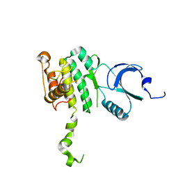
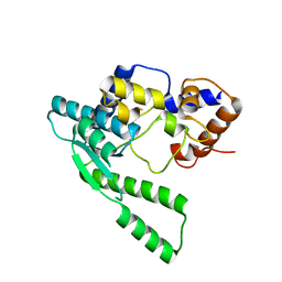
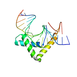
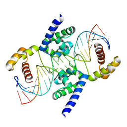
1PZW
 
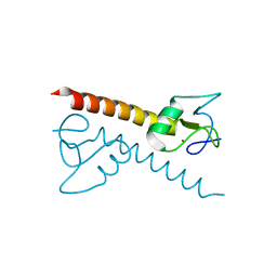 | | Crystal structure of the zinc finger associated domain of the Drosophila transcription factor Grauzone | | 分子名称: | Transcription factor grauzone, ZINC ION | | 著者 | Jauch, R, Bourenkov, G.P, Chung, H.-R, Urlaub, H, Reidt, U, Jaeckle, H, Wahl, M.C. | | 登録日 | 2003-07-14 | | 公開日 | 2003-11-04 | | 最終更新日 | 2024-02-14 | | 実験手法 | X-RAY DIFFRACTION (2 Å) | | 主引用文献 | The zinc finger-associated domain of the Drosophila transcription factor grauzone is a novel zinc-coordinating protein-protein interaction module
STRUCTURE, 11, 2003
|
|
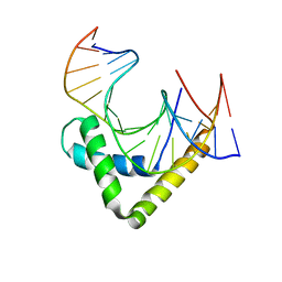
2XSD
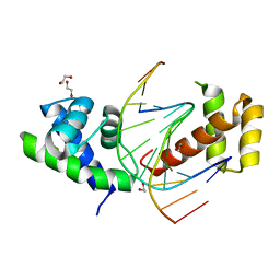 | | Crystal Structure of the dimeric Oct-6 (Pou3f1) POU domain bound to palindromic MORE DNA | | 分子名称: | 1,2-ETHANEDIOL, 5'-D(*AP*TP*GP*CP*AP*TP*GP*AP*GP*GP*AP)-3', 5'-D(*CP*CP*TP*CP*AP*TP*GP*CP*AP*TP*AP)-3', ... | | 著者 | Jauch, R, Choo, S.H, Ng, C.K.L, Kolatkar, P.R. | | 登録日 | 2010-09-28 | | 公開日 | 2010-10-20 | | 最終更新日 | 2023-12-20 | | 実験手法 | X-RAY DIFFRACTION (2.049 Å) | | 主引用文献 | Crystal Structure of the Dimeric Oct6 (POU3F1) POU Domain Bound to Palindromic More DNA.
Proteins, 79, 2011
|
|
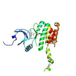
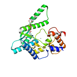
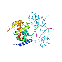
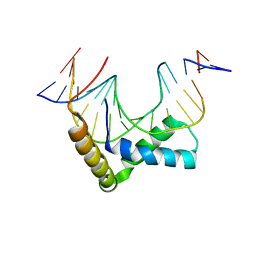
2VI6
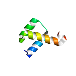 | | Crystal Structure of the Nanog Homeodomain | | 分子名称: | HOMEOBOX PROTEIN NANOG | | 著者 | Jauch, R, Ng, C.K.L, Saitakendu, K.S, Stevens, R.C, Kolatkar, P.R. | | 登録日 | 2007-11-28 | | 公開日 | 2008-01-15 | | 最終更新日 | 2023-12-13 | | 実験手法 | X-RAY DIFFRACTION (2.6 Å) | | 主引用文献 | Crystal Structure and DNA Binding of the Homeodomain of the Stem Cell Transcription Factor Nanog.
J.Mol.Biol., 376, 2008
|
|
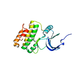
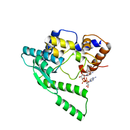
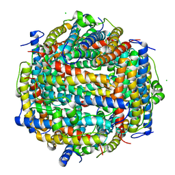
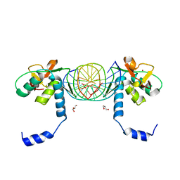
2AC3
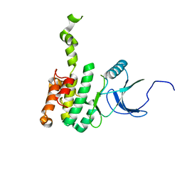 | | Structure of human Mnk2 Kinase Domain | | 分子名称: | MAP kinase-interacting serine/threonine kinase 2, ZINC ION | | 著者 | Jauch, R, Wahl, M.C, Netter, C, Jakel, S, Schreiter, K, Aicher, B, Jackle, H. | | 登録日 | 2005-07-18 | | 公開日 | 2005-10-04 | | 最終更新日 | 2024-03-13 | | 実験手法 | X-RAY DIFFRACTION (2.1 Å) | | 主引用文献 | Crystal structures of the Mnk2 kinase domain reveal an inhibitory conformation and a zinc binding site.
Structure, 13, 2005
|
|
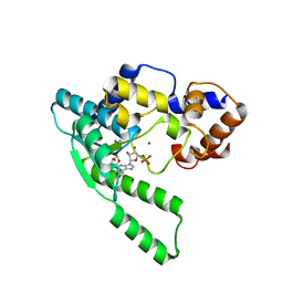
2AC5
 | | Structure of human Mnk2 Kinase Domain mutant D228G | | 分子名称: | MAP kinase-interacting serine/threonine kinase 2, ZINC ION | | 著者 | Jauch, R, Wahl, M.C, Jakel, S, Schreiter, K, Aicher, B, Jackle, H. | | 登録日 | 2005-07-18 | | 公開日 | 2005-10-04 | | 最終更新日 | 2024-05-29 | | 実験手法 | X-RAY DIFFRACTION (3.2 Å) | | 主引用文献 | Crystal structures of the Mnk2 kinase domain reveal an inhibitory conformation and a zinc binding site.
Structure, 13, 2005
|
|
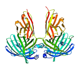
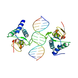
3U2B
 | | Structure of the Sox4 HMG domain bound to DNA | | 分子名称: | DNA (5'-D(*CP*CP*AP*GP*GP*AP*CP*AP*AP*TP*AP*GP*AP*GP*AP*C)-3'), DNA (5'-D(*GP*TP*CP*TP*CP*TP*AP*TP*TP*GP*TP*CP*CP*TP*GP*G)-3'), Transcription factor SOX-4 | | 著者 | Jauch, R, Ng, C.K.L, Kolatkar, P.R. | | 登録日 | 2011-10-03 | | 公開日 | 2011-12-28 | | 最終更新日 | 2024-03-20 | | 実験手法 | X-RAY DIFFRACTION (2.402 Å) | | 主引用文献 | The crystal structure of the Sox4 HMG domain-DNA complex suggests a mechanism for positional interdependence in DNA recognition
Biochem.J., 443, 2012
|
|
2HW7
 | |
2HW6
 | | Crystal structure of Mnk1 catalytic domain | | 分子名称: | MAP kinase-interacting serine/threonine-protein kinase 1, SULFATE ION | | 著者 | Jauch, R, Wahl, M.C. | | 登録日 | 2006-08-01 | | 公開日 | 2006-08-29 | | 最終更新日 | 2023-08-30 | | 実験手法 | X-RAY DIFFRACTION (2.5 Å) | | 主引用文献 | Mitogen-activated protein kinases interacting kinases are autoinhibited by a reprogrammed activation segment.
Embo J., 25, 2006
|
|
1WXI
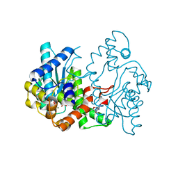 | | E.coli NAD Synthetase, AMP.PP | | 分子名称: | ADENOSINE MONOPHOSPHATE, DIPHOSPHATE, MAGNESIUM ION, ... | | 著者 | Jauch, R, Humm, A, Huber, R, Wahl, M.C. | | 登録日 | 2005-01-23 | | 公開日 | 2005-02-15 | | 最終更新日 | 2024-03-13 | | 実験手法 | X-RAY DIFFRACTION (1.7 Å) | | 主引用文献 | Structures of Escherichia coli NAD Synthetase with Substrates and Products Reveal Mechanistic Rearrangements
J.Biol.Chem., 280, 2005
|
|
1WXG
 | | E.coli NAD Synthetase, DND | | 分子名称: | MAGNESIUM ION, NH(3)-dependent NAD(+) synthetase, NICOTINIC ACID ADENINE DINUCLEOTIDE | | 著者 | Jauch, R, Humm, A, Huber, R, Wahl, M.C. | | 登録日 | 2005-01-23 | | 公開日 | 2005-02-15 | | 最終更新日 | 2024-03-13 | | 実験手法 | X-RAY DIFFRACTION (1.9 Å) | | 主引用文献 | Structures of Escherichia coli NAD Synthetase with Substrates and Products Reveal Mechanistic Rearrangements
J.Biol.Chem., 280, 2005
|
|
1WXH
 | | E.coli NAD Synthetase, NAD | | 分子名称: | NH(3)-dependent NAD(+) synthetase, NICOTINAMIDE-ADENINE-DINUCLEOTIDE | | 著者 | Jauch, R, Humm, A, Huber, R, Wahl, M.C. | | 登録日 | 2005-01-23 | | 公開日 | 2005-02-15 | | 最終更新日 | 2024-03-13 | | 実験手法 | X-RAY DIFFRACTION (1.9 Å) | | 主引用文献 | Structures of Escherichia coli NAD Synthetase with Substrates and Products Reveal Mechanistic Rearrangements
J.Biol.Chem., 280, 2005
|
|
1WXE
 | | E.coli NAD Synthetase, AMP | | 分子名称: | ADENOSINE MONOPHOSPHATE, MAGNESIUM ION, NH(3)-dependent NAD(+) synthetase | | 著者 | Jauch, R, Humm, A, Huber, R, Wahl, M.C. | | 登録日 | 2005-01-23 | | 公開日 | 2005-02-15 | | 最終更新日 | 2024-03-13 | | 実験手法 | X-RAY DIFFRACTION (1.9 Å) | | 主引用文献 | Structures of Escherichia coli NAD Synthetase with Substrates and Products Reveal Mechanistic Rearrangements
J.Biol.Chem., 280, 2005
|
|
1WXF
 | | E.coli NAD Synthetase | | 分子名称: | NH(3)-dependent NAD(+) synthetase | | 著者 | Jauch, R, Humm, A, Huber, R, Wahl, M.C. | | 登録日 | 2005-01-23 | | 公開日 | 2005-02-15 | | 最終更新日 | 2024-03-13 | | 実験手法 | X-RAY DIFFRACTION (2.3 Å) | | 主引用文献 | Structures of Escherichia coli NAD Synthetase with Substrates and Products Reveal Mechanistic Rearrangements
J.Biol.Chem., 280, 2005
|
|
4PFE
 | | Crystal structure of vsfGFP-0 | | 分子名称: | Green fluorescent protein | | 著者 | Jauch, R, Chen, S.L. | | 登録日 | 2014-04-29 | | 公開日 | 2015-06-24 | | 最終更新日 | 2023-11-15 | | 実験手法 | X-RAY DIFFRACTION (2.603 Å) | | 主引用文献 | Rational Structure-Based Design of Bright GFP-Based Complexes with Tunable Dimerization.
Angew.Chem.Int.Ed.Engl., 54, 2015
|
|
7XRC
 | |
5XGO
 | | The Ferritin E-Domain: Toward Understanding Its Role in Protein Cage Assembly Through the Crystal Structure of a Maxi-/Mini-Ferritin Chimera | | 分子名称: | CHLORIDE ION, DNA protection during starvation protein,Bacterioferritin | | 著者 | Cornell, T.A, Srivastava, Y, Jauch, R, Fan, R, Orner, B.P. | | 登録日 | 2017-04-14 | | 公開日 | 2018-02-28 | | 最終更新日 | 2024-03-27 | | 実験手法 | X-RAY DIFFRACTION (1.99 Å) | | 主引用文献 | The Crystal Structure of a Maxi/Mini-Ferritin Chimera Reveals Guiding Principles for the Assembly of Protein Cages.
Biochemistry, 56, 2017
|
|
4Y60
 | | Structure of SOX18-HMG/PROX1-DNA | | 分子名称: | DNA (5'-D(*CP*AP*CP*TP*AP*GP*CP*AP*TP*TP*GP*TP*CP*TP*GP*GP*G)-3'), DNA (5'-D(*GP*CP*CP*CP*AP*GP*AP*CP*AP*AP*TP*GP*CP*TP*AP*GP*T)-3'), Transcription factor SOX-18 | | 著者 | Narasimhan, K, Prokoph, N, Kolatkar, P, Robinson, H, Jauch, R. | | 登録日 | 2015-02-12 | | 公開日 | 2016-03-02 | | 最終更新日 | 2024-03-20 | | 実験手法 | X-RAY DIFFRACTION (1.75 Å) | | 主引用文献 | Structure and decoy-mediated inhibition of the SOX18/Prox1-DNA interaction.
Nucleic Acids Res., 44, 2016
|
|
3F27
 | | Structure of Sox17 Bound to DNA | | 分子名称: | DNA (5'-D(*DCP*DCP*DAP*DGP*DGP*DAP*DCP*DAP*DAP*DTP*DAP*DGP*DAP*DGP*DAP*DC)-3'), DNA (5'-D(*DGP*DTP*DCP*DTP*DCP*DTP*DAP*DTP*DTP*DGP*DTP*DCP*DCP*DTP*DGP*DG)-3'), Transcription factor SOX-17 | | 著者 | Palasingam, P, Jauch, R, Ng, C.K.L, Kolatkar, P.R. | | 登録日 | 2008-10-29 | | 公開日 | 2009-04-07 | | 最終更新日 | 2023-11-08 | | 実験手法 | X-RAY DIFFRACTION (2.75 Å) | | 主引用文献 | The Structure of Sox17 Bound to DNA Reveals a Conserved Bending Topology but Selective Protein Interaction Platforms
J.Mol.Biol., 388, 2009
|
|
3KMP
 | | Crystal Structure of SMAD1-MH1/DNA complex | | 分子名称: | 5'-D(P*AP*TP*CP*AP*GP*TP*CP*TP*AP*GP*AP*CP*AP*TP*A)-3', 5'-D(P*GP*TP*AP*TP*GP*TP*CP*TP*AP*GP*AP*CP*TP*GP*A)-3', GLYCEROL, ... | | 著者 | Baburajendran, N, Palasingam, P, Narasimhan, K, Jauch, R, Kolatkar, P.R. | | 登録日 | 2009-11-11 | | 公開日 | 2010-02-23 | | 最終更新日 | 2024-03-20 | | 実験手法 | X-RAY DIFFRACTION (2.7 Å) | | 主引用文献 | Structure of Smad1 MH1/DNA complex reveals distinctive rearrangements of BMP and TGF-beta effectors.
Nucleic Acids Res., 38, 2010
|
|
3QSV
 | |
3L1P
 | | POU protein:DNA complex | | 分子名称: | DNA (5'-D(*AP*TP*CP*CP*AP*TP*TP*TP*GP*CP*CP*TP*TP*TP*CP*AP*AP*AP*TP*GP*TP*GP*G)-3'), DNA (5'-D(*TP*CP*CP*AP*CP*AP*TP*TP*TP*GP*AP*AP*AP*GP*GP*CP*AP*AP*AP*TP*GP*GP*A)-3'), POU domain, ... | | 著者 | Vahokoski, J, Groves, M.R, Pogenberg, V, Wilmanns, M. | | 登録日 | 2009-12-14 | | 公開日 | 2010-12-15 | | 最終更新日 | 2023-11-01 | | 実験手法 | X-RAY DIFFRACTION (2.8 Å) | | 主引用文献 | A unique Oct4 interface is crucial for reprogramming to pluripotency
Nat.Cell Biol., 15, 2013
|
|
4A3N
 | | Crystal Structure of HMG-BOX of Human SOX17 | | 分子名称: | TRANSCRIPTION FACTOR SOX-17, ZINC ION | | 著者 | Gao, N, Gao, H, Qian, H, Si, S, Xie, Y. | | 登録日 | 2011-10-01 | | 公開日 | 2011-12-28 | | 最終更新日 | 2023-12-20 | | 実験手法 | X-RAY DIFFRACTION (2.4 Å) | | 主引用文献 | Structural Basis of Human Transcription Factor Sry-Related Box 17 Binding to DNA.
Protein Pept.Lett., 20, 2013
|
|


