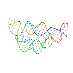
9BUN
 
 | |
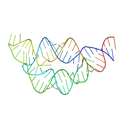
3LQX
 | |
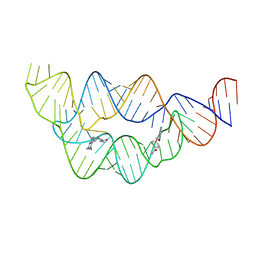
1DUL
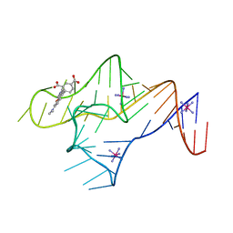
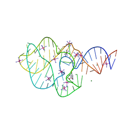
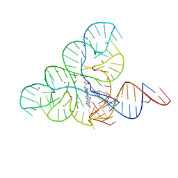
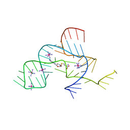
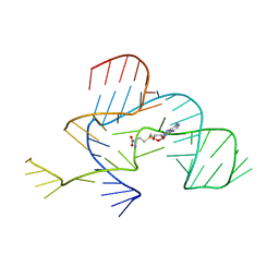
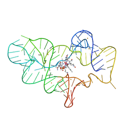
 | | STRUCTURE OF THE RIBONUCLEOPROTEIN CORE OF THE E. COLI SIGNAL RECOGNITION PARTICLE | | 分子名称: | 4.5 S RNA DOMAIN IV, MAGNESIUM ION, POTASSIUM ION, ... | | 著者 | Batey, R.T, Rambo, R.P, Lucast, L, Rha, B, Doudna, J.A. | | 登録日 | 2000-01-17 | | 公開日 | 2000-02-28 | | 最終更新日 | 2020-10-14 | | 実験手法 | X-RAY DIFFRACTION (1.8 Å) | | 主引用文献 | Crystal structure of the ribonucleoprotein core of the signal recognition particle.
Science, 287, 2000
|
|
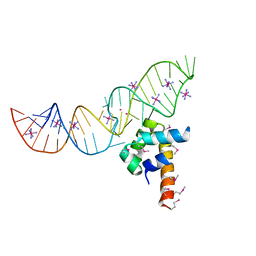
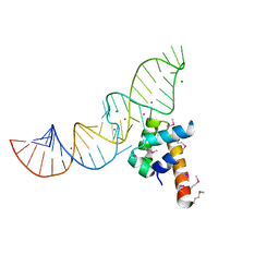
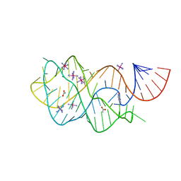
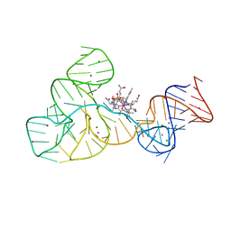
1HQ1
 | | STRUCTURAL AND ENERGETIC ANALYSIS OF RNA RECOGNITION BY A UNIVERSALLY CONSERVED PROTEIN FROM THE SIGNAL RECOGNITION PARTICLE | | 分子名称: | 4.5S RNA DOMAIN IV, MAGNESIUM ION, POTASSIUM ION, ... | | 著者 | Batey, R.T, Sagar, M.B, Doudna, J.A. | | 登録日 | 2000-12-13 | | 公開日 | 2001-01-03 | | 最終更新日 | 2023-08-09 | | 実験手法 | X-RAY DIFFRACTION (1.52 Å) | | 主引用文献 | Structural and energetic analysis of RNA recognition by a universally conserved protein from the signal recognition particle.
J.Mol.Biol., 307, 2001
|
|
5KPY
 | | Structure of a 5-hydroxytryptophan aptamer | | 分子名称: | 5-hydroxy-L-tryptophan, 5-hydroxytryptophan RNA aptamer, IRIDIUM HEXAMMINE ION, ... | | 著者 | Batey, R.T, Porter, E, Merck, M. | | 登録日 | 2016-07-05 | | 公開日 | 2017-01-11 | | 最終更新日 | 2024-03-06 | | 実験手法 | X-RAY DIFFRACTION (2 Å) | | 主引用文献 | Recurrent RNA motifs as scaffolds for genetically encodable small-molecule biosensors.
Nat. Chem. Biol., 13, 2017
|
|
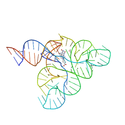
4FE5
 | | Crystal structure of the xpt-pbuX guanine riboswitch aptamer domain in complex with hypoxanthine | | 分子名称: | ACETATE ION, COBALT HEXAMMINE(III), HYPOXANTHINE, ... | | 著者 | Stoddard, C.D, Trausch, J.J, Widmann, J, Marcano, J, Knight, R, Batey, R.T. | | 登録日 | 2012-05-29 | | 公開日 | 2012-06-27 | | 最終更新日 | 2024-02-28 | | 実験手法 | X-RAY DIFFRACTION (1.32 Å) | | 主引用文献 | Structure of a natural guanine-responsive riboswitch complexed with the metabolite hypoxanthine.
Nature, 432, 2004
|
|
3D0X
 | |
6DN2
 | | CRYSTAL STRUCTURE OF THE FMN RIBOSWITCH BOUND TO BRX1354 SPLIT RNA | | 分子名称: | 4-{benzyl[2-(7,8-dimethyl-2,4-dioxo-3,4-dihydrobenzo[g]pteridin-10(2H)-yl)ethyl]amino}butanoic acid, MAGNESIUM ION, POTASSIUM ION, ... | | 著者 | Vicens, Q, Mondragon, E, Reyes, F.E, Berman, J, Kaur, H, Kells, K, Wickens, P, Wilson, J, Gadwood, R, Schostarez, H, Suto, R.K, Coish, P, Blount, K.F, Batey, R.T. | | 登録日 | 2018-06-05 | | 公開日 | 2018-09-05 | | 最終更新日 | 2023-10-11 | | 実験手法 | X-RAY DIFFRACTION (2.88 Å) | | 主引用文献 | Structure-Activity Relationship of Flavin Analogues That Target the Flavin Mononucleotide Riboswitch.
ACS Chem. Biol., 13, 2018
|
|
6DN3
 | | CRYSTAL STRUCTURE OF THE FMN RIBOSWITCH BOUND TO BRX1555 SPLIT RNA | | 分子名称: | 7,8-dimethyl-2,4-dioxo-10-(3-phenylpropyl)-1,2,3,4-tetrahydrobenzo[g]pteridin-10-ium, CHLORIDE ION, MAGNESIUM ION, ... | | 著者 | Vicens, Q, Mondragon, E, Reyes, F.E, Berman, J, Kaur, H, Kells, K, Wickens, P, Wilson, J, Gadwood, R, Schostarez, H, Suto, R.K, Coish, P, Blount, K.F, Batey, R.T. | | 登録日 | 2018-06-05 | | 公開日 | 2018-09-05 | | 最終更新日 | 2023-10-11 | | 実験手法 | X-RAY DIFFRACTION (2.8 Å) | | 主引用文献 | Structure-Activity Relationship of Flavin Analogues That Target the Flavin Mononucleotide Riboswitch.
ACS Chem. Biol., 13, 2018
|
|
6DN1
 | | CRYSTAL STRUCTURE OF THE FMN RIBOSWITCH BOUND TO BRX1151 SPLIT RNA | | 分子名称: | 10-(6-carboxyhexyl)-8-(cyclopentylamino)-2,4-dihydroxy-7-methylbenzo[g]pteridin-10-ium, MAGNESIUM ION, POTASSIUM ION, ... | | 著者 | Vicens, Q, Mondragon, E, Reyes, F.E, Berman, J, Kaur, H, Kells, K, Wickens, P, Wilson, J, Gadwood, R, Schostarez, H, Suto, R.K, Coish, P, Blount, K.F, Batey, R.T. | | 登録日 | 2018-06-05 | | 公開日 | 2018-09-05 | | 最終更新日 | 2023-10-11 | | 実験手法 | X-RAY DIFFRACTION (3.03 Å) | | 主引用文献 | Structure-Activity Relationship of Flavin Analogues That Target the Flavin Mononucleotide Riboswitch.
ACS Chem. Biol., 13, 2018
|
|
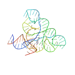
3SD3
 | | The structure of the tetrahydrofolate riboswitch containing a U25C mutation | | 分子名称: | IRIDIUM HEXAMMINE ION, N-[4-({[(6S)-2-amino-5-formyl-4-oxo-3,4,5,6,7,8-hexahydropteridin-6-yl]methyl}amino)benzoyl]-L-glutamic acid, Tetrahydrofolate riboswitch | | 著者 | Reyes, F.E, Trausch, J.J, Ceres, P, Batey, R.T. | | 登録日 | 2011-06-08 | | 公開日 | 2011-09-21 | | 最終更新日 | 2024-02-28 | | 実験手法 | X-RAY DIFFRACTION (1.95 Å) | | 主引用文献 | The structure of a tetrahydrofolate-sensing riboswitch reveals two ligand binding sites in a single aptamer.
Structure, 19, 2011
|
|
4LVV
 | | Structure of the THF riboswitch | | 分子名称: | N-[4-({[(6S)-2-amino-5-formyl-4-oxo-3,4,5,6,7,8-hexahydropteridin-6-yl]methyl}amino)benzoyl]-L-glutamic acid, THF riboswitch | | 著者 | Trausch, J.J, Batey, R.T. | | 登録日 | 2013-07-26 | | 公開日 | 2014-03-19 | | 最終更新日 | 2024-02-28 | | 実験手法 | X-RAY DIFFRACTION (2.1 Å) | | 主引用文献 | A Disconnect between High-Affinity Binding and Efficient Regulation by Antifolates and Purines in the Tetrahydrofolate Riboswitch.
Chem.Biol., 21, 2014
|
|
4LW0
 | |
4LVW
 | |
4LVX
 | |
4LVZ
 | |
4LVY
 | | Structure of the THF riboswitch bound to pemetrexed | | 分子名称: | 2-{4-[2-(2-AMINO-4-OXO-4,7-DIHYDRO-3H-PYRROLO[2,3-D]PYRIMIDIN-5-YL)-ETHYL]-BENZOYLAMINO}-PENTANEDIOIC ACID, THF riboswitch | | 著者 | Trausch, J.J, Batey, R.T. | | 登録日 | 2013-07-26 | | 公開日 | 2014-03-19 | | 最終更新日 | 2024-02-28 | | 実験手法 | X-RAY DIFFRACTION (2 Å) | | 主引用文献 | A Disconnect between High-Affinity Binding and Efficient Regulation by Antifolates and Purines in the Tetrahydrofolate Riboswitch.
Chem.Biol., 21, 2014
|
|
3NPQ
 | |
3NPN
 | |
6P2H
 | |
4XWF
 | |
4XW7
 | |
4FRN
 | | Crystal structure of the cobalamin riboswitch regulatory element | | 分子名称: | BARIUM ION, Cobalamin riboswitch aptamer domain, Hydroxocobalamin | | 著者 | Reyes, F.E, Johnson, J.E, Polaski, J.T, Batey, R.T. | | 登録日 | 2012-06-26 | | 公開日 | 2012-10-17 | | 最終更新日 | 2023-09-13 | | 実験手法 | X-RAY DIFFRACTION (3.43 Å) | | 主引用文献 | B12 cofactors directly stabilize an mRNA regulatory switch.
Nature, 492, 2012
|
|
4FRG
 | | Crystal structure of the cobalamin riboswitch aptamer domain | | 分子名称: | Hydroxocobalamin, IRIDIUM (III) ION, MAGNESIUM ION, ... | | 著者 | Reyes, F.E, Johnson, J.E, Polaski, J.T, Batey, R.T. | | 登録日 | 2012-06-26 | | 公開日 | 2012-10-17 | | 最終更新日 | 2024-02-28 | | 実験手法 | X-RAY DIFFRACTION (2.95 Å) | | 主引用文献 | B12 cofactors directly stabilize an mRNA regulatory switch.
Nature, 492, 2012
|
|
4GMA
 | | Crystal structure of the adenosylcobalamin riboswitch | | 分子名称: | Adenosylcobalamin, Adenosylcobalamin riboswitch | | 著者 | Reyes, F.E, Johnson, J.E, Polaski, J.T, Batey, R.T. | | 登録日 | 2012-08-15 | | 公開日 | 2012-10-17 | | 最終更新日 | 2024-02-28 | | 実験手法 | X-RAY DIFFRACTION (3.94 Å) | | 主引用文献 | B12 cofactors directly stabilize an mRNA regulatory switch.
Nature, 492, 2012
|
|


