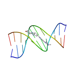
1D9R
 
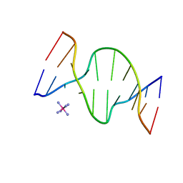 | | CRYSTAL STRUCTURE OF DNA SHEARED TANDEM G-A BASE PAIRS | | 分子名称: | 5'-D(*CP*CP*GP*AP*AP*(BRU)P*GP*AP*GP*G)-3', COBALT HEXAMMINE(III) | | 著者 | Gao, Y.-G, Robinson, H, Sanishvili, R, Joachimiak, A, Wang, A.H.-J. | | 登録日 | 1999-10-29 | | 公開日 | 1999-11-05 | | 最終更新日 | 2024-02-07 | | 実験手法 | X-RAY DIFFRACTION (1.5 Å) | | 主引用文献 | Structure and recognition of sheared tandem G x A base pairs associated with human centromere DNA sequence at atomic resolution.
Biochemistry, 38, 1999
|
|
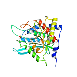
2ZED
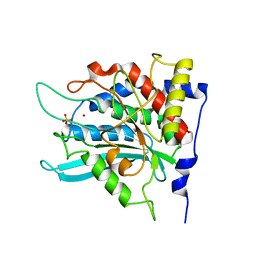
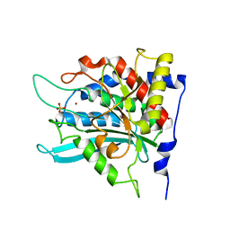
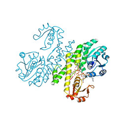
 | | Crystal structure of the human glutaminyl cyclase mutant S160A at 1.7 angstrom resolution | | 分子名称: | Glutaminyl-peptide cyclotransferase, SULFATE ION, ZINC ION | | 著者 | Huang, K.F, Wang, Y.R, Chang, E.C, Chou, T.L, Wang, A.H. | | 登録日 | 2007-12-12 | | 公開日 | 2008-04-22 | | 最終更新日 | 2023-11-01 | | 実験手法 | X-RAY DIFFRACTION (1.7 Å) | | 主引用文献 | A conserved hydrogen-bond network in the catalytic centre of animal glutaminyl cyclases is critical for catalysis.
Biochem.J., 411, 2008
|
|
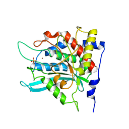
2ZEH
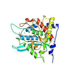
 | | Crystal structure of the human glutaminyl cyclase mutant E201Q at 1.8 angstrom resolution | | 分子名称: | Glutaminyl-peptide cyclotransferase, SULFATE ION, ZINC ION | | 著者 | Huang, K.F, Wang, Y.R, Chang, E.C, Chou, T.L, Wang, A.H. | | 登録日 | 2007-12-12 | | 公開日 | 2008-04-22 | | 最終更新日 | 2023-11-01 | | 実験手法 | X-RAY DIFFRACTION (1.8 Å) | | 主引用文献 | A conserved hydrogen-bond network in the catalytic centre of animal glutaminyl cyclases is critical for catalysis.
Biochem.J., 411, 2008
|
|
1DNZ
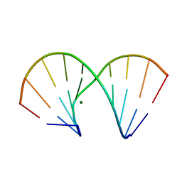
 | | A-DNA DECAMER ACCGGCCGGT WITH MAGNESIUM BINDING SITES | | 分子名称: | DNA (5'-D(*AP*CP*CP*GP*GP*CP*CP*GP*GP*T)-3'), MAGNESIUM ION | | 著者 | Robinson, H, Gao, Y.-G, Sanishvili, R, Joachimiak, A, Wang, A.H.-J. | | 登録日 | 1999-12-17 | | 公開日 | 2000-04-10 | | 最終更新日 | 2024-02-07 | | 実験手法 | X-RAY DIFFRACTION (1.6 Å) | | 主引用文献 | Hexahydrated magnesium ions bind in the deep major groove and at the outer mouth of A-form nucleic acid duplexes.
Nucleic Acids Res., 28, 2000
|
|
2ZEN
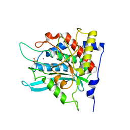
 | | Crystal structure of the human glutaminyl cyclase mutant D305A at 1.78 angstrom resolution | | 分子名称: | Glutaminyl-peptide cyclotransferase, SULFATE ION, ZINC ION | | 著者 | Huang, K.F, Wang, Y.R, Chang, E.C, Chou, T.L, Wang, A.H. | | 登録日 | 2007-12-13 | | 公開日 | 2008-04-22 | | 最終更新日 | 2023-11-01 | | 実験手法 | X-RAY DIFFRACTION (1.78 Å) | | 主引用文献 | A conserved hydrogen-bond network in the catalytic centre of animal glutaminyl cyclases is critical for catalysis.
Biochem.J., 411, 2008
|
|
1D10
 | | STRUCTURAL COMPARISON OF ANTICANCER DRUG-DNA COMPLEXES. ADRIAMYCIN AND DAUNOMYCIN | | 分子名称: | DAUNOMYCIN, DNA (5'-D(*CP*GP*AP*TP*CP*G)-3'), SODIUM ION, ... | | 著者 | Frederick, C.A, Williams, L.D, Ughetto, G, Van Der Marel, G.A, Van Boom, J.H, Rich, A, Wang, A.H.-J. | | 登録日 | 1989-10-20 | | 公開日 | 1990-10-15 | | 最終更新日 | 2024-02-07 | | 実験手法 | X-RAY DIFFRACTION (1.5 Å) | | 主引用文献 | Structural comparison of anticancer drug-DNA complexes: adriamycin and daunomycin.
Biochemistry, 29, 1990
|
|
1DNT
 | | RNA/DNA DODECAMER R(GC)D(GTATACGC) WITH MAGNESIUM BINDING SITES | | 分子名称: | DNA/RNA (5'-R(*GP*CP)-D(*GP*TP*AP*TP*AP*CP*GP*C)-3'), MAGNESIUM ION | | 著者 | Robinson, H, Gao, Y.-G, Sanishvili, R, Joachimiak, A, Wang, A.H.-J. | | 登録日 | 1999-12-16 | | 公開日 | 2000-04-10 | | 最終更新日 | 2024-02-07 | | 実験手法 | X-RAY DIFFRACTION (1.7 Å) | | 主引用文献 | Hexahydrated magnesium ions bind in the deep major groove and at the outer mouth of A-form nucleic acid duplexes.
Nucleic Acids Res., 28, 2000
|
|
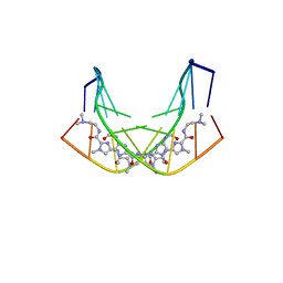
1DNH
 | | THE MOLECULAR STRUCTURE OF THE COMPLEX OF HOECHST 33258 AND THE DNA DODECAMER D(CGCGAATTCGCG) | | 分子名称: | 2'-(4-HYDROXYPHENYL)-5-(4-METHYL-1-PIPERAZINYL)-2,5'-BI-BENZIMIDAZOLE, DNA (5'-D(*CP*GP*CP*GP*AP*AP*TP*TP*CP*GP*CP*G)-3') | | 著者 | Teng, M.-K, Usman, N, Frederick, C.A, Wang, A.H.-J. | | 登録日 | 1988-02-16 | | 公開日 | 1989-01-09 | | 最終更新日 | 2024-02-07 | | 実験手法 | X-RAY DIFFRACTION (2.25 Å) | | 主引用文献 | The molecular structure of the complex of Hoechst 33258 and the DNA dodecamer d(CGCGAATTCGCG).
Nucleic Acids Res., 16, 1988
|
|
1DNX
 | | RNA/DNA DODECAMER R(G)D(CGTATACGC) WITH MAGNESIUM BINDING SITES | | 分子名称: | DNA/RNA (5'-R(*GP)-D(*CP*GP*TP*AP*TP*AP*CP*GP*C)-3'), MAGNESIUM ION | | 著者 | Robinson, H, Gao, Y.-G, Sanishvili, R, Joachimiak, A, Wang, A.H.-J. | | 登録日 | 1999-12-16 | | 公開日 | 2000-04-10 | | 最終更新日 | 2024-02-07 | | 実験手法 | X-RAY DIFFRACTION (1.7 Å) | | 主引用文献 | Hexahydrated magnesium ions bind in the deep major groove and at the outer mouth of A-form nucleic acid duplexes.
Nucleic Acids Res., 28, 2000
|
|
1AO4
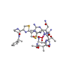
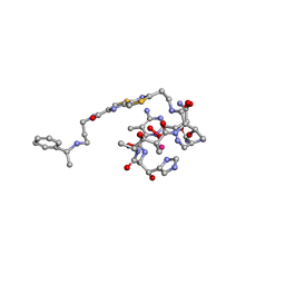 | | COBALT(III)-PEPLOMYCIN COMPLEX DETERMINED BY NMR STUDIES | | 分子名称: | 3-O-carbamoyl-alpha-D-mannopyranose-(1-2)-alpha-L-gulopyranose, AGLYCON OF PEPLOMYCIN, COBALT (III) ION, ... | | 著者 | Caceres-Cortes, J, Sugiyama, H, Ikudome, K, Saito, I, Wang, A.H.-J. | | 登録日 | 1997-07-16 | | 公開日 | 1999-07-30 | | 最終更新日 | 2023-12-27 | | 実験手法 | SOLUTION NMR | | 主引用文献 | Structures of cobalt(III)-pepleomycin and cobalt(III)-deglycopepleomycin (green forms) determined by NMR studies.
Eur.J.Biochem., 244, 1997
|
|
1B0S
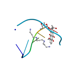
 | | BINDING OF AR-1-144, A TRI-IMIDAZOLE DNA MINOR GROOVE BINDER, TO CCGG SEQUENCE ANALYZED BY NMR SPECTROSCOPY | | 分子名称: | (2-{[4-({4-[(4-FORMYLAMINO-1-METHYL-1H-IMIDAZOLE-2-CARBONYL)-AMINO]-1-METHYL-1H-IMIDAZOLE-2-CARBONYL}-AMINO)-1-METHYL-1 H-IMIDAZOLE-2-CARBONYL]-AMINO}-ETHYL)-DIMETHYL-AMMONIUM, DNA (5'-D(*GP*AP*AP*CP*CP*GP*GP*TP*TP*C)-3') | | 著者 | Yang, X.-L, Kaenzig, C, Lee, M, Wang, A.H.-J. | | 登録日 | 1998-11-12 | | 公開日 | 1999-08-31 | | 最終更新日 | 2023-12-27 | | 実験手法 | SOLUTION NMR | | 主引用文献 | Binding of AR-1-144, a tri-imidazole DNA minor groove binder, to CCGG sequence analyzed by NMR spectroscopy.
Eur.J.Biochem., 263, 1999
|
|
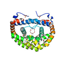
3W1O
 | | Neisseria DNA mimic protein DMP12 | | 分子名称: | DNA mimic protein DMP12, MAGNESIUM ION | | 著者 | Wang, H.C, Ko, T.P, Wu, M.L, Wang, A.H.J. | | 登録日 | 2012-11-19 | | 公開日 | 2013-04-10 | | 最終更新日 | 2024-03-20 | | 実験手法 | X-RAY DIFFRACTION (1.85 Å) | | 主引用文献 | Neisseria conserved hypothetical protein DMP12 is a DNA mimic that binds to histone-like HU protein
Nucleic Acids Res., 41, 2013
|
|
1AO2
 | | cobalt(III)-deglycopepleomycin determined by NMR studies | | 分子名称: | AGLYCON OF PEPLOMYCIN, COBALT (III) ION, HYDROGEN PEROXIDE | | 著者 | Caceres-Cortes, J, Sugiyama, H, Ikudome, K, Saito, I, Wang, A.H.-J. | | 登録日 | 1997-07-16 | | 公開日 | 1999-07-30 | | 最終更新日 | 2024-03-13 | | 実験手法 | SOLUTION NMR | | 主引用文献 | Structures of cobalt(III)-pepleomycin and cobalt(III)-deglycopepleomycin (green forms) determined by NMR studies.
Eur.J.Biochem., 244, 1997
|
|
2ZEM
 | | Crystal structure of the human glutaminyl cyclase mutant D248Q at 2.18 angstrom resolution | | 分子名称: | Glutaminyl-peptide cyclotransferase, SULFATE ION, ZINC ION | | 著者 | Huang, K.F, Wang, Y.R, Chang, E.C, Chou, T.L, Wang, A.H. | | 登録日 | 2007-12-13 | | 公開日 | 2008-04-22 | | 最終更新日 | 2023-11-01 | | 実験手法 | X-RAY DIFFRACTION (2.18 Å) | | 主引用文献 | A conserved hydrogen-bond network in the catalytic centre of animal glutaminyl cyclases is critical for catalysis.
Biochem.J., 411, 2008
|
|
2ZEP
 | | Crystal structure of the human glutaminyl cyclase mutant H319L at 2.1 angstrom resolution | | 分子名称: | Glutaminyl-peptide cyclotransferase, SULFATE ION, ZINC ION | | 著者 | Huang, K.F, Wang, Y.R, Chang, E.C, Chou, T.L, Wang, A.H. | | 登録日 | 2007-12-13 | | 公開日 | 2008-04-22 | | 最終更新日 | 2023-11-01 | | 実験手法 | X-RAY DIFFRACTION (2.1 Å) | | 主引用文献 | A conserved hydrogen-bond network in the catalytic centre of animal glutaminyl cyclases is critical for catalysis.
Biochem.J., 411, 2008
|
|
2ZEE
 | | Crystal structure of the human glutaminyl cyclase mutant S160G at 1.99 angstrom resolution | | 分子名称: | Glutaminyl-peptide cyclotransferase, SULFATE ION, ZINC ION | | 著者 | Huang, K.F, Wang, Y.R, Chang, E.C, Chou, T.L, Wang, A.H. | | 登録日 | 2007-12-12 | | 公開日 | 2008-04-22 | | 最終更新日 | 2023-11-01 | | 実験手法 | X-RAY DIFFRACTION (1.99 Å) | | 主引用文献 | A conserved hydrogen-bond network in the catalytic centre of animal glutaminyl cyclases is critical for catalysis.
Biochem.J., 411, 2008
|
|
2ZEO
 | | Crystal structure of the human glutaminyl cyclase mutant D305E at 1.66 angstrom resolution | | 分子名称: | Glutaminyl-peptide cyclotransferase, SULFATE ION, ZINC ION | | 著者 | Huang, K.F, Wang, Y.R, Chang, E.C, Chou, T.L, Wang, A.H. | | 登録日 | 2007-12-13 | | 公開日 | 2008-04-22 | | 最終更新日 | 2023-11-01 | | 実験手法 | X-RAY DIFFRACTION (1.66 Å) | | 主引用文献 | A conserved hydrogen-bond network in the catalytic centre of animal glutaminyl cyclases is critical for catalysis.
Biochem.J., 411, 2008
|
|
2ZEF
 | | Crystal structure of the human glutaminyl cyclase mutant E201D at 1.67 angstrom resolution | | 分子名称: | Glutaminyl-peptide cyclotransferase, SULFATE ION, ZINC ION | | 著者 | Huang, K.F, Wang, Y.R, Chang, E.C, Chou, T.L, Wang, A.H. | | 登録日 | 2007-12-12 | | 公開日 | 2008-04-22 | | 最終更新日 | 2023-11-01 | | 実験手法 | X-RAY DIFFRACTION (1.67 Å) | | 主引用文献 | A conserved hydrogen-bond network in the catalytic centre of animal glutaminyl cyclases is critical for catalysis.
Biochem.J., 411, 2008
|
|
2ZEL
 | | Crystal structure of the human glutaminyl cyclase mutant D248A at 1.97 angstrom resolution | | 分子名称: | Glutaminyl-peptide cyclotransferase, SULFATE ION, ZINC ION | | 著者 | Huang, K.F, Wang, Y.R, Chang, E.C, Chou, T.L, Wang, A.H. | | 登録日 | 2007-12-13 | | 公開日 | 2008-04-22 | | 最終更新日 | 2023-11-01 | | 実験手法 | X-RAY DIFFRACTION (1.97 Å) | | 主引用文献 | A conserved hydrogen-bond network in the catalytic centre of animal glutaminyl cyclases is critical for catalysis.
Biochem.J., 411, 2008
|
|
2ZEG
 | | Crystal structure of the human glutaminyl cyclase mutant E201L at 2.08 angstrom resolution | | 分子名称: | Glutaminyl-peptide cyclotransferase, IMIDAZOLE, SULFATE ION, ... | | 著者 | Huang, K.F, Wang, Y.R, Chang, E.C, Chou, T.L, Wang, A.H. | | 登録日 | 2007-12-12 | | 公開日 | 2008-04-22 | | 最終更新日 | 2023-11-01 | | 実験手法 | X-RAY DIFFRACTION (2.08 Å) | | 主引用文献 | A conserved hydrogen-bond network in the catalytic centre of animal glutaminyl cyclases is critical for catalysis.
Biochem.J., 411, 2008
|
|
3WUR
 | | Structure of DMP19 Complex with 18-crown-6 | | 分子名称: | 1,2-ETHANEDIOL, 1,4,7,10,13,16-HEXAOXACYCLOOCTADECANE, L(+)-TARTARIC ACID, ... | | 著者 | Lee, C.C, Wang, H.C, Wang, A.H.J. | | 登録日 | 2014-05-02 | | 公開日 | 2014-10-15 | | 最終更新日 | 2024-05-29 | | 実験手法 | X-RAY DIFFRACTION (1.45 Å) | | 主引用文献 | Crowning proteins: modulating the protein surface properties using crown ethers.
Angew.Chem.Int.Ed.Engl., 53, 2014
|
|
3PID
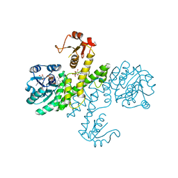
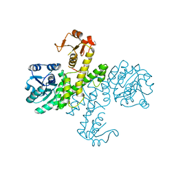 | | The apo-form UDP-glucose 6-dehydrogenase with a C-terminal six-histidine tag | | 分子名称: | UDP-glucose 6-dehydrogenase | | 著者 | Chen, Y.-Y, Ko, T.-P, Lin, C.-H, Chen, W.-H, Wang, A.H.-J. | | 登録日 | 2010-11-06 | | 公開日 | 2011-09-28 | | 最終更新日 | 2023-11-01 | | 実験手法 | X-RAY DIFFRACTION (1.4 Å) | | 主引用文献 | Conformational change upon product binding to Klebsiella pneumoniae UDP-glucose dehydrogenase: a possible inhibition mechanism for the key enzyme in polymyxin resistance.
J.Struct.Biol., 175, 2011
|
|
3PJG
 | | Crystal structure of UDP-glucose dehydrogenase from Klebsiella pneumoniae complexed with product UDP-glucuronic acid | | 分子名称: | 3-CYCLOHEXYL-1-PROPYLSULFONIC ACID, UDP-glucose 6-dehydrogenase, URIDINE-5'-DIPHOSPHATE-GLUCURONIC ACID | | 著者 | Chen, Y.-Y, Ko, T.-P, Lin, C.-H, Chen, W.-H, Wang, A.H.-J. | | 登録日 | 2010-11-10 | | 公開日 | 2011-09-28 | | 最終更新日 | 2023-11-01 | | 実験手法 | X-RAY DIFFRACTION (2.7 Å) | | 主引用文献 | Conformational change upon product binding to Klebsiella pneumoniae UDP-glucose dehydrogenase: a possible inhibition mechanism for the key enzyme in polymyxin resistance.
J.Struct.Biol., 175, 2011
|
|
3PLR
 | | Crystal structure of Klebsiella pneumoniae UDP-glucose 6-dehydrogenase complexed with NADH and UDP-glucose | | 分子名称: | 1,4-DIHYDRONICOTINAMIDE ADENINE DINUCLEOTIDE, UDP-glucose 6-dehydrogenase, URIDINE-5'-MONOPHOSPHATE | | 著者 | Chen, Y.-Y, Ko, T.-P, Lin, C.-H, Chen, W.-H, Wang, A.H.-J. | | 登録日 | 2010-11-15 | | 公開日 | 2011-09-28 | | 最終更新日 | 2023-11-01 | | 実験手法 | X-RAY DIFFRACTION (1.7 Å) | | 主引用文献 | Conformational change upon product binding to Klebsiella pneumoniae UDP-glucose dehydrogenase: a possible inhibition mechanism for the key enzyme in polymyxin resistance.
J.Struct.Biol., 175, 2011
|
|
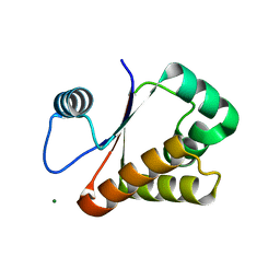
3VOP
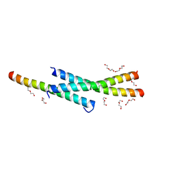 | | Structure of Vaccinia virus A27 | | 分子名称: | DI(HYDROXYETHYL)ETHER, Protein A27 | | 著者 | Chang, T.H, Ko, T.P, Hsieh, F.L, Wang, A.H.J. | | 登録日 | 2012-01-31 | | 公開日 | 2013-03-06 | | 最終更新日 | 2024-03-20 | | 実験手法 | X-RAY DIFFRACTION (2.2 Å) | | 主引用文献 | Crystal structure of vaccinia viral A27 protein reveals a novel structure critical for its function and complex formation with A26 protein.
Plos Pathog., 9, 2013
|
|


