4O8U
 
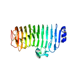
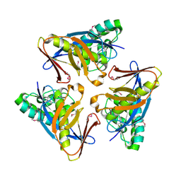 | | Structure of PF2046 | | 分子名称: | Uncharacterized protein PF2046 | | 著者 | Su, J, Liu, Z.-J. | | 登録日 | 2013-12-30 | | 公開日 | 2014-04-30 | | 実験手法 | X-RAY DIFFRACTION (2.345 Å) | | 主引用文献 | Crystal structure of a novel non-Pfam protein PF2046 solved using low resolution B-factor sharpening and multi-crystal averaging methods
Protein Cell, 1, 2010
|
|
6KVE
 | |
6AKK
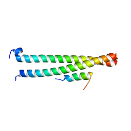
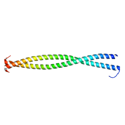
 | | Crystal structure of the second Coiled-coil domain of SIKE1 | | 分子名称: | GLYCEROL, Suppressor of IKBKE 1 | | 著者 | Zhou, L, Chen, M, Zhou, Z.C. | | 登録日 | 2018-09-02 | | 公開日 | 2019-01-16 | | 最終更新日 | 2024-03-27 | | 実験手法 | X-RAY DIFFRACTION (1.5 Å) | | 主引用文献 | Architecture, substructures, and dynamic assembly of STRIPAK complexes in Hippo signaling.
Cell Discov, 5, 2019
|
|
6AKL
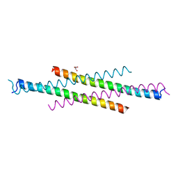
 | | Crystal structure of Striatin3 in complex with SIKE1 Coiled-coil domain | | 分子名称: | Striatin-3, Suppressor of IKBKE 1 | | 著者 | Zhou, L, Chen, M, Zhou, Z.C. | | 登録日 | 2018-09-02 | | 公開日 | 2019-01-16 | | 最終更新日 | 2023-11-22 | | 実験手法 | X-RAY DIFFRACTION (1.75 Å) | | 主引用文献 | Architecture, substructures, and dynamic assembly of STRIPAK complexes in Hippo signaling.
Cell Discov, 5, 2019
|
|
6KVH
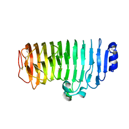
 | | The mutant crystal structure of endo-polygalacturonase (T284A) from Talaromyces leycettanus JCM 12802 | | 分子名称: | 2-acetamido-2-deoxy-beta-D-glucopyranose, alpha-D-mannopyranose, endo-polygalacturonase | | 著者 | Tu, T, Wang, Z, Luo, H, Yao, B. | | 登録日 | 2019-09-04 | | 公開日 | 2020-09-09 | | 最終更新日 | 2023-11-22 | | 実験手法 | X-RAY DIFFRACTION (1.2 Å) | | 主引用文献 | Structural Insights into the Mechanisms Underlying the Kinetic Stability of GH28 Endo-Polygalacturonase.
J.Agric.Food Chem., 69, 2021
|
|
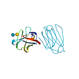
4KTE
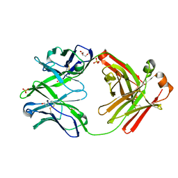 | | Fab fragment of HIV vaccine-elicited CD4bs-directed antibody, GE148, from non-human primate | | 分子名称: | GE148 Heavy Chain Fab, GE148 Light Chain Fab, GLYCEROL, ... | | 著者 | Poulsen, C, Tran, K, Stanfield, R, Wyatt, R.T. | | 登録日 | 2013-05-20 | | 公開日 | 2014-02-05 | | 最終更新日 | 2019-12-25 | | 実験手法 | X-RAY DIFFRACTION (1.8 Å) | | 主引用文献 | Vaccine-elicited primate antibodies use a distinct approach to the HIV-1 primary receptor binding site informing vaccine redesign.
Proc.Natl.Acad.Sci.USA, 111, 2014
|
|
5YF1
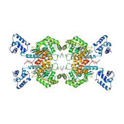
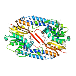 | | Crystal structure of CARNMT1 bound to carnosine and SFG | | 分子名称: | (2~{S})-2-(3-azanylpropanoylamino)-3-(1~{H}-imidazol-4-yl)propanoic acid, 1,2-ETHANEDIOL, ACETATE ION, ... | | 著者 | Cao, R, Li, H. | | 登録日 | 2017-09-20 | | 公開日 | 2018-08-01 | | 最終更新日 | 2024-03-27 | | 実験手法 | X-RAY DIFFRACTION (2.399 Å) | | 主引用文献 | Molecular basis for histidine N1 position-specific methylation by CARNMT1.
Cell Res., 28, 2018
|
|
5YF0
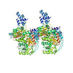
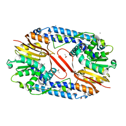 | | Crystal structure of CARNMT1 bound to SAM | | 分子名称: | 1,2-ETHANEDIOL, ACETATE ION, CALCIUM ION, ... | | 著者 | Cao, R, Zhang, X, Li, H. | | 登録日 | 2017-09-20 | | 公開日 | 2018-08-01 | | 最終更新日 | 2024-03-27 | | 実験手法 | X-RAY DIFFRACTION (2.25 Å) | | 主引用文献 | Molecular basis for histidine N1 position-specific methylation by CARNMT1.
Cell Res., 28, 2018
|
|
5YT6
 | |
5YF2
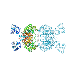
 | | Crystal structure of CARNMT1 bound to anserine and SAH | | 分子名称: | (2~{S})-2-(3-azanylpropanoylamino)-3-(3-methylimidazol-4-yl)propanoic acid, CALCIUM ION, Carnosine N-methyltransferase, ... | | 著者 | Cao, R, Li, H. | | 登録日 | 2017-09-20 | | 公開日 | 2018-08-01 | | 最終更新日 | 2024-03-27 | | 実験手法 | X-RAY DIFFRACTION (2.802 Å) | | 主引用文献 | Molecular basis for histidine N1 position-specific methylation by CARNMT1.
Cell Res., 28, 2018
|
|
6JTP
 | | Crystal structure of HLA-C08 in complex with a tumor mut9m peptide | | 分子名称: | 9-mer peptide, Beta-2-microglobulin, HLA class I antigen, ... | | 著者 | Bai, P, Zhou, Q, Wei, P, Lei, Y. | | 登録日 | 2019-04-11 | | 公開日 | 2020-04-15 | | 最終更新日 | 2021-02-17 | | 実験手法 | X-RAY DIFFRACTION (1.9 Å) | | 主引用文献 | Immune-based mutation classification enables neoantigen prioritization and immune feature discovery in cancer immunotherapy.
Oncoimmunology, 10, 2021
|
|
3BAT
 | |
5WCH
 | | Crystal structure of the catalytic domain of human USP9X | | 分子名称: | Probable ubiquitin carboxyl-terminal hydrolase FAF-X, UNKNOWN ATOM OR ION, ZINC ION | | 著者 | Dong, A, Zhang, Q, Walker, J.R, Bountra, C, Arrowsmith, C.H, Edwards, A.M, Tong, Y, Structural Genomics Consortium (SGC) | | 登録日 | 2017-06-30 | | 公開日 | 2018-07-04 | | 最終更新日 | 2024-03-13 | | 実験手法 | X-RAY DIFFRACTION (2.5 Å) | | 主引用文献 | Crystal structure and activity-based labeling reveal the mechanisms for linkage-specific substrate recognition by deubiquitinase USP9X.
Proc. Natl. Acad. Sci. U.S.A., 116, 2019
|
|
3BAS
 | |
6UJG
 | |
6UME
 | |
6UMD
 | |
3BZH
 | | Crystal structure of human ubiquitin-conjugating enzyme E2 E1 | | 分子名称: | GLYCEROL, Ubiquitin-conjugating enzyme E2 E1 | | 著者 | Walker, J.R, Avvakumov, G.V, Xue, S, Li, Y, Weigelt, J, Arrowsmith, C.H, Edwards, A.M, Bochkarev, A, Dhe-Paganon, S, Structural Genomics Consortium (SGC) | | 登録日 | 2008-01-18 | | 公開日 | 2008-02-26 | | 最終更新日 | 2023-08-30 | | 実験手法 | X-RAY DIFFRACTION (1.6 Å) | | 主引用文献 | A human ubiquitin conjugating enzyme (E2)-HECT E3 ligase structure-function screen.
Mol Cell Proteomics, 11, 2012
|
|
6UMC
 | |
6A62
 | | Placental protein 13/galectin-13 variant R53HH57RD33G with Lactose | | 分子名称: | Galactoside-binding soluble lectin 13, beta-D-galactopyranose-(1-4)-beta-D-glucopyranose | | 著者 | Su, J. | | 登録日 | 2018-06-26 | | 公開日 | 2018-12-26 | | 最終更新日 | 2020-07-29 | | 実験手法 | X-RAY DIFFRACTION (2.03 Å) | | 主引用文献 | Resetting the ligand binding site of placental protein 13/galectin-13 recovers its ability to bind lactose
Biosci. Rep., 38, 2018
|
|
6A1T
 | | Charcot-Leyden crystal protein/Galectin-10 variant E33A with lactose | | 分子名称: | Galectin-10, beta-D-galactopyranose-(1-4)-beta-D-glucopyranose | | 著者 | Su, J. | | 登録日 | 2018-06-08 | | 公開日 | 2018-12-26 | | 最終更新日 | 2024-03-27 | | 実験手法 | X-RAY DIFFRACTION (1.97 Å) | | 主引用文献 | Identification of key amino acid residues determining ligand binding specificity, homodimerization and cellular distribution of human galectin-10
Glycobiology, 29, 2019
|
|
6A4C
 | |
6ULJ
 | |
6UMF
 | |
6UK6
 | |


