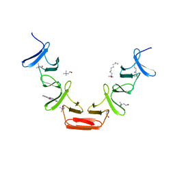
2PKR
 
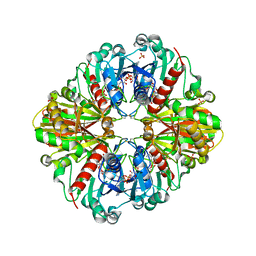 | | Crystal structure of (A+CTE)4 chimeric form of photosyntetic glyceraldehyde-3-phosphate dehydrogenase, complexed with NADP | | 分子名称: | Glyceraldehyde-3-phosphate dehydrogenase Aor, NADPH DIHYDRO-NICOTINAMIDE-ADENINE-DINUCLEOTIDE PHOSPHATE, SULFATE ION | | 著者 | Fermani, S, Falini, G, Ripamonti, A. | | 登録日 | 2007-04-18 | | 公開日 | 2007-06-19 | | 最終更新日 | 2024-03-13 | | 実験手法 | X-RAY DIFFRACTION (2.4 Å) | | 主引用文献 | Molecular mechanism of thioredoxin regulation in photosynthetic A2B2-glyceraldehyde-3-phosphate dehydrogenase.
Proc.Natl.Acad.Sci.Usa, 104, 2007
|
|
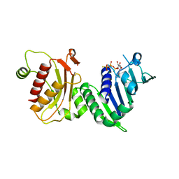
1NHQ
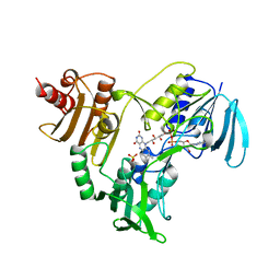 | | CRYSTALLOGRAPHIC ANALYSES OF NADH PEROXIDASE CYS42ALA AND CYS42SER MUTANTS: ACTIVE SITE STRUCTURE, MECHANISTIC IMPLICATIONS, AND AN UNUSUAL ENVIRONMENT OF ARG303 | | 分子名称: | FLAVIN-ADENINE DINUCLEOTIDE, NADH PEROXIDASE, SULFATE ION | | 著者 | Mande, S.S, Claiborne, A, Hol, W.G.J. | | 登録日 | 1994-12-09 | | 公開日 | 1995-02-14 | | 最終更新日 | 2024-02-14 | | 実験手法 | X-RAY DIFFRACTION (2 Å) | | 主引用文献 | Crystallographic analyses of NADH peroxidase Cys42Ala and Cys42Ser mutants: active site structures, mechanistic implications, and an unusual environment of Arg 303.
Biochemistry, 34, 1995
|
|
8F68
 | | E. coli cytochrome bo3 ubiquinol oxidase monomer | | 分子名称: | 1,2-Distearoyl-sn-glycerophosphoethanolamine, COPPER (II) ION, Cytochrome bo(3) ubiquinol oxidase subunit 1, ... | | 著者 | Guo, Y, Karimullina, E, Borek, D, Savchenko, A. | | 登録日 | 2022-11-16 | | 公開日 | 2022-11-30 | | 最終更新日 | 2024-05-22 | | 実験手法 | ELECTRON MICROSCOPY (3.15 Å) | | 主引用文献 | Monomer and dimer structures of cytochrome bo 3 ubiquinol oxidase from Escherichia coli.
Protein Sci., 32, 2023
|
|
8F6C
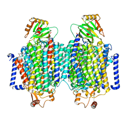 | | E. coli cytochrome bo3 ubiquinol oxidase dimer | | 分子名称: | 1,2-Distearoyl-sn-glycerophosphoethanolamine, COPPER (II) ION, Cytochrome bo(3) ubiquinol oxidase subunit 1, ... | | 著者 | Guo, Y, Karimullina, E, Borek, D, Savchenko, A. | | 登録日 | 2022-11-16 | | 公開日 | 2022-11-30 | | 最終更新日 | 2024-05-22 | | 実験手法 | ELECTRON MICROSCOPY (3.46 Å) | | 主引用文献 | Monomer and dimer structures of cytochrome bo 3 ubiquinol oxidase from Escherichia coli.
Protein Sci., 32, 2023
|
|
1N7L
 | |
1H3O
 | |
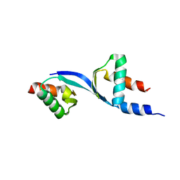
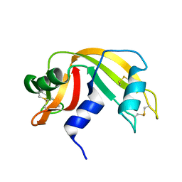
1H5O
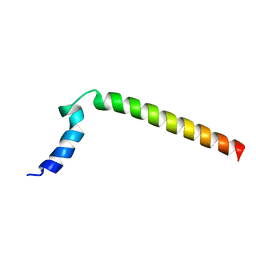
 | | Solution structure of Crotamine, a neurotoxin from Crotalus durissus terrificus | | 分子名称: | MYOTOXIN | | 著者 | Nicastro, G, Franzoni, L, De Chiara, C, Mancin, C.A, Giglio, J.R, Spisni, A. | | 登録日 | 2001-05-23 | | 公開日 | 2003-05-09 | | 最終更新日 | 2013-07-24 | | 実験手法 | SOLUTION NMR | | 主引用文献 | Solution Structure of Crotamine, a Na+ Channel Affecting Toxin from Crotalus Durissus Terrificus Venom
Eur.J.Biochem., 270, 2003
|
|
1N8P
 | | Crystal Structure of cystathionine gamma-lyase from yeast | | 分子名称: | Cystathionine gamma-lyase, PYRIDOXAL-5'-PHOSPHATE | | 著者 | Messerschmidt, A, Worbs, M, Steegborn, C, Wahl, M.C, Huber, R, Clausen, T. | | 登録日 | 2002-11-21 | | 公開日 | 2002-12-04 | | 最終更新日 | 2023-08-16 | | 実験手法 | X-RAY DIFFRACTION (2.6 Å) | | 主引用文献 | Determinants of Enzymatic Specificity in the Cys-Met-Metabolism PLP-Dependent Enzymes Family: Crystal Structure of Cystathionine gamma-lyase from Yeast and Intrafamiliar Structural Comparison
BIOL.CHEM., 384, 2003
|
|
8EOZ
 | |
1H8V
 | | The X-ray Crystal Structure of the Trichoderma reesei Family 12 Endoglucanase 3, Cel12A, at 1.9 A Resolution | | 分子名称: | 2-acetamido-2-deoxy-beta-D-glucopyranose, ENDO-BETA-1,4-GLUCANASE | | 著者 | Sandgren, M, Shaw, A, Ropp, T.H, Wu, S, Bott, R, Cameron, A.D, Stahlberg, J, Mitchinson, C, Jones, T.A. | | 登録日 | 2001-02-16 | | 公開日 | 2001-04-24 | | 最終更新日 | 2020-07-29 | | 実験手法 | X-RAY DIFFRACTION (1.9 Å) | | 主引用文献 | The X-Ray Crystal Structure of the Trichoderma Reesei Family 12 Endoglucanase 3, Cel12A, at 1.9 A Resolution
J.Mol.Biol., 308, 2001
|
|
1HE9
 | | Crystal structure of the GAP domain of the Pseudomonas aeruginosa ExoS toxin | | 分子名称: | EXOENZYME S | | 著者 | Wurtele, M, Renault, L, Barbieri, J.T, Wittinghofer, A, Wolf, E. | | 登録日 | 2000-11-21 | | 公開日 | 2001-03-19 | | 最終更新日 | 2011-07-13 | | 実験手法 | X-RAY DIFFRACTION (2.4 Å) | | 主引用文献 | Structure of the Exos Gtpase Activating Domain
FEBS Lett., 491, 2001
|
|
1H3F
 | | Tyrosyl-tRNA synthetase from Thermus thermophilus complexed with tyrosinol | | 分子名称: | 4-[(2S)-2-amino-3-hydroxypropyl]phenol, SULFATE ION, TYROSYL-TRNA SYNTHETASE | | 著者 | Cusack, S, Yaremchuk, A, Kriklivyi, I, Tukalo, M. | | 登録日 | 2002-08-28 | | 公開日 | 2002-09-12 | | 最終更新日 | 2024-05-01 | | 実験手法 | X-RAY DIFFRACTION (2 Å) | | 主引用文献 | Class I Tyrosyl-tRNA Synthetase Has a Class II Mode or tRNA Recognition
Embo J., 21, 2002
|
|
1KF3
 | | Atomic Resolution Structure of RNase A at pH 5.9 | | 分子名称: | SULFATE ION, pancreatic ribonuclease | | 著者 | Berisio, R, Sica, F, Lamzin, V.S, Wilson, K.S, Zagari, A, Mazzarella, L. | | 登録日 | 2001-11-19 | | 公開日 | 2001-12-19 | | 最終更新日 | 2023-08-16 | | 実験手法 | X-RAY DIFFRACTION (1.05 Å) | | 主引用文献 | Atomic resolution structures of ribonuclease A at six pH values.
Acta Crystallogr.,Sect.D, 58, 2002
|
|
1XZI
 | |
5SZW
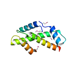
 | | NMR solution structure of the RRM1 domain of the post-transcriptional regulator HuR | | 分子名称: | ELAV-like protein 1 | | 著者 | Lixa, C, Mujo, A, Jendiroba, K.A, Almeida, F.C.L, Lima, L.M.T.R, Pinheiro, A.S. | | 登録日 | 2016-08-15 | | 公開日 | 2017-09-06 | | 最終更新日 | 2024-05-01 | | 実験手法 | SOLUTION NMR | | 主引用文献 | Oligomeric transition and dynamics of RNA binding by the HuR RRM1 domain in solution.
J. Biomol. NMR, 72, 2018
|
|
1KJ1
 | | MANNOSE-SPECIFIC AGGLUTININ (LECTIN) FROM GARLIC (ALLIUM SATIVUM) BULBS COMPLEXED WITH ALPHA-D-MANNOSE | | 分子名称: | alpha-D-mannopyranose, lectin I, lectin II | | 著者 | Ramachandraiah, G, Chandra, N.R, Surolia, A, Vijayan, M. | | 登録日 | 2001-12-04 | | 公開日 | 2002-02-22 | | 最終更新日 | 2023-08-16 | | 実験手法 | X-RAY DIFFRACTION (2.2 Å) | | 主引用文献 | Re-refinement using reprocessed data to improve the quality of the structure: a case study involving garlic lectin.
Acta Crystallogr.,Sect.D, 58, 2002
|
|
1GQU
 | |
1GZF
 | | Structure of the Clostridium botulinum C3 exoenzyme (wild-type) in complex with NAD | | 分子名称: | 3-(AMINOCARBONYL)-1-[(3R,4S,5R)-3,4-DIHYDROXY-5-METHYLTETRAHYDRO-2-FURANYL]PYRIDINIUM, ADENOSINE-5'-DIPHOSPHATE, MONO-ADP-RIBOSYLTRANSFERASE C3, ... | | 著者 | Menetrey, J, Flatau, G, Stura, E.A, Charbonnier, J.B, Gas, F, Teulon, J.M, Le Du, M.H, Boquet, P, Menez, A. | | 登録日 | 2002-05-21 | | 公開日 | 2002-08-29 | | 最終更新日 | 2024-05-08 | | 実験手法 | X-RAY DIFFRACTION (1.95 Å) | | 主引用文献 | Nad Binding Induces Conformational Changes in Rho Adp-Ribosylating Clostridium Botulinum C3 Exoenzyme
J.Biol.Chem., 277, 2002
|
|
1H6K
 | | nuclear Cap Binding Complex | | 分子名称: | 20 KDA NUCLEAR CAP BINDING PROTEIN, CBP80 | | 著者 | Mazza, C, Ohno, M, Segref, A, Mattaj, I.W, Cusack, S. | | 登録日 | 2001-06-18 | | 公開日 | 2001-09-13 | | 最終更新日 | 2024-05-08 | | 実験手法 | X-RAY DIFFRACTION (2 Å) | | 主引用文献 | Crystal Structure of the Human Nuclear CAP Binding Complex
Mol.Cell, 8, 2001
|
|
1HCX
 | | Choline binding domain of the major autolysin (C-LytA) from Streptococcus pneumoniae | | 分子名称: | 2,2':6',2''-TERPYRIDINE PLATINUM(II) Chloride, CHOLINE ION, DECYLAMINE-N,N-DIMETHYL-N-OXIDE, ... | | 著者 | Fernandez-Tornero, C, Lopez, R, Garcia, E, Gimenez-Gallego, G, Romero, A. | | 登録日 | 2001-05-10 | | 公開日 | 2001-11-29 | | 最終更新日 | 2024-05-08 | | 実験手法 | X-RAY DIFFRACTION (2.6 Å) | | 主引用文献 | A Novel Solenoid Fold in the Cell Wall Anchoring Domain of the Pneumococcal Virulence Factor Lyta
Nat.Struct.Biol., 8, 2001
|
|
1H7U
 | | hPMS2-ATPgS | | 分子名称: | MAGNESIUM ION, MISMATCH REPAIR ENDONUCLEASE PMS2, PHOSPHOTHIOPHOSPHORIC ACID-ADENYLATE ESTER | | 著者 | Guarne, A, Junop, M.S, Yang, W. | | 登録日 | 2001-07-10 | | 公開日 | 2001-11-27 | | 最終更新日 | 2024-05-08 | | 実験手法 | X-RAY DIFFRACTION (2.7 Å) | | 主引用文献 | Structure and Function of the N-Terminal 40 kDa Fragment of Human Pms2: A Monomeric Ghl ATPase
Embo J., 20, 2001
|
|
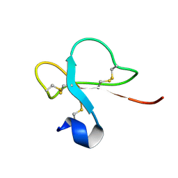
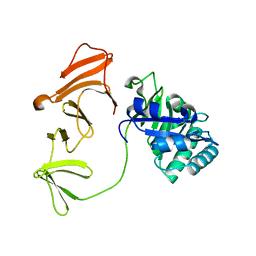
3DOM
 | | Crystal Structure of the complex between Tfb5 and the C-terminal domain of Tfb2 | | 分子名称: | RNA polymerase II transcription factor B subunit 2, RNA polymerase II transcription factor B subunit 5 | | 著者 | Kainov, D.E, Cavarelli, J, Egly, J.M, Poterszman, A. | | 登録日 | 2008-07-04 | | 公開日 | 2008-08-19 | | 最終更新日 | 2024-02-21 | | 実験手法 | X-RAY DIFFRACTION (2.6 Å) | | 主引用文献 | Structural basis for group A trichothiodystrophy
Nat.Struct.Mol.Biol., 15, 2008
|
|
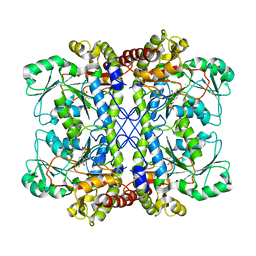
8ERN
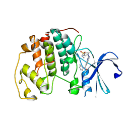 | | Cyclin-free CDK2 in complex with Cpd21 | | 分子名称: | Cyclin-dependent kinase 2, GLYCEROL, N,3-dimethyl-4-({7-[(2S)-2-methylpyrrolidine-1-carbonyl]quinazolin-2-yl}amino)benzene-1-sulfonamide | | 著者 | Murray, J.M, Oh, A. | | 登録日 | 2022-10-12 | | 公開日 | 2023-10-18 | | 実験手法 | X-RAY DIFFRACTION (1.64 Å) | | 主引用文献 | Cyclin-free CDK2 in complex with Cpd21
To Be Published
|
|
1KF5
 | | Atomic Resolution Structure of RNase A at pH 7.1 | | 分子名称: | pancreatic ribonuclease | | 著者 | Berisio, R, Sica, F, Lamzin, V.S, Wilson, K.S, Zagari, A, Mazzarella, L. | | 登録日 | 2001-11-19 | | 公開日 | 2001-12-19 | | 最終更新日 | 2023-08-16 | | 実験手法 | X-RAY DIFFRACTION (1.15 Å) | | 主引用文献 | Atomic resolution structures of ribonuclease A at six pH values.
Acta Crystallogr.,Sect.D, 58, 2002
|
|
1H09
 | | Multimodular Pneumococcal Cell Wall Endolysin from phage Cp-1 | | 分子名称: | LYSOZYME | | 著者 | Hermoso, J.A, Monterroso, B, Albert, A, Garcia, P, Menendez, M, Martinez-Ripoll, M, Garcia, J.L. | | 登録日 | 2002-06-12 | | 公開日 | 2003-06-26 | | 最終更新日 | 2024-05-08 | | 実験手法 | X-RAY DIFFRACTION (2.1 Å) | | 主引用文献 | Structural Basis for Selective Recognition of Pneumococcal Cell Wall by Modular Endolysin from Phage Cp-1
Structure, 11, 2003
|
|


