8ILZ
 
 | |
6CDW
 | |
6CGK
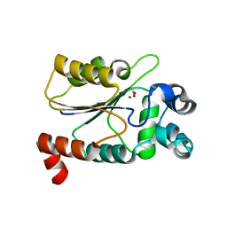
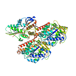
 | | Structure of the HAD domain of effector protein Lem4 (lpg1101) from Legionella pneumophila (inactive mutant)with phosphate bound in the active site | | 分子名称: | GLYCEROL, MAGNESIUM ION, PHOSPHATE ION, ... | | 著者 | Beyrakhova, K.A, Xu, C, Cygler, M. | | 登録日 | 2018-02-20 | | 公開日 | 2018-07-18 | | 最終更新日 | 2023-10-04 | | 実験手法 | X-RAY DIFFRACTION (1.668 Å) | | 主引用文献 | Legionella pneumophilaeffector Lem4 is a membrane-associated protein tyrosine phosphatase.
J. Biol. Chem., 293, 2018
|
|
6CNQ
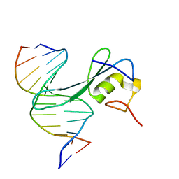 | | MBD2 in complex with methylated DNA | | 分子名称: | DNA (5'-D(*GP*CP*CP*AP*AP*(5CM)P*GP*TP*TP*GP*GP*C)-3'), Methyl-CpG-binding domain protein 2, UNKNOWN ATOM OR ION | | 著者 | Liu, K, Xu, C, Min, J, Structural Genomics Consortium (SGC) | | 登録日 | 2018-03-08 | | 公開日 | 2018-03-28 | | 最終更新日 | 2023-10-04 | | 実験手法 | X-RAY DIFFRACTION (2.151 Å) | | 主引用文献 | Structural basis for the ability of MBD domains to bind methyl-CG and TG sites in DNA.
J. Biol. Chem., 293, 2018
|
|
6CGJ
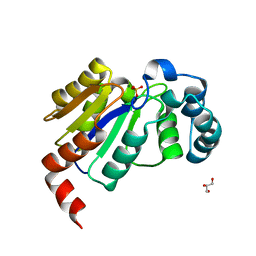
 | | Structure of the HAD domain of effector protein Lem4 (lpg1101) from Legionella pneumophila | | 分子名称: | ACETATE ION, GLYCEROL, MAGNESIUM ION, ... | | 著者 | Beyrakhova, K.A, Xu, C, Cygler, M. | | 登録日 | 2018-02-20 | | 公開日 | 2018-07-18 | | 最終更新日 | 2024-03-13 | | 実験手法 | X-RAY DIFFRACTION (1.75 Å) | | 主引用文献 | Legionella pneumophilaeffector Lem4 is a membrane-associated protein tyrosine phosphatase.
J. Biol. Chem., 293, 2018
|
|
6CT6
 | | Crystal structure of lactate dehydrogenase from Eimeria maxima with NADH and oxamate | | 分子名称: | 1,4-DIHYDRONICOTINAMIDE ADENINE DINUCLEOTIDE, Lactate dehydrogenase, OXAMIC ACID, ... | | 著者 | Wirth, J.D, Xu, C, Theobald, D.L. | | 登録日 | 2018-03-22 | | 公開日 | 2019-03-27 | | 最終更新日 | 2023-10-04 | | 実験手法 | X-RAY DIFFRACTION (1.705 Å) | | 主引用文献 | The Mechanistic, Structural, and Evolutionary Origin of Lactate Dehydrogenase Substrate Specificity in Apicomplexa
To Be Published
|
|
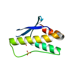
1YOP
 | | The solution structure of Kti11p | | 分子名称: | Kti11p, ZINC ION | | 著者 | Sun, J, Zhang, J, Wu, F, Xu, C, Li, S, Zhao, W, Wu, Z, Wu, J, Zhou, C.-Z, Shi, Y. | | 登録日 | 2005-01-28 | | 公開日 | 2005-04-05 | | 最終更新日 | 2024-05-29 | | 実験手法 | SOLUTION NMR | | 主引用文献 | Solution structure of Kti11p from Saccharomyces cerevisiae reveals a novel zinc-binding module.
Biochemistry, 44, 2005
|
|
3N2C
 | | Crystal structure of prolidase eah89906 complexed with n-methylphosphonate-l-proline | | 分子名称: | 1-[(R)-hydroxy(methyl)phosphoryl]-L-proline, PROLIDASE, ZINC ION | | 著者 | Patskovsky, Y, Xu, C, Sauder, J.M, Burley, S.K, Raushel, F.M, Almo, S.C, New York SGX Research Center for Structural Genomics (NYSGXRC) | | 登録日 | 2010-05-17 | | 公開日 | 2010-06-02 | | 最終更新日 | 2023-11-22 | | 実験手法 | X-RAY DIFFRACTION (2.81 Å) | | 主引用文献 | Functional identification and structure determination of two novel prolidases from cog1228 in the amidohydrolase superfamily .
Biochemistry, 49, 2010
|
|
3PD7
 | |
6MEW
 | | RFXANK ankyrin repeats in complex with a RFX7 peptide | | 分子名称: | DNA-binding protein RFXANK, RFX7 peptide, UNKNOWN ATOM OR ION | | 著者 | Tempel, W, Xu, C, Dong, A, Li, Y, Bountra, C, Arrowsmith, C.H, Edwards, A.M, Min, J, Structural Genomics Consortium (SGC) | | 登録日 | 2018-09-07 | | 公開日 | 2018-10-03 | | 最終更新日 | 2023-10-11 | | 実験手法 | X-RAY DIFFRACTION (1.78 Å) | | 主引用文献 | RFXANK ankyrin repeats in complex with a RFX7 peptide
to be published
|
|
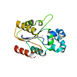
3R93
 | | Crystal structure of the chromo domain of M-phase phosphoprotein 8 bound to H3K9Me3 peptide | | 分子名称: | H3K9Me3 peptide, M-phase phosphoprotein 8, UNKNOWN ATOM OR ION | | 著者 | Li, J, Li, Z, Ruan, J, Xu, C, Tong, Y, Pan, P.W, Tempel, W, Crombet, L, Min, J, Zang, J, Structural Genomics Consortium (SGC) | | 登録日 | 2011-03-24 | | 公開日 | 2011-04-06 | | 最終更新日 | 2023-09-13 | | 実験手法 | X-RAY DIFFRACTION (2.057 Å) | | 主引用文献 | Structural basis for specific binding of human MPP8 chromodomain to histone H3 methylated at lysine 9.
Plos One, 6, 2011
|
|
3J8X
 | | High-resolution structure of no-nucleotide kinesin on microtubules | | 分子名称: | GUANOSINE-5'-DIPHOSPHATE, GUANOSINE-5'-TRIPHOSPHATE, Kinesin-1 heavy chain, ... | | 著者 | Shang, Z, Zhou, K, Xu, C, Csencsits, R, Cochran, J.C, Sindelar, C.V. | | 登録日 | 2014-11-20 | | 公開日 | 2014-12-10 | | 最終更新日 | 2024-02-21 | | 実験手法 | ELECTRON MICROSCOPY (5 Å) | | 主引用文献 | High-resolution structures of kinesin on microtubules provide a basis for nucleotide-gated force-generation.
Elife, 3, 2014
|
|
3J8Y
 | | High-resolution structure of ATP analog-bound kinesin on microtubules | | 分子名称: | ADENOSINE-5'-TRIPHOSPHATE, GUANOSINE-5'-DIPHOSPHATE, GUANOSINE-5'-TRIPHOSPHATE, ... | | 著者 | Shang, Z, Zhou, K, Xu, C, Csencsits, R, Cochran, J.C, Sindelar, C.V. | | 登録日 | 2014-11-20 | | 公開日 | 2014-12-10 | | 最終更新日 | 2024-02-21 | | 実験手法 | ELECTRON MICROSCOPY (5 Å) | | 主引用文献 | High-resolution structures of kinesin on microtubules provide a basis for nucleotide-gated force-generation.
Elife, 3, 2014
|
|
3EOP
 | | Crystal Structure of the DUF55 domain of human thymocyte nuclear protein 1 | | 分子名称: | SULFATE ION, Thymocyte nuclear protein 1 | | 著者 | Yu, F, Song, A, Xu, C, Sun, L, Li, L, Tang, L, Hu, H, He, J. | | 登録日 | 2008-09-29 | | 公開日 | 2009-09-29 | | 最終更新日 | 2023-11-01 | | 実験手法 | X-RAY DIFFRACTION (2.3 Å) | | 主引用文献 | Determining the DUF55-domain structure of human thymocyte nuclear protein 1 from crystals partially twinned by tetartohedry
Acta Crystallogr.,Sect.D, 65, 2009
|
|
3LWE
 | | The crystal structure of MPP8 | | 分子名称: | M-phase phosphoprotein 8 | | 著者 | Li, Z, Li, Z, Ruan, J, Xu, C, Tong, Y, Pan, P.W, Tempel, W, Crombet, L, Min, J, Zang, J, Structural Genomics Consortium (SGC) | | 登録日 | 2010-02-23 | | 公開日 | 2010-03-02 | | 最終更新日 | 2023-09-06 | | 実験手法 | X-RAY DIFFRACTION (2.05 Å) | | 主引用文献 | Structural basis for specific binding of human MPP8 chromodomain to histone H3 methylated at lysine 9.
Plos One, 6, 2011
|
|
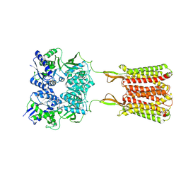
4PYP
 | | Crystal structure of the human glucose transporter GLUT1 | | 分子名称: | Solute carrier family 2, facilitated glucose transporter member 1, nonyl beta-D-glucopyranoside | | 著者 | Deng, D, Yan, C.Y, Xu, C, Wu, J.P, Sun, P.C, Hu, M.X, Yan, N. | | 登録日 | 2014-03-27 | | 公開日 | 2014-05-21 | | 最終更新日 | 2023-11-08 | | 実験手法 | X-RAY DIFFRACTION (3.166 Å) | | 主引用文献 | Crystal structure of the human glucose transporter GLUT1
Nature, 510, 2014
|
|
5YBJ
 | | Structure of apo KANK1 ankyrin domain | | 分子名称: | GLYCEROL, KN motif and ankyrin repeat domain-containing protein 1 | | 著者 | Guo, Q, Liao, S, Min, J, Xu, C, Structural Genomics Consortium (SGC) | | 登録日 | 2017-09-05 | | 公開日 | 2017-12-06 | | 最終更新日 | 2024-03-27 | | 実験手法 | X-RAY DIFFRACTION (2.341 Å) | | 主引用文献 | Structural basis for the recognition of kinesin family member 21A (KIF21A) by the ankyrin domains of KANK1 and KANK2 proteins.
J. Biol. Chem., 293, 2018
|
|
5YBV
 | | The structure of the KANK2 ankyrin domain with the KIF21A peptide | | 分子名称: | GLYCEROL, KN motif and ankyrin repeat domain-containing protein 2, Kinesin-like protein KIF21A, ... | | 著者 | Guo, Q, Liao, S, Min, J, Xu, C, Structural Genomics Consortium (SGC) | | 登録日 | 2017-09-05 | | 公開日 | 2017-12-06 | | 最終更新日 | 2023-11-22 | | 実験手法 | X-RAY DIFFRACTION (2.12 Å) | | 主引用文献 | Structural basis for the recognition of kinesin family member 21A (KIF21A) by the ankyrin domains of KANK1 and KANK2 proteins.
J. Biol. Chem., 293, 2018
|
|
5YBU
 | | Structure of the KANK1 ankyrin domain in complex with KIF21A peptide | | 分子名称: | KN motif and ankyrin repeat domain-containing protein 1, Kinesin-like protein KIF21A | | 著者 | Guo, Q, Liao, S, Min, J, Xu, C, Structural Genomics Consortium (SGC) | | 登録日 | 2017-09-05 | | 公開日 | 2017-12-06 | | 最終更新日 | 2023-11-22 | | 実験手法 | X-RAY DIFFRACTION (1.89 Å) | | 主引用文献 | Structural basis for the recognition of kinesin family member 21A (KIF21A) by the ankyrin domains of KANK1 and KANK2 proteins.
J. Biol. Chem., 293, 2018
|
|
5ZAZ
 | |
5ZN9
 | | Crystal structure of PX domain | | 分子名称: | SULFATE ION, Sorting nexin-27 | | 著者 | Li, Y, Zhu, Z, Li, F, Liao, S, Xu, C. | | 登録日 | 2018-04-08 | | 公開日 | 2019-04-10 | | 最終更新日 | 2023-11-22 | | 実験手法 | X-RAY DIFFRACTION (1.776 Å) | | 主引用文献 | Crystal structure of PX domain
To Be Published
|
|
2H60
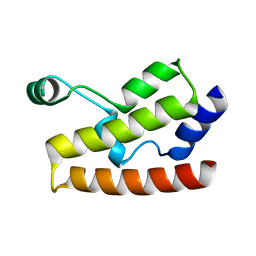 | | Solution Structure of Human Brg1 Bromodomain | | 分子名称: | Probable global transcription activator SNF2L4 | | 著者 | Shen, W, Xu, C, Zhang, J, Wu, J, Shi, Y. | | 登録日 | 2006-05-30 | | 公開日 | 2007-02-13 | | 最終更新日 | 2024-05-29 | | 実験手法 | SOLUTION NMR | | 主引用文献 | Solution structure of human Brg1 bromodomain and its specific binding to acetylated histone tails
Biochemistry, 46, 2007
|
|
2HTG
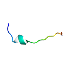 | | Structural and functional characterization of TM VII of the NHE1 isoform of the Na+/H+ exchanger | | 分子名称: | NHE1 isoform of Na+/H+ exchanger 1 | | 著者 | Rainey, J.K, Ding, J, Xu, C, Fliegel, L, Sykes, B.D. | | 登録日 | 2006-07-25 | | 公開日 | 2006-08-08 | | 最終更新日 | 2022-03-09 | | 実験手法 | SOLUTION NMR | | 主引用文献 | Structural and functional characterization of transmembrane segment VII of the Na(+)/H(+) exchanger isoform 1
J.Biol.Chem., 281, 2006
|
|
2K9X
 | | Solution structure of Urm1 from Trypanosoma brucei | | 分子名称: | Uncharacterized protein | | 著者 | Zhang, W, Zhang, J, Xu, C, Wang, T, Zhang, X, Tu, X. | | 登録日 | 2008-10-27 | | 公開日 | 2009-03-10 | | 最終更新日 | 2024-05-29 | | 実験手法 | SOLUTION NMR | | 主引用文献 | Solution structure of Urm1 from Trypanosoma brucei
Proteins, 75, 2009
|
|
7C7Q
 | | Cryo-EM structure of the baclofen/BHFF-bound human GABA(B) receptor in active state | | 分子名称: | (3S)-5,7-ditert-butyl-3-oxidanyl-3-(trifluoromethyl)-1-benzofuran-2-one, 2-acetamido-2-deoxy-beta-D-glucopyranose, Gamma-aminobutyric acid type B receptor subunit 1, ... | | 著者 | Mao, C, Shen, C, Li, C, Shen, D, Xu, C, Zhang, S, Zhou, R, Shen, Q, Chen, L, Jiang, Z, Liu, J, Zhang, Y. | | 登録日 | 2020-05-26 | | 公開日 | 2020-07-01 | | 最終更新日 | 2020-07-29 | | 実験手法 | ELECTRON MICROSCOPY (3 Å) | | 主引用文献 | Cryo-EM structures of inactive and active GABABreceptor.
Cell Res., 30, 2020
|
|


