4LFK
 
 | |
4LFL
 | |
4LFN
 | |
4DMD
 | |
6PV1
 | |
4NIZ
 | |
4NJ2
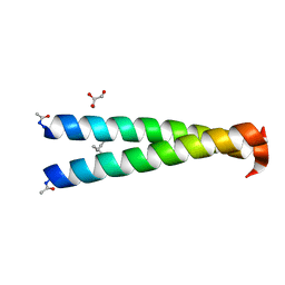
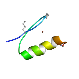
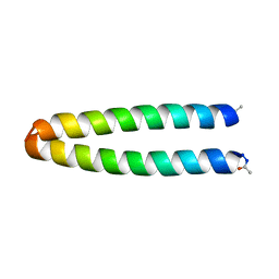
 | | GCN4-p1 triple Val9, 23,30 to Ile mutant | | 分子名称: | GLYCEROL, General control protein GCN4 | | 著者 | Oshaben, K.M, Horne, W.S. | | 登録日 | 2013-11-08 | | 公開日 | 2014-08-20 | | 最終更新日 | 2023-09-20 | | 実験手法 | X-RAY DIFFRACTION (2.2 Å) | | 主引用文献 | Tuning assembly size in Peptide-based supramolecular polymers by modulation of subunit association affinity.
Biomacromolecules, 15, 2014
|
|
6PV3
 | |
4OZB
 | | Backbone Modifications in the Protein GB1 Helix: beta-ACPC24, beta-3-Lys28, beta-3-Lys31, beta-ACPC35 | | 分子名称: | GLYCEROL, Streptococcal Protein GB1 Backbone Modified Variant: beta-ACPC24, beta-3-Lys28, ... | | 著者 | Reinert, Z.E, Horne, W.S. | | 登録日 | 2014-02-14 | | 公開日 | 2014-07-16 | | 最終更新日 | 2023-11-15 | | 実験手法 | X-RAY DIFFRACTION (1.8 Å) | | 主引用文献 | Folding Thermodynamics of Protein-Like Oligomers with Heterogeneous Backbones.
Chem Sci, 5, 2014
|
|
4CAK
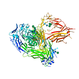 | | Three-dimensional reconstruction of intact human integrin alphaIIbbeta3 in a phospholipid bilayer nanodisc | | 分子名称: | 2-acetamido-2-deoxy-beta-D-glucopyranose, 2-acetamido-2-deoxy-beta-D-glucopyranose-(1-4)-2-acetamido-2-deoxy-beta-D-glucopyranose, Integrin alpha-IIb, ... | | 著者 | Choi, W.S, Rice, W.J, Stokes, D.L, Coller, B.S. | | 登録日 | 2013-10-08 | | 公開日 | 2013-10-30 | | 最終更新日 | 2020-07-29 | | 実験手法 | ELECTRON MICROSCOPY (20.5 Å) | | 主引用文献 | Three-Dimensional Reconstruction of Intact Human Integrin Alphaiibbeta3; New Implications for Activation-Dependent Ligand Binding.
Blood, 122, 2013
|
|
6PV2
 | |
6PV0
 | |
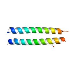
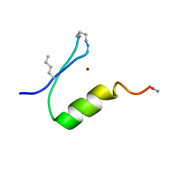
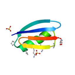
4DME
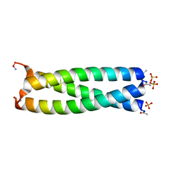 | | GCN4 leucine zipper domain in a trimeric oligomerization state | | 分子名称: | GCN4-p1 leucine zipper domain, SULFATE ION | | 著者 | Oshaben, K.M, Salari, R, Chong, L.T, Horne, W.S. | | 登録日 | 2012-02-07 | | 公開日 | 2012-11-14 | | 最終更新日 | 2023-09-13 | | 実験手法 | X-RAY DIFFRACTION (2.2 Å) | | 主引用文献 | The Native GCN4 Leucine-Zipper Domain Does Not Uniquely Specify a Dimeric Oligomerization State.
Biochemistry, 51, 2012
|
|
4OZC
 | | Backbone Modifications in the Protein GB1 Helix and Loops: beta-ACPC21, beta-ACPC24, beta-3-Lys28, beta-3-Lys31, beta-ACPC35, beta-ACPC40 | | 分子名称: | GLYCEROL, SULFATE ION, Streptococcal Protein GB1 Backbone Modified Variant: beta-ACPC21, ... | | 著者 | Reinert, Z.E, Horne, W.S. | | 登録日 | 2014-02-14 | | 公開日 | 2014-07-16 | | 最終更新日 | 2023-11-15 | | 実験手法 | X-RAY DIFFRACTION (2.301 Å) | | 主引用文献 | Folding Thermodynamics of Protein-Like Oligomers with Heterogeneous Backbones.
Chem Sci, 5, 2014
|
|
4OZA
 | | Backbone Modifications in the Protein GB1 Helix: beta-3-Ala24, beta-3-Lys28, beta-3-Gln32, beta-3-Asp36 | | 分子名称: | ISOPROPYL ALCOHOL, Streptococcal Protein GB1 Backbone Modified Variant: beta-3-Ala24, beta-3-Lys28, ... | | 著者 | Reinert, Z.E, Horne, W.S. | | 登録日 | 2014-02-14 | | 公開日 | 2014-07-16 | | 最終更新日 | 2024-07-10 | | 実験手法 | X-RAY DIFFRACTION (2.201 Å) | | 主引用文献 | Folding Thermodynamics of Protein-Like Oligomers with Heterogeneous Backbones.
Chem Sci, 5, 2014
|
|
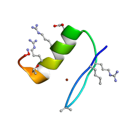
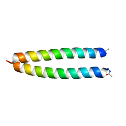
4NJ0
 | | GCN4-p1 single Val9 to Ile mutant | | 分子名称: | General control protein GCN4 | | 著者 | Oshaben, K.M, Horne, W.S. | | 登録日 | 2013-11-08 | | 公開日 | 2014-08-20 | | 最終更新日 | 2023-09-20 | | 実験手法 | X-RAY DIFFRACTION (1.9 Å) | | 主引用文献 | Tuning assembly size in Peptide-based supramolecular polymers by modulation of subunit association affinity.
Biomacromolecules, 15, 2014
|
|
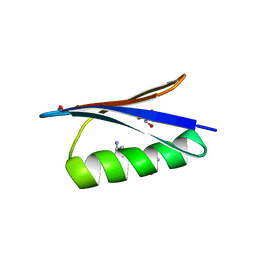
4NJ1
 | | GCN4-p1 double Val9, 23 to Ile mutant | | 分子名称: | General control protein GCN4 | | 著者 | Oshaben, K.M, Horne, W.S. | | 登録日 | 2013-11-08 | | 公開日 | 2014-08-20 | | 最終更新日 | 2023-09-20 | | 実験手法 | X-RAY DIFFRACTION (2 Å) | | 主引用文献 | Tuning assembly size in Peptide-based supramolecular polymers by modulation of subunit association affinity.
Biomacromolecules, 15, 2014
|
|
4NX9
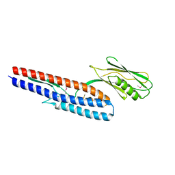 | |
4FZV
 | | Crystal structure of the human MTERF4:NSUN4:SAM ternary complex | | 分子名称: | 1,2-ETHANEDIOL, FORMIC ACID, Putative methyltransferase NSUN4, ... | | 著者 | Guja, K.E, Yakubovskaya, E, Mejia, E, Castano, S, Hambardjieva, E, Choi, W.S, Garcia-Diaz, M. | | 登録日 | 2012-07-08 | | 公開日 | 2012-10-03 | | 最終更新日 | 2012-11-28 | | 実験手法 | X-RAY DIFFRACTION (1.9996 Å) | | 主引用文献 | Structure of the Essential MTERF4:NSUN4 Protein Complex Reveals How an MTERF Protein Collaborates to Facilitate rRNA Modification.
Structure, 20, 2012
|
|
5YCL
 | |
3F87
 | |
3F50
 | |
3F4Z
 | |
3F86
 | |
3F4Y
 | |


