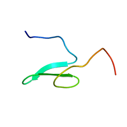
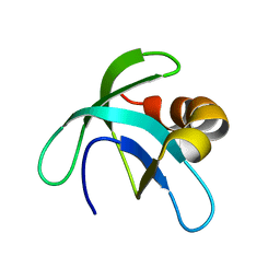
7ZWU
 
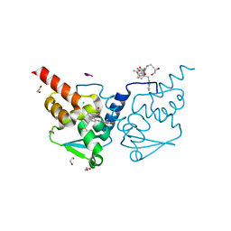 | | Crystal structure of human BCL6 BTB domain in complex with compound 15 | | 分子名称: | 1,2-ETHANEDIOL, ALA-TRP-VAL-ILE-PRO-ALA, B-cell lymphoma 6 protein, ... | | 著者 | Collie, G.W, Le Bihan, Y.-V, van Montfort, R.L.M. | | 登録日 | 2022-05-19 | | 公開日 | 2022-11-16 | | 最終更新日 | 2024-01-31 | | 実験手法 | X-RAY DIFFRACTION (1.56 Å) | | 主引用文献 | Discovering cell-active BCL6 inhibitors: effectively combining biochemical HTS with multiple biophysical techniques, X-ray crystallography and cell-based assays.
Sci Rep, 12, 2022
|
|
1C8P
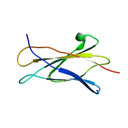 | | NMR STRUCTURE OF THE LIGAND BINDING DOMAIN OF THE COMMON BETA-CHAIN IN THE GM-CSF, IL-3 AND IL-5 RECEPTORS | | 分子名称: | CYTOKINE RECEPTOR COMMON BETA CHAIN | | 著者 | Mulhern, T.D, D'Andrea, R.J, Gaunt, C, Vandeleur, L, Vadas, M.A, Lopez, A.F, Booker, G.W, Bagley, C.J. | | 登録日 | 1999-10-05 | | 公開日 | 2000-06-15 | | 最終更新日 | 2023-12-27 | | 実験手法 | SOLUTION NMR | | 主引用文献 | The solution structure of the cytokine-binding domain of the common beta-chain of the receptors for granulocyte-macrophage colony-stimulating factor, interleukin-3 and interleukin-5.
J.Mol.Biol., 297, 2000
|
|
1B64
 | | SOLUTION STRUCTURE OF THE GUANINE NUCLEOTIDE EXCHANGE FACTOR DOMAIN FROM HUMAN ELONGATION FACTOR-ONE BETA, NMR, 20 STRUCTURES | | 分子名称: | ELONGATION FACTOR 1-BETA | | 著者 | Perez, J.M.J, Siegal, G, Kriek, J, Hard, K, Dijk, J, Canters, G.W, Moller, W. | | 登録日 | 1999-01-20 | | 公開日 | 1999-05-18 | | 最終更新日 | 2024-05-22 | | 実験手法 | SOLUTION NMR | | 主引用文献 | The solution structure of the guanine nucleotide exchange domain of human elongation factor 1beta reveals a striking resemblance to that of EF-Ts from Escherichia coli.
Structure Fold.Des., 7, 1999
|
|
1CLX
 | | CATALYTIC CORE OF XYLANASE A | | 分子名称: | CALCIUM ION, XYLANASE A | | 著者 | Harris, G.W, Jenkins, J.A, Connerton, I, Pickersgill, R.W. | | 登録日 | 1995-08-31 | | 公開日 | 1996-06-20 | | 最終更新日 | 2011-07-13 | | 実験手法 | X-RAY DIFFRACTION (1.8 Å) | | 主引用文献 | Refined crystal structure of the catalytic domain of xylanase A from Pseudomonas fluorescens at 1.8 A resolution.
Acta Crystallogr.,Sect.D, 52, 1996
|
|
6JIT
 | | Complex structure of an imine reductase at 2.05 Angstrom resolution | | 分子名称: | 1-(2-phenylethyl)-3,4-dihydroisoquinoline, 6-phosphogluconate dehydrogenase NAD-binding protein, CHLORIDE ION, ... | | 著者 | Li, H, Wu, L, Zheng, G.W, Zhou, J.H. | | 登録日 | 2019-02-23 | | 公開日 | 2020-02-26 | | 最終更新日 | 2023-11-22 | | 実験手法 | X-RAY DIFFRACTION (2.052 Å) | | 主引用文献 | Complex structure of an imine reductase at 2.05 Angstrom resolution
To Be Published
|
|
6JIZ
 | | Apo structure of an imine reductase at 1.76 Angstrom resolution | | 分子名称: | 1,2-ETHANEDIOL, 3-ethylheptane, 6-phosphogluconate dehydrogenase NAD-binding protein, ... | | 著者 | Li, H, Wu, L, Zheng, G.W, Zhou, J.H. | | 登録日 | 2019-02-24 | | 公開日 | 2020-02-26 | | 最終更新日 | 2024-03-27 | | 実験手法 | X-RAY DIFFRACTION (1.763 Å) | | 主引用文献 | Apo structure of an imine reductase at 1.76 Angstrom resolution
To Be Published
|
|
1GG8
 | | DESIGN OF INHIBITORS OF GLYCOGEN PHOSPHORYLASE: A STUDY OF ALPHA-AND BETA-C-GLUCOSIDES AND 1-THIO-BETA-D-GLUCOSE COMPOUNDS | | 分子名称: | ALPHA-D-GLUCOPYRANOSYL-2-CARBOXYLIC ACID AMIDE, INOSINIC ACID, PROTEIN (GLYCOGEN PHOSPHORYLASE), ... | | 著者 | Watson, K.A, Mitchell, E.P, Johnson, L.N, Son, J.C, Bichard, C.J, Orchard, M.G, Fleet, G.W, Oikonomakos, N.G, Leonidas, D.D, Kontou, M, Papageorgiou, A.C. | | 登録日 | 2000-07-30 | | 公開日 | 2000-08-23 | | 最終更新日 | 2023-08-09 | | 実験手法 | X-RAY DIFFRACTION (2.31 Å) | | 主引用文献 | Design of inhibitors of glycogen phosphorylase: a study of alpha- and beta-C-glucosides and 1-thio-beta-D-glucose compounds.
Biochemistry, 33, 1994
|
|
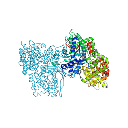
1GWZ
 | | CRYSTAL STRUCTURE OF THE CATALYTIC DOMAIN OF THE PROTEIN TYROSINE PHOSPHATASE SHP-1 | | 分子名称: | SHP-1 | | 著者 | Yang, J, Liang, X, Niu, T, Meng, W, Zhao, Z, Zhou, G.W. | | 登録日 | 1998-08-22 | | 公開日 | 1999-08-22 | | 最終更新日 | 2024-04-03 | | 実験手法 | X-RAY DIFFRACTION (2.5 Å) | | 主引用文献 | Crystal structure of the catalytic domain of protein-tyrosine phosphatase SHP-1.
J.Biol.Chem., 273, 1998
|
|
6LI1
 | | Crystal structure of GPR52 ligand free form with flavodoxin fusion | | 分子名称: | (2R)-2,3-dihydroxypropyl (9Z)-octadec-9-enoate, Chimera of G-protein coupled receptor 52 and Flavodoxin, DI(HYDROXYETHYL)ETHER, ... | | 著者 | Luo, Z.P, Lin, X, Xu, F, Han, G.W. | | 登録日 | 2019-12-10 | | 公開日 | 2020-02-26 | | 最終更新日 | 2024-04-03 | | 実験手法 | X-RAY DIFFRACTION (2.9 Å) | | 主引用文献 | Structural basis of ligand recognition and self-activation of orphan GPR52.
Nature, 579, 2020
|
|
1WLX
 | |
1WR4
 | |
1WR7
 | |
1WR3
 | |
1WMV
 | |
1YOB
 | | C69A Flavodoxin II from Azotobacter vinelandii | | 分子名称: | FLAVIN MONONUCLEOTIDE, Flavodoxin 2, SULFATE ION | | 著者 | Alagaratnam, S, van Pouderoyen, G, Pijning, T, Dijkstra, B.W, Cavazzini, D, Rossi, G.L, Canters, G.W. | | 登録日 | 2005-01-27 | | 公開日 | 2005-10-18 | | 最終更新日 | 2023-10-25 | | 実験手法 | X-RAY DIFFRACTION (2.25 Å) | | 主引用文献 | A crystallographic study of Cys69Ala flavodoxin II from Azotobacter vinelandii: structural determinants of redox potential
Protein Sci., 14, 2005
|
|
1ZDQ
 | | Crystal Structure of Met150Gly AfNiR with Methylsulfanyl Methane Bound | | 分子名称: | (METHYLSULFANYL)METHANE, COPPER (II) ION, Copper-containing nitrite reductase | | 著者 | Wijma, H.J, MacPherson, I.S, Alexandre, M, Diederix, R.E.M, Canters, G.W, Murphy, M.E.P, Verbeet, M.P. | | 登録日 | 2005-04-14 | | 公開日 | 2006-03-28 | | 最終更新日 | 2024-02-14 | | 実験手法 | X-RAY DIFFRACTION (1.8 Å) | | 主引用文献 | A rearranging ligand enables allosteric control of catalytic activity in copper-containing nitrite reductase.
J.Mol.Biol., 358, 2006
|
|
1XYS
 | | CATALYTIC CORE OF XYLANASE A E246C MUTANT | | 分子名称: | CALCIUM ION, XYLANASE A | | 著者 | Harris, G.W, Jenkins, J.A, Connerton, I, Pickersgill, R.W. | | 登録日 | 1994-09-02 | | 公開日 | 1995-07-10 | | 最終更新日 | 2024-02-14 | | 実験手法 | X-RAY DIFFRACTION (2.5 Å) | | 主引用文献 | Structure of the catalytic core of the family F xylanase from Pseudomonas fluorescens and identification of the xylopentaose-binding sites.
Structure, 2, 1994
|
|
1ZDS
 | | Crystal Structure of Met150Gly AfNiR with Acetamide Bound | | 分子名称: | ACETAMIDE, COPPER (II) ION, Copper-containing nitrite reductase | | 著者 | Wijma, H.J, MacPherson, I.S, Alexandre, M, Diederix, R.E.M, Canters, G.W, Murphy, M.E.P, Verbeet, M.P. | | 登録日 | 2005-04-14 | | 公開日 | 2006-03-28 | | 最終更新日 | 2024-02-14 | | 実験手法 | X-RAY DIFFRACTION (1.55 Å) | | 主引用文献 | A rearranging ligand enables allosteric control of catalytic activity in copper-containing nitrite reductase.
J.Mol.Biol., 358, 2006
|
|
2A7Y
 | | Solution Structure of the Conserved Hypothetical Protein Rv2302 from the Bacterium Mycobacterium tuberculosis | | 分子名称: | Hypothetical protein Rv2302/MT2359 | | 著者 | Buchko, G.W, Kim, C.-Y, Terwilliger, T.C, Kennedy, M.A, TB Structural Genomics Consortium (TBSGC) | | 登録日 | 2005-07-06 | | 公開日 | 2005-08-23 | | 最終更新日 | 2024-05-22 | | 実験手法 | SOLUTION NMR | | 主引用文献 | Solution structure of the conserved hypothetical protein Rv2302 from Mycobacterium tuberculosis.
J.Bacteriol., 188, 2006
|
|
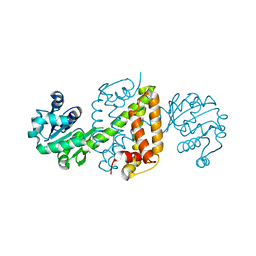
1Y2M
 | | Crystal structure of phenylalanine ammonia-lyase from yeast Rhododporidium toruloides | | 分子名称: | Phenylalanine ammonia-lyase | | 著者 | Wang, L, Gamez, A, Sarkissian, C.N, Straub, M, Patch, M.G, Han, G.W, Scriver, C.R, Stevens, R.C. | | 登録日 | 2004-11-22 | | 公開日 | 2005-11-01 | | 最終更新日 | 2011-07-13 | | 実験手法 | X-RAY DIFFRACTION (1.6 Å) | | 主引用文献 | Structure-based chemical modification strategy for enzyme replacement treatment of phenylketonuria.
Mol.Genet.Metab., 86, 2005
|
|
2B3R
 | | Crystal structure of the C2 domain of class II phosphatidylinositide 3-kinase C2 | | 分子名称: | Phosphatidylinositol-4-phosphate 3-kinase C2 domain-containing alpha polypeptide, SULFATE ION | | 著者 | Liu, L, Song, X, He, D, Komma, C, Kita, A, Verbasius, J.V, Bellamy, H, Miki, K, Czech, M.P, Zhou, G.W. | | 登録日 | 2005-09-20 | | 公開日 | 2005-12-13 | | 最終更新日 | 2024-02-14 | | 実験手法 | X-RAY DIFFRACTION (2.3 Å) | | 主引用文献 | Crystal structure of the C2 domain of class II phosphatidylinositide 3-kinase C2alpha.
J.Biol.Chem., 281, 2006
|
|
2B08
 | | Reduced acetamide-bound M150G Nitrite Reductase from Alcaligenes faecalis | | 分子名称: | ACETAMIDE, COPPER (I) ION, Copper-containing nitrite reductase | | 著者 | Wijma, H.J, MacPherson, I.S, Farver, O, Tocheva, E.I, Pecht, I, Verbeet, M.Ph, Murphy, M.E.P, Canters, G.W. | | 登録日 | 2005-09-13 | | 公開日 | 2006-09-26 | | 最終更新日 | 2024-02-14 | | 実験手法 | X-RAY DIFFRACTION (1.9 Å) | | 主引用文献 | Effect of the methionine ligand on the reorganization energy of the type-1 copper site of nitrite reductase.
J.Am.Chem.Soc., 129, 2007
|
|
4XNW
 | | The human P2Y1 receptor in complex with MRS2500 | | 分子名称: | P2Y purinoceptor 1,Rubredoxin,P2Y purinoceptor 1, ZINC ION, [(1R,2S,4S,5S)-4-[2-iodo-6-(methylamino)-9H-purin-9-yl]-2-(phosphonooxy)bicyclo[3.1.0]hex-1-yl]methyl dihydrogen phosphate | | 著者 | Zhang, D, Gao, Z, Jacobson, K, Han, G.W, Stevens, R, Zhao, Q, Wu, B, GPCR Network (GPCR) | | 登録日 | 2015-01-16 | | 公開日 | 2015-04-01 | | 最終更新日 | 2020-02-05 | | 実験手法 | X-RAY DIFFRACTION (2.7 Å) | | 主引用文献 | Two disparate ligand-binding sites in the human P2Y1 receptor
Nature, 520, 2015
|
|
4XYX
 | | NanB plus Optactamide | | 分子名称: | Optactamide, PHOSPHATE ION, Sialidase B | | 著者 | Rogers, G.W, Brear, P, Yang, L, Taylor, G.L, Westwood, N.J. | | 登録日 | 2015-02-03 | | 公開日 | 2016-02-10 | | 最終更新日 | 2024-01-10 | | 実験手法 | X-RAY DIFFRACTION (2.1 Å) | | 主引用文献 | The Hunt for Serendipitous Allosteric Sites: Discovery of a novel allosteric inhibitor of the bacterial sialidase NanB
To Be Published
|
|
4XNV
 | | The human P2Y1 receptor in complex with BPTU | | 分子名称: | (2R)-2,3-dihydroxypropyl (9Z)-octadec-9-enoate, 1-[2-(2-tert-butylphenoxy)pyridin-3-yl]-3-[4-(trifluoromethoxy)phenyl]urea, CHOLESTEROL, ... | | 著者 | Zhang, D, Gao, Z, Jacobson, K, Han, G.W, Stevens, R, Zhao, Q, Wu, B, GPCR Network (GPCR) | | 登録日 | 2015-01-16 | | 公開日 | 2015-04-01 | | 最終更新日 | 2020-02-05 | | 実験手法 | X-RAY DIFFRACTION (2.2 Å) | | 主引用文献 | Two disparate ligand-binding sites in the human P2Y1 receptor
Nature, 520, 2015
|
|


