6U6P
 
 | |
6U6R
 | |
6U6S
 | |
6U6Q
 | |
7QBY
 | |
4KHT
 | |
4KHX
 | |
1PGX
 | |
3EZM
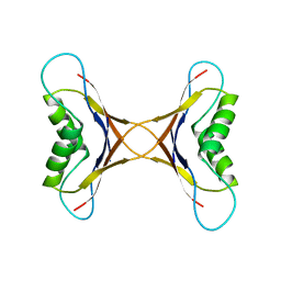
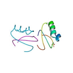
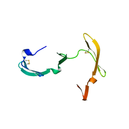 | | CYANOVIRIN-N | | 分子名称: | PROTEIN (CYANOVIRIN-N) | | 著者 | Yang, F, Wlodawer, A. | | 登録日 | 1998-12-15 | | 公開日 | 1998-12-23 | | 最終更新日 | 2024-11-06 | | 実験手法 | X-RAY DIFFRACTION (1.5 Å) | | 主引用文献 | Crystal structure of cyanovirin-N, a potent HIV-inactivating protein, shows unexpected domain swapping.
J.Mol.Biol., 288, 1999
|
|
1PGA
 | |
1PGB
 | |
2Y1S
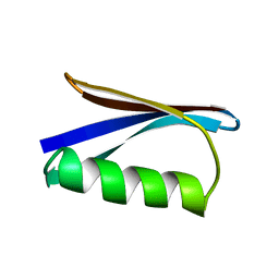
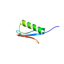
 | | Microvirin lectin | | 分子名称: | Mannan-binding lectin | | 著者 | Bewley, C.A, Hussan, S. | | 登録日 | 2010-12-10 | | 公開日 | 2011-04-06 | | 最終更新日 | 2024-10-23 | | 実験手法 | SOLUTION NMR | | 主引用文献 | Solution structure of the monovalent lectin microvirin in complex with Man(alpha)(1-2)Man provides a basis for anti-HIV activity with low toxicity.
J. Biol. Chem., 286, 2011
|
|
2SXL
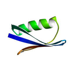
 | | SEX-LETHAL RBD1, NMR, MINIMIZED AVERAGE STRUCTURE | | 分子名称: | SEX-LETHAL PROTEIN | | 著者 | Inoue, M, Muto, Y, Sakamoto, H, Kigawa, T, Takio, K, Shimura, Y, Yokoyama, S, RIKEN Structural Genomics/Proteomics Initiative (RSGI) | | 登録日 | 1997-07-16 | | 公開日 | 1998-07-22 | | 最終更新日 | 2024-05-22 | | 実験手法 | SOLUTION NMR | | 主引用文献 | A characteristic arrangement of aromatic amino acid residues in the solution structure of the amino-terminal RNA-binding domain of Drosophila sex-lethal.
J.Mol.Biol., 272, 1997
|
|
1MVK
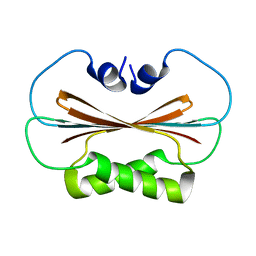
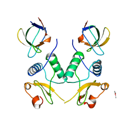
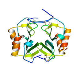
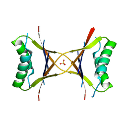 | | X-ray structure of the tetrameric mutant of the B1 domain of streptococcal protein G | | 分子名称: | Immunoglobulin G binding protein G, SULFATE ION | | 著者 | Frank, M.K, Dyda, F, Dobrodumov, A, Gronenborn, A.M. | | 登録日 | 2002-09-25 | | 公開日 | 2002-10-30 | | 最終更新日 | 2024-02-14 | | 実験手法 | X-RAY DIFFRACTION (2.5 Å) | | 主引用文献 | Core mutations switch monomeric protein GB1 into an intertwined tetramer.
Nat.Struct.Biol., 9, 2002
|
|
1NAP
 | |
1MPE
 | |
1IGC
 | |
1IGD
 | |
1EFN
 | |
1F5J
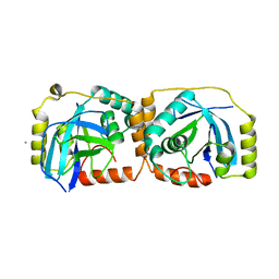
 | | CRYSTAL STRUCTURE OF XYNB, A HIGHLY THERMOSTABLE BETA-1,4-XYLANASE FROM DICTYOGLOMUS THERMOPHILUM RT46B.1, AT 1.8 A RESOLUTION | | 分子名称: | BETA-1,4-XYLANASE, SULFATE ION | | 著者 | McCarthy, A.A, Baker, E.N. | | 登録日 | 2000-07-26 | | 公開日 | 2000-11-15 | | 最終更新日 | 2024-02-07 | | 実験手法 | X-RAY DIFFRACTION (1.8 Å) | | 主引用文献 | Structure of XynB, a highly thermostable beta-1,4-xylanase from Dictyoglomus thermophilum Rt46B.1, at 1.8 A resolution.
Acta Crystallogr.,Sect.D, 56, 2000
|
|
1FL1
 | | KSHV PROTEASE | | 分子名称: | POTASSIUM ION, PROTEASE | | 著者 | Reiling, K.K, Pray, T.R, Craik, C.S, Stroud, R.M. | | 登録日 | 2000-08-11 | | 公開日 | 2000-11-22 | | 最終更新日 | 2024-02-07 | | 実験手法 | X-RAY DIFFRACTION (2.2 Å) | | 主引用文献 | Functional consequences of the Kaposi's sarcoma-associated herpesvirus protease structure: regulation of activity and dimerization by conserved structural elements.
Biochemistry, 39, 2000
|
|
1CQ4
 | |
1TVX
 | |
1UL9
 | | CGL2 ligandfree | | 分子名称: | galectin-2 | | 著者 | Walser, P.J, Haebel, P.W, Kuenzler, M, Kues, U, Aebi, M, Ban, N. | | 登録日 | 2003-09-12 | | 公開日 | 2004-04-20 | | 最終更新日 | 2024-04-03 | | 実験手法 | X-RAY DIFFRACTION (2.22 Å) | | 主引用文献 | Structure and Functional Analysis of the Fungal Galectin CGL2
STRUCTURE, 12, 2004
|
|
1ULC
 | | CGL2 in complex with lactose | | 分子名称: | beta-D-galactopyranose-(1-4)-beta-D-glucopyranose, galectin-2 | | 著者 | Walser, P.J, Haebel, P.W, Kuenzler, M, Kues, U, Aebi, M, Ban, N. | | 登録日 | 2003-09-12 | | 公開日 | 2004-04-20 | | 最終更新日 | 2023-12-27 | | 実験手法 | X-RAY DIFFRACTION (2.6 Å) | | 主引用文献 | Structure and Functional Analysis of the Fungal Galectin CGL2
STRUCTURE, 12, 2004
|
|


