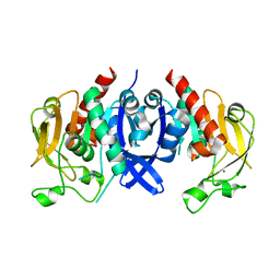
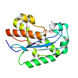
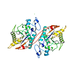
6B5H
 
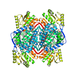 | |
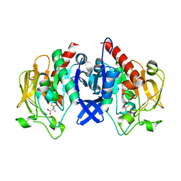
6ALJ
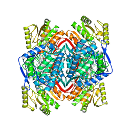
 | | ALDH1A2 liganded with NAD and compound WIN18,446 | | 分子名称: | Aldehyde dehydrogenase 1A2, N,N'-(octane-1,8-diyl)bis(2,2-dichloroacetamide), NICOTINAMIDE-ADENINE-DINUCLEOTIDE | | 著者 | Chen, Y, Zhu, J.-Y, Schonbrunn, E. | | 登録日 | 2017-08-08 | | 公開日 | 2018-01-10 | | 最終更新日 | 2023-10-04 | | 実験手法 | X-RAY DIFFRACTION (1.89 Å) | | 主引用文献 | Structural Basis of ALDH1A2 Inhibition by Irreversible and Reversible Small Molecule Inhibitors.
ACS Chem. Biol., 13, 2018
|
|
1FLO
 | | FLP Recombinase-Holliday Junction Complex I | | 分子名称: | FLP RECOMBINASE, PHOSPHONIC ACID, SYMMETRIZED FRT DNA SITES | | 著者 | Chen, Y, Narendra, U, Iype, L.E, Cox, M.M, Rice, P.A. | | 登録日 | 2000-08-14 | | 公開日 | 2000-09-04 | | 最終更新日 | 2024-02-07 | | 実験手法 | X-RAY DIFFRACTION (2.65 Å) | | 主引用文献 | Crystal structure of a Flp recombinase-Holliday junction complex: assembly of an active oligomer by helix swapping.
Mol.Cell, 6, 2000
|
|
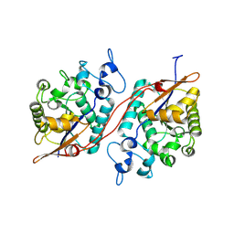
7D54
 | | Crstal structure MsGATase with Gln | | 分子名称: | GLUTAMINE, Glutamine amidotransferase class-I | | 著者 | Chen, Y, Zhang, Q, Bartlam, M. | | 登録日 | 2020-09-24 | | 公開日 | 2021-10-06 | | 最終更新日 | 2023-11-29 | | 実験手法 | X-RAY DIFFRACTION (1.85 Å) | | 主引用文献 | Structure and mechanism of the gamma-glutamyl-gamma-aminobutyrate hydrolase SpuA from Pseudomonas aeruginosa.
Acta Crystallogr D Struct Biol, 77, 2021
|
|
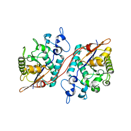
7D4R
 | | SpuA native structure | | 分子名称: | MAGNESIUM ION, Probable glutamine amidotransferase | | 著者 | Chen, Y, Zhang, Q, Bartlam, M. | | 登録日 | 2020-09-24 | | 公開日 | 2021-10-06 | | 最終更新日 | 2023-11-29 | | 実験手法 | X-RAY DIFFRACTION (1.6 Å) | | 主引用文献 | Structure and mechanism of the gamma-glutamyl-gamma-aminobutyrate hydrolase SpuA from Pseudomonas aeruginosa.
Acta Crystallogr D Struct Biol, 77, 2021
|
|
7D50
 | | SpuA mutant - H221N with glutamyl-thioester | | 分子名称: | MAGNESIUM ION, Probable glutamine amidotransferase | | 著者 | Chen, Y, Zhang, Q, Bartlam, M. | | 登録日 | 2020-09-24 | | 公開日 | 2021-10-06 | | 最終更新日 | 2023-11-29 | | 実験手法 | X-RAY DIFFRACTION (1.77 Å) | | 主引用文献 | Structure and mechanism of the gamma-glutamyl-gamma-aminobutyrate hydrolase SpuA from Pseudomonas aeruginosa.
Acta Crystallogr D Struct Biol, 77, 2021
|
|
7D53
 | | SpuA mutant - H221N with Glu | | 分子名称: | GLUTAMIC ACID, MAGNESIUM ION, Probable glutamine amidotransferase | | 著者 | Chen, Y, Zhang, Q, Bartlam, M. | | 登録日 | 2020-09-24 | | 公開日 | 2021-10-06 | | 最終更新日 | 2023-11-29 | | 実験手法 | X-RAY DIFFRACTION (1.6 Å) | | 主引用文献 | Structure and mechanism of the gamma-glutamyl-gamma-aminobutyrate hydrolase SpuA from Pseudomonas aeruginosa.
Acta Crystallogr D Struct Biol, 77, 2021
|
|
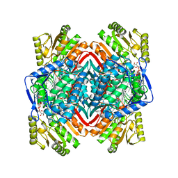
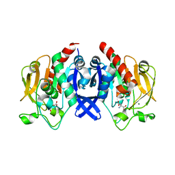
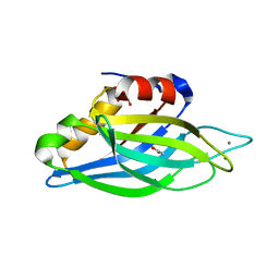
6B5G
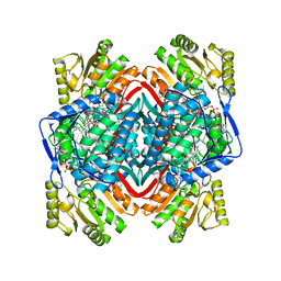 | |
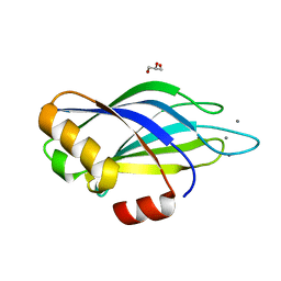
6B5I
 | |
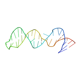
1ZBU
 | | crystal structure of full-length 3'-exonuclease | | 分子名称: | 3'-5' exonuclease ERI1, ADENOSINE MONOPHOSPHATE, MAGNESIUM ION | | 著者 | Cheng, Y, Patel, D.J. | | 登録日 | 2005-04-08 | | 公開日 | 2006-09-19 | | 最終更新日 | 2011-07-13 | | 実験手法 | X-RAY DIFFRACTION (2.998 Å) | | 主引用文献 | Structural basis for 3'-end specific recognition of histone mRNA stem-loop by 3'-exonuclease, a human nuclease that also targets siRNA
To be Published
|
|
2D28
 | | Structure of the N-terminal domain of XpsE (crystal form P43212) | | 分子名称: | CACODYLATE ION, type II secretion ATPase XpsE | | 著者 | Chen, Y, Shiue, S.-J, Huang, C.-W, Chang, J.-L, Chien, Y.-L, Hu, N.-T, Chan, N.-L. | | 登録日 | 2005-09-03 | | 公開日 | 2005-09-20 | | 最終更新日 | 2021-11-10 | | 実験手法 | X-RAY DIFFRACTION (2 Å) | | 主引用文献 | Structure and Function of the XpsE N-Terminal Domain, an Essential Component of the Xanthomonas campestris Type II Secretion System
J.Biol.Chem., 280, 2005
|
|
3PUM
 | | Crystal structure of P domain dimer of Norovirus VA207 | | 分子名称: | Capsid | | 著者 | Chen, Y, Tan, M, Xia, M, Hao, N, Zhang, X.C, Huang, P, Jiang, X, Li, X, Rao, Z. | | 登録日 | 2010-12-06 | | 公開日 | 2011-08-03 | | 最終更新日 | 2023-11-01 | | 実験手法 | X-RAY DIFFRACTION (2.252 Å) | | 主引用文献 | Crystallography of a Lewis-binding norovirus, elucidation of strain-specificity to the polymorphic human histo-blood group antigens
Plos Pathog., 7, 2011
|
|
3PVD
 | | Crystal structure of P domain dimer of Norovirus VA207 complexed with 3'-sialyl-Lewis x tetrasaccharide | | 分子名称: | Capsid, N-acetyl-alpha-neuraminic acid-(2-3)-beta-D-galactopyranose-(1-4)-[alpha-L-fucopyranose-(1-3)]2-acetamido-2-deoxy-alpha-D-glucopyranose | | 著者 | Chen, Y, Tan, M, Xia, M, Hao, N, Zhang, X.C, Huang, P, Jiang, X, Li, X, Rao, Z. | | 登録日 | 2010-12-06 | | 公開日 | 2011-08-03 | | 最終更新日 | 2023-11-01 | | 実験手法 | X-RAY DIFFRACTION (1.9 Å) | | 主引用文献 | Crystallography of a Lewis-binding norovirus, elucidation of strain-specificity to the polymorphic human histo-blood group antigens
Plos Pathog., 7, 2011
|
|
3PUN
 | | Crystal structure of P domain dimer of Norovirus VA207 with Lewis y tetrasaccharide | | 分子名称: | Capsid, alpha-L-fucopyranose-(1-2)-beta-D-galactopyranose-(1-4)-[alpha-L-fucopyranose-(1-3)]2-acetamido-2-deoxy-alpha-D-glucopyranose | | 著者 | Chen, Y, Tan, M, Xia, M, Hao, N, Zhang, X.C, Huang, P, Jiang, X, Li, X, Rao, Z. | | 登録日 | 2010-12-06 | | 公開日 | 2011-08-03 | | 最終更新日 | 2023-11-01 | | 実験手法 | X-RAY DIFFRACTION (2.05 Å) | | 主引用文献 | Crystallography of a Lewis-binding norovirus, elucidation of strain-specificity to the polymorphic human histo-blood group antigens
Plos Pathog., 7, 2011
|
|
2D27
 | | Structure of the N-terminal domain of XpsE (crystal form I4122) | | 分子名称: | type II secretion ATPase XpsE | | 著者 | Chen, Y, Shiue, S.-J, Huang, C.-W, Chang, J.-L, Chien, Y.-L, Hu, N.-T, Chan, N.-L. | | 登録日 | 2005-09-03 | | 公開日 | 2005-09-20 | | 最終更新日 | 2011-07-13 | | 実験手法 | X-RAY DIFFRACTION (2.21 Å) | | 主引用文献 | Structure and Function of the XpsE N-Terminal Domain, an Essential Component of the Xanthomonas campestris Type II Secretion System
J.Biol.Chem., 280, 2005
|
|
1W0H
 | | Crystallographic structure of the nuclease domain of 3'hExo, a DEDDh family member, bound to rAMP | | 分子名称: | 3'-5' EXONUCLEASE ERI1, ADENOSINE MONOPHOSPHATE, MAGNESIUM ION | | 著者 | Cheng, Y, Patel, D. | | 登録日 | 2004-06-04 | | 公開日 | 2004-09-30 | | 最終更新日 | 2011-07-13 | | 実験手法 | X-RAY DIFFRACTION (1.59 Å) | | 主引用文献 | Crystallographic Structure of the Nuclease Domain of 3'Hexo, a Deddh Family Member, Bound to Ramp
J.Mol.Biol., 343, 2004
|
|
1UOW
 | | Calcium binding domain C2B | | 分子名称: | ACETATE ION, CALCIUM ION, GLYCEROL, ... | | 著者 | Cheng, Y, Sequeira, S.M, Sollner, T.H, Patel, D.J. | | 登録日 | 2003-09-24 | | 公開日 | 2004-09-16 | | 最終更新日 | 2023-12-13 | | 実験手法 | X-RAY DIFFRACTION (1.04 Å) | | 主引用文献 | Crystallographic Identification of Ca2+ and Sr2+ Coordination Sites in Synaptotagmin I C2B Domain
Protein Sci., 13, 2004
|
|
1UOV
 | | Calcium binding domain C2B | | 分子名称: | CALCIUM ION, GLYCEROL, SYNAPTOTAGMIN I | | 著者 | Cheng, Y, Sequeira, S.M, Sollner, T.H, Patel, D.J. | | 登録日 | 2003-09-24 | | 公開日 | 2004-09-09 | | 最終更新日 | 2023-12-13 | | 実験手法 | X-RAY DIFFRACTION (1.65 Å) | | 主引用文献 | Crystallographic Identification of Ca2+ and Sr2+ Coordination Sites in Synaptotagmin I C2B Domain
Protein Sci., 13, 2004
|
|
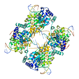
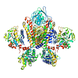
3KMD
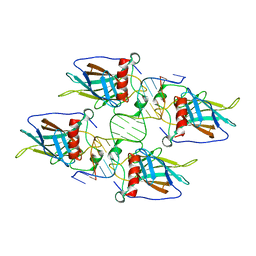 | |
2FEY
 | | The structure of stem loop IV of Tetrahymena telomerase RNA | | 分子名称: | stem-loop IV of Tetrahymena telomerase RNA | | 著者 | Chen, Y, Fender, J, Legassie, J.D, Jarstfer, M.B, Bryan, T.M, Varani, G. | | 登録日 | 2005-12-16 | | 公開日 | 2006-06-27 | | 最終更新日 | 2024-05-29 | | 実験手法 | SOLUTION NMR | | 主引用文献 | Structure of stem-loop IV of Tetrahymena telomerase RNA.
Embo J., 25, 2006
|
|
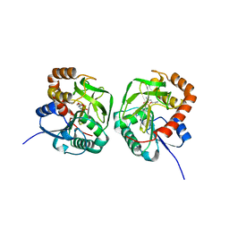
3D4I
 | |
3D6A
 | | Crystal structure of the 2H-phosphatase domain of Sts-2 in complex with tungstate. | | 分子名称: | MAGNESIUM ION, SODIUM ION, Sts-2 protein, ... | | 著者 | Chen, Y, Carpino, N, Nassar, N. | | 登録日 | 2008-05-19 | | 公開日 | 2009-03-03 | | 最終更新日 | 2024-03-13 | | 実験手法 | X-RAY DIFFRACTION (2.25 Å) | | 主引用文献 | Structural and functional characterization of the 2H-phosphatase domain of Sts-2 reveals an acid-dependent phosphatase activity.
Biochemistry, 48, 2009
|
|
3M9E
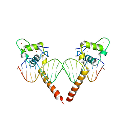 | | Thyroid hormone beta DNA binding domain homodimer with inverted palindrome TRE | | 分子名称: | DNA (5'-D(*AP*TP*TP*GP*AP*CP*CP*TP*CP*AP*GP*CP*TP*GP*AP*GP*GP*TP*CP*AP*AP*T)-3'), Thyroid hormone receptor beta, ZINC ION | | 著者 | Chen, Y. | | 登録日 | 2010-03-22 | | 公開日 | 2010-08-18 | | 最終更新日 | 2023-09-06 | | 実験手法 | X-RAY DIFFRACTION (2.406 Å) | | 主引用文献 | Structure of a thyroid hormone receptor DNA-binding domain homodimer bound to an inverted palindrome DNA response element.
Mol.Endocrinol., 24, 2010
|
|
3C7T
 | | Crystal structure of the ecdysone phosphate phosphatase, EPPase, from Bombix mori in complex with tungstate | | 分子名称: | CHLORIDE ION, Ecdysteroid-phosphate phosphatase, IODIDE ION, ... | | 著者 | Chen, Y, Carpino, N, Nassar, N. | | 登録日 | 2008-02-08 | | 公開日 | 2009-03-24 | | 最終更新日 | 2024-02-21 | | 実験手法 | X-RAY DIFFRACTION (1.76 Å) | | 主引用文献 | Structural and functional characterization of the c-terminal domain of the ecdysteroid phosphate phosphatase from Bombyx mori reveals a new enzymatic activity.
Biochemistry, 47, 2008
|
|
1SUV
 | | Structure of Human Transferrin Receptor-Transferrin Complex | | 分子名称: | CARBONATE ION, FE (III) ION, Serotransferrin, ... | | 著者 | Cheng, Y, Zak, O, Aisen, P, Harrison, S.C, Walz, T. | | 登録日 | 2004-03-26 | | 公開日 | 2004-04-13 | | 最終更新日 | 2011-07-13 | | 実験手法 | ELECTRON MICROSCOPY (7.5 Å) | | 主引用文献 | Structure of the Human Transferrin Receptor-Transferrin Complex
Cell(Cambridge,Mass.), 116, 2004
|
|


