3ASJ
 
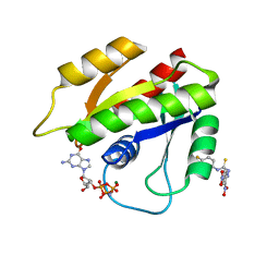
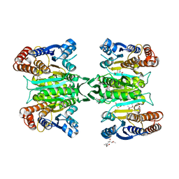 | | Crystal structure of homoisocitrate dehydrogenase in complex with a designed inhibitor | | 分子名称: | (2Z)-3-[(carboxymethyl)sulfanyl]-2-hydroxyprop-2-enoic acid, (4S)-2-METHYL-2,4-PENTANEDIOL, 3-[(carboxymethyl)sulfanyl]-2-oxopropanoic acid, ... | | 著者 | Nango, E, Kumasaka, T, Eguchi, T. | | 登録日 | 2010-12-13 | | 公開日 | 2011-09-21 | | 最終更新日 | 2023-11-01 | | 実験手法 | X-RAY DIFFRACTION (2.6 Å) | | 主引用文献 | Structure of Thermus thermophilus homoisocitrate dehydrogenase in complex with a designed inhibitor
J.Biochem., 150, 2011
|
|
1GCJ
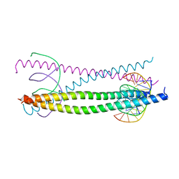
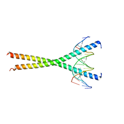
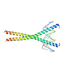
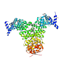 | | N-TERMINAL FRAGMENT OF IMPORTIN-BETA | | 分子名称: | IMPORTIN BETA | | 著者 | Lee, S.J, Imamoto, N, Sakai, H, Nakagawa, A, Kose, S, Koike, M, Yamamoto, M, Kumasaka, T, Yoneda, Y, Tsukihara, T. | | 登録日 | 2000-07-31 | | 公開日 | 2000-10-18 | | 最終更新日 | 2024-10-16 | | 実験手法 | X-RAY DIFFRACTION (2.6 Å) | | 主引用文献 | The adoption of a twisted structure of importin-beta is essential for the protein-protein interaction required for nuclear transport.
J.Mol.Biol., 302, 2000
|
|
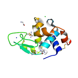
3AGG
 | | X-ray analysis of lysozyme in the absence of Arg | | 分子名称: | ACETATE ION, CHLORIDE ION, Lysozyme C, ... | | 著者 | Ito, L, Shiraki, K, Hasegawa, K, Baba, S, Kumasaka, T. | | 登録日 | 2010-03-31 | | 公開日 | 2011-03-23 | | 最終更新日 | 2024-11-06 | | 実験手法 | X-RAY DIFFRACTION (1.6 Å) | | 主引用文献 | High-resolution X-ray analysis reveals binding of arginine to aromatic residues of lysozyme surface: implication of suppression of protein aggregation by arginine
Protein Eng.Des.Sel., 24, 2011
|
|
3AGH
 | | X-ray analysis of lysozyme in the presence of 200 mM Arg | | 分子名称: | ACETATE ION, CHLORIDE ION, Lysozyme C, ... | | 著者 | Ito, L, Shiraki, K, Hasegawa, K, Baba, S, Kumasaka, T. | | 登録日 | 2010-03-31 | | 公開日 | 2011-03-23 | | 最終更新日 | 2024-10-30 | | 実験手法 | X-RAY DIFFRACTION (1.49 Å) | | 主引用文献 | High-resolution X-ray analysis reveals binding of arginine to aromatic residues of lysozyme surface: implication of suppression of protein aggregation by arginine
Protein Eng.Des.Sel., 24, 2011
|
|
3AGI
 | | High resolution X-ray analysis of Arg-lysozyme complex in the presence of 500 mM Arg | | 分子名称: | ACETATE ION, ARGININE, CHLORIDE ION, ... | | 著者 | Ito, L, Shiraki, K, Hasegawa, K, Baba, S, Kumasaka, T. | | 登録日 | 2010-03-31 | | 公開日 | 2011-03-23 | | 最終更新日 | 2024-10-30 | | 実験手法 | X-RAY DIFFRACTION (1.2 Å) | | 主引用文献 | High-resolution X-ray analysis reveals binding of arginine to aromatic residues of lysozyme surface: implication of suppression of protein aggregation by arginine
Protein Eng.Des.Sel., 24, 2011
|
|
3AJN
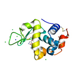
 | | Structural basis of glycine amide on suppression of protein aggregation by high resolution X-ray analysis | | 分子名称: | AMINOMETHYLAMIDE, CHLORIDE ION, Lysozyme C, ... | | 著者 | Ito, L, Shiraki, K, Hasegawa, K, Kumasaka, T. | | 登録日 | 2010-06-09 | | 公開日 | 2011-02-16 | | 最終更新日 | 2024-10-23 | | 実験手法 | X-RAY DIFFRACTION (1.05 Å) | | 主引用文献 | Glycine amide shielding on the aromatic surfaces of lysozyme: Implication for suppression of protein aggregation
Febs Lett., 585, 2011
|
|
2ZW2
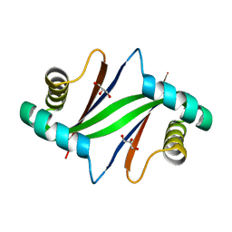 | | Crystal Structure of Formylglycinamide Ribonucleotide Amidotransferase III from SULFOLOBUS TOKODAII (STPURS) | | 分子名称: | GLYCEROL, Putative uncharacterized protein STS178 | | 著者 | Suzuki, S, Tamura, S, Okada, K, Baba, S, Kumasaka, T, Nakagawa, N, Masui, R, Kuramitsu, S, Sampei, G, Kawai, G. | | 登録日 | 2008-11-27 | | 公開日 | 2009-11-17 | | 最終更新日 | 2023-11-01 | | 実験手法 | X-RAY DIFFRACTION (1.55 Å) | | 主引用文献 | Crystal Structure of Formylglycinamide Ribonucleotide Amidotransferase III from SULFOLOBUS TOKODAII (STPURS)
To be Published
|
|
3ATO
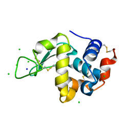
 | | Glycine ethyl ester shielding on the aromatic surfaces of lysozyme: Implication for suppression of protein aggregation | | 分子名称: | CHLORIDE ION, Lysozyme C, SODIUM ION, ... | | 著者 | Ito, L, Shiraki, K, Hasegawa, K, Kumasaka, T. | | 登録日 | 2011-01-06 | | 公開日 | 2012-03-07 | | 最終更新日 | 2024-10-09 | | 実験手法 | X-RAY DIFFRACTION (1.17 Å) | | 主引用文献 | Glycine ethyl ester shielding on the aromatic surfaces of lysozyme: Implication for suppression of protein aggregation
To be Published
|
|
3ATN
 | |
3CE1
 | | Crystal Structure of the Cu/Zn Superoxide Dismutase from Cryptococcus liquefaciens Strain N6 | | 分子名称: | ACETATE ION, COPPER (II) ION, Superoxide dismutase [Cu-Zn], ... | | 著者 | Teh, A.H, Kanamasa, S, Kajiwara, S, Kumasaka, T. | | 登録日 | 2008-02-27 | | 公開日 | 2008-08-26 | | 最終更新日 | 2024-10-16 | | 実験手法 | X-RAY DIFFRACTION (1.2 Å) | | 主引用文献 | Structure of Cu/Zn superoxide dismutase from the heavy-metal-tolerant yeast Cryptococcus liquefaciens strain N6.
Biochem.Biophys.Res.Commun., 374, 2008
|
|
1IOO
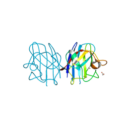
 | | CRYSTAL STRUCTURE OF NICOTIANA ALATA GEMETOPHYTIC SELF-INCOMPATIBILITY ASSOCIATED SF11-RNASE | | 分子名称: | 2-acetamido-2-deoxy-beta-D-glucopyranose-(1-2)-alpha-D-mannopyranose-(1-3)-[beta-D-xylopyranose-(1-2)][alpha-D-mannopyranose-(1-6)]beta-D-mannopyranose-(1-4)-2-acetamido-2-deoxy-beta-D-glucopyranose-(1-4)-2-acetamido-2-deoxy-beta-D-glucopyranose, SF11-RNASE, beta-D-xylopyranose-(1-2)-[alpha-D-mannopyranose-(1-6)]beta-D-mannopyranose-(1-4)-2-acetamido-2-deoxy-beta-D-glucopyranose-(1-4)-2-acetamido-2-deoxy-beta-D-glucopyranose | | 著者 | Ida, K, Sato, M, Sakiyama, F, Norioka, S, Yamamoto, M, Kumasaka, T, Yamashita, E. | | 登録日 | 2001-03-26 | | 公開日 | 2002-05-08 | | 最終更新日 | 2023-12-27 | | 実験手法 | X-RAY DIFFRACTION (1.55 Å) | | 主引用文献 | The 1.55 A resolution structure of Nicotiana alata S(F11)-RNase associated with gametophytic self-incompatibility.
J.Mol.Biol., 314, 2001
|
|
1IO4
 | |
1DDZ
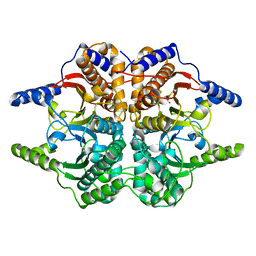 | | X-RAY STRUCTURE OF A BETA-CARBONIC ANHYDRASE FROM THE RED ALGA, PORPHYRIDIUM PURPUREUM R-1 | | 分子名称: | CARBONIC ANHYDRASE, ZINC ION | | 著者 | Mitsuhashi, S, Mizushima, T, Yamashita, E, Miyachi, S, Tsukihara, T. | | 登録日 | 1999-11-12 | | 公開日 | 2000-03-08 | | 最終更新日 | 2024-02-07 | | 実験手法 | X-RAY DIFFRACTION (2.2 Å) | | 主引用文献 | X-ray structure of beta-carbonic anhydrase from the red alga, Porphyridium purpureum, reveals a novel catalytic site for CO(2) hydration.
J.Biol.Chem., 275, 2000
|
|
1GTW
 | |
1GU4
 | |
1GU5
 | |
1H8A
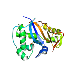
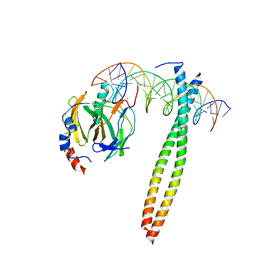
 | | CRYSTAL STRUCTURE OF TERNARY PROTEIN-DNA COMPLEX3 | | 分子名称: | CAAT/ENHANCER BINDING PROTEIN BETA, DNA(5'-(*CP*CP*AP*GP*TP*CP*CP*GP*TP*TP*AP* AP*GP*GP*AP*TP*TP*GP*CP*GP*CP*CP*AP*CP*AP*T)-3'), DNA(5'-(*GP*AP*TP*GP*TP*GP*GP*CP*GP*CP*AP* AP*TP*CP*CP*TP*TP*AP*AP*CP*GP*GP*AP*CP*TP*G)-3'), ... | | 著者 | Tahirov, T.H, Ogata, K. | | 登録日 | 2001-01-31 | | 公開日 | 2002-01-28 | | 最終更新日 | 2023-12-13 | | 実験手法 | X-RAY DIFFRACTION (2.23 Å) | | 主引用文献 | Mechanism of C-Myb-C/Ebpbeta Cooperation from Separated Sites on a Promoter
Cell(Cambridge,Mass.), 108, 2002
|
|
1H89
 | | CRYSTAL STRUCTURE OF TERNARY PROTEIN-DNA COMPLEX2 | | 分子名称: | CAAT/ENHANCER BINDING PROTEIN BETA, DNA(5'-(*CP*CP*AP*GP*TP*CP*CP*GP*TP*TP*AP* AP*GP*GP*AP*TP*TP*GP*CP*GP*CP*CP*AP*CP*AP*T)-3'), DNA(5'-(*GP*AP*TP*GP*TP*GP*GP*CP*GP*CP*AP* AP*TP*CP*CP*TP*TP*AP*AP*CP*GP*GP*AP*CP*TP*G)-3'), ... | | 著者 | Tahirov, T.H, Ogata, K. | | 登録日 | 2001-01-30 | | 公開日 | 2002-01-28 | | 最終更新日 | 2023-12-13 | | 実験手法 | X-RAY DIFFRACTION (2.45 Å) | | 主引用文献 | Mechanism of C-Myb-C/Ebpbeta Cooperation from Separated Sites on a Promoter
Cell(Cambridge,Mass.), 108, 2002
|
|
8Y1W
 | | Crystal structure of Thermococcus pacificus dUTPase complexed with dUMP. | | 分子名称: | 2'-DEOXYURIDINE 5'-MONOPHOSPHATE, GLYCEROL, PHOSPHATE ION, ... | | 著者 | Fukui, K, Murakawa, T, Yano, T. | | 登録日 | 2024-01-25 | | 公開日 | 2024-11-06 | | 実験手法 | X-RAY DIFFRACTION (1.36 Å) | | 主引用文献 | dUTP pyrophosphatases from hyperthermophilic eubacterium and archaeon: Structural and functional examinations on the suitability for PCR application.
Protein Sci., 33, 2024
|
|
8Y1Q
 | |
2LWI
 | | Solution structure of H-RasT35S mutant protein in complex with Kobe2601 | | 分子名称: | 2-(2,4-dinitrophenyl)-N-(4-fluorophenyl)hydrazinecarbothioamide, GTPase HRas, MAGNESIUM ION, ... | | 著者 | Araki, M, Tamura, A, Shima, F, Kataoka, T. | | 登録日 | 2012-08-01 | | 公開日 | 2013-05-22 | | 最終更新日 | 2024-05-15 | | 実験手法 | SOLUTION NMR | | 主引用文献 | In silico discovery of small-molecule Ras inhibitors that display antitumor activity by blocking the Ras-effector interaction.
Proc.Natl.Acad.Sci.USA, 110, 2013
|
|
6JLM
 | | XFEL structure of cyanobacterial photosystem II (dark state, dataset2) | | 分子名称: | 1,2-DI-O-ACYL-3-O-[6-DEOXY-6-SULFO-ALPHA-D-GLUCOPYRANOSYL]-SN-GLYCEROL, 1,2-DIPALMITOYL-PHOSPHATIDYL-GLYCEROLE, 1,2-DISTEAROYL-MONOGALACTOSYL-DIGLYCERIDE, ... | | 著者 | Suga, M, Shen, J.R. | | 登録日 | 2019-03-06 | | 公開日 | 2019-10-16 | | 最終更新日 | 2024-11-06 | | 実験手法 | X-RAY DIFFRACTION (2.35 Å) | | 主引用文献 | An oxyl/oxo mechanism for oxygen-oxygen coupling in PSII revealed by an x-ray free-electron laser.
Science, 366, 2019
|
|
6JLK
 | | XFEL structure of cyanobacterial photosystem II (1F state, dataset1) | | 分子名称: | 1,2-DI-O-ACYL-3-O-[6-DEOXY-6-SULFO-ALPHA-D-GLUCOPYRANOSYL]-SN-GLYCEROL, 1,2-DIPALMITOYL-PHOSPHATIDYL-GLYCEROLE, 1,2-DISTEAROYL-MONOGALACTOSYL-DIGLYCERIDE, ... | | 著者 | Suga, M, Shen, J.R. | | 登録日 | 2019-03-06 | | 公開日 | 2019-10-16 | | 最終更新日 | 2023-11-22 | | 実験手法 | X-RAY DIFFRACTION (2.15 Å) | | 主引用文献 | An oxyl/oxo mechanism for oxygen-oxygen coupling in PSII revealed by an x-ray free-electron laser.
Science, 366, 2019
|
|
6JLO
 | | XFEL structure of cyanobacterial photosystem II (2F state, dataset2) | | 分子名称: | 1,2-DI-O-ACYL-3-O-[6-DEOXY-6-SULFO-ALPHA-D-GLUCOPYRANOSYL]-SN-GLYCEROL, 1,2-DIPALMITOYL-PHOSPHATIDYL-GLYCEROLE, 1,2-DISTEAROYL-MONOGALACTOSYL-DIGLYCERIDE, ... | | 著者 | Suga, M, Shen, J.R. | | 登録日 | 2019-03-06 | | 公開日 | 2019-10-16 | | 最終更新日 | 2024-10-23 | | 実験手法 | X-RAY DIFFRACTION (2.4 Å) | | 主引用文献 | An oxyl/oxo mechanism for oxygen-oxygen coupling in PSII revealed by an x-ray free-electron laser.
Science, 366, 2019
|
|
6JLL
 | | XFEL structure of cyanobacterial photosystem II (2F state, dataset1) | | 分子名称: | 1,2-DI-O-ACYL-3-O-[6-DEOXY-6-SULFO-ALPHA-D-GLUCOPYRANOSYL]-SN-GLYCEROL, 1,2-DIPALMITOYL-PHOSPHATIDYL-GLYCEROLE, 1,2-DISTEAROYL-MONOGALACTOSYL-DIGLYCERIDE, ... | | 著者 | Suga, M, Shen, J.R. | | 登録日 | 2019-03-06 | | 公開日 | 2019-10-16 | | 最終更新日 | 2024-10-23 | | 実験手法 | X-RAY DIFFRACTION (2.15 Å) | | 主引用文献 | An oxyl/oxo mechanism for oxygen-oxygen coupling in PSII revealed by an x-ray free-electron laser.
Science, 366, 2019
|
|


