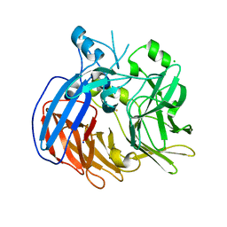
5MOR
 
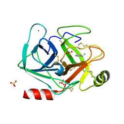 | | Joint X-ray/neutron structure of cationic trypsin in complex with benzylamine | | 分子名称: | (phenylmethyl)azanium, CALCIUM ION, Cationic trypsin, ... | | 著者 | Schiebel, J, Schrader, T.E, Ostermann, A, Heine, A, Klebe, G. | | 登録日 | 2016-12-14 | | 公開日 | 2018-02-28 | | 最終更新日 | 2024-05-01 | | 実験手法 | NEUTRON DIFFRACTION (0.98 Å), X-RAY DIFFRACTION | | 主引用文献 | Joint X-ray/neutron structure of cationic trypsin in complex with benzylamine
to be published
|
|
2MUC
 | | MUCONATE CYCLOISOMERASE VARIANT F329I | | 分子名称: | MANGANESE (II) ION, PROTEIN (MUCONATE CYCLOISOMERASE) | | 著者 | Schell, U, Helin, S, Kajander, T, Schlomann, M, Goldman, A. | | 登録日 | 1998-10-26 | | 公開日 | 1999-12-22 | | 最終更新日 | 2023-12-27 | | 実験手法 | X-RAY DIFFRACTION (2.3 Å) | | 主引用文献 | Structural basis for the activity of two muconate cycloisomerase variants toward substituted muconates.
Proteins, 34, 1999
|
|
7TH0
 | |
7BQ6
 | | Crystal structure of Pennisetum glaucum monodehydroascorbate reductase | | 分子名称: | 2-AMINO-2-HYDROXYMETHYL-PROPANE-1,3-DIOL, ACETATE ION, FLAVIN-ADENINE DINUCLEOTIDE, ... | | 著者 | Sonkar, K.S, Arulandu, A, Achary, M.M, Reddy, M.K. | | 登録日 | 2020-03-24 | | 公開日 | 2021-03-24 | | 最終更新日 | 2023-11-29 | | 実験手法 | X-RAY DIFFRACTION (1.89 Å) | | 主引用文献 | Biochemical and structural characterization of a robust and thermostable ascorbate recycling monodehydroascorbate reductase (MDHAR) from stress adapted pearl millet.
Biochem.Biophys.Res.Commun., 662, 2023
|
|
2MXG
 | |
7BLE
 | | Co-crystal structure of Human Nicotinamide N-methyltransferase (NNMT) with the tricyclic inhibitor (3) | | 分子名称: | 3-ethyl-1,3-diazatricyclo[6.3.1.0^{4,12}]dodeca-4,6,8(12)-trien-2-imine, Nicotinamide N-methyltransferase, S-ADENOSYL-L-HOMOCYSTEINE | | 著者 | Schreuder, H.A, Liesum, A. | | 登録日 | 2021-01-18 | | 公開日 | 2021-03-17 | | 最終更新日 | 2024-01-31 | | 実験手法 | X-RAY DIFFRACTION (2.809 Å) | | 主引用文献 | Novel Inhibitors of Nicotinamide- N -Methyltransferase for the Treatment of Metabolic Disorders.
Molecules, 26, 2021
|
|
7SWG
 | | cTnC-TnI chimera complexed with A1 | | 分子名称: | 4-(3-cyano-3-methylazetidine-1-carbonyl)-N-[(3S)-7-fluoro-6-methyl-3,4-dihydro-2H-1-benzopyran-3-yl]-5-methyl-1H-pyrrole-2-sulfonamide, Troponin C, slow skeletal and cardiac muscles,Troponin I, ... | | 著者 | Poppe, L, Hartman, J.J, Romero, A, Reagan, J.D. | | 登録日 | 2021-11-19 | | 公開日 | 2022-04-06 | | 最終更新日 | 2024-05-15 | | 実験手法 | SOLUTION NMR | | 主引用文献 | Structural and Thermodynamic Model for the Activation of Cardiac Troponin.
Biochemistry, 61, 2022
|
|
7SXD
 | | NMR solution structure TnC-TnI chimera | | 分子名称: | Troponin C, slow skeletal and cardiac muscles,Troponin I, cardiac muscle chimera | | 著者 | Poppe, L, Hartman, J.J, Romero, A, Reagan, J.D. | | 登録日 | 2021-11-22 | | 公開日 | 2022-04-06 | | 最終更新日 | 2024-05-15 | | 実験手法 | SOLUTION NMR | | 主引用文献 | Structural and Thermodynamic Model for the Activation of Cardiac Troponin.
Biochemistry, 61, 2022
|
|
6VCF
 | | Crystal structure of Nitrosotalea devanaterra carotenoid cleavage dioxygenase, iron form | | 分子名称: | BICARBONATE ION, CHLORIDE ION, FE (II) ION, ... | | 著者 | Daruwalla, A, Shi, W, Kiser, P.D. | | 登録日 | 2019-12-20 | | 公開日 | 2020-07-08 | | 最終更新日 | 2023-10-11 | | 実験手法 | X-RAY DIFFRACTION (2.687 Å) | | 主引用文献 | Structural basis for carotenoid cleavage by an archaeal carotenoid dioxygenase.
Proc.Natl.Acad.Sci.USA, 117, 2020
|
|
7SXC
 | | cTnC-TnI chimera complexed with calcium | | 分子名称: | CALCIUM ION, Troponin C, slow skeletal and cardiac muscles,Troponin I, ... | | 著者 | Poppe, L, Hartman, J.J, Romero, A, Reagan, J.D. | | 登録日 | 2021-11-22 | | 公開日 | 2022-04-06 | | 最終更新日 | 2024-05-15 | | 実験手法 | SOLUTION NMR | | 主引用文献 | Structural and Thermodynamic Model for the Activation of Cardiac Troponin.
Biochemistry, 61, 2022
|
|
7SXB
 | |
2N63
 | | Structure of C4VG16KRKP | | 分子名称: | PENTANOIC ACID, antimicrobial peptide C4VG16KRKP | | 著者 | Bhunia, A, Datta, A. | | 登録日 | 2015-08-11 | | 公開日 | 2016-03-23 | | 最終更新日 | 2024-01-24 | | 実験手法 | SOLUTION NMR | | 主引用文献 | Designing potent antimicrobial peptides by disulphide linked dimerization and N-terminal lipidation to increase antimicrobial activity and membrane perturbation: Structural insights into lipopolysaccharide binding.
J Colloid Interface Sci, 461, 2016
|
|
6VC5
 | |
6V88
 | |
7THH
 | | SUD-C and Ubl2 domains of SARS CoV-2 Nsp3 protein | | 分子名称: | 1,2-ETHANEDIOL, CHLORIDE ION, HEXAETHYLENE GLYCOL, ... | | 著者 | Osipiuk, J, Jedrzejczak, R, Endres, M, Wydorski, P, Joachimiak, L, Joachimiak, A, Center for Structural Genomics of Infectious Diseases (CSGID) | | 登録日 | 2022-01-11 | | 公開日 | 2022-01-19 | | 最終更新日 | 2023-10-18 | | 実験手法 | X-RAY DIFFRACTION (1.32 Å) | | 主引用文献 | SUD-C and Ubl2 domains of SARS CoV-2 Nsp3 protein
to be published
|
|
2MZ3
 | |
6VEK
 | | Contact-dependent growth inhibition toxin-immunity protein complex from from E. coli 3006, full-length | | 分子名称: | contact-dependent immunity protein CdiI, contact-dependent toxin CdiA | | 著者 | Michalska, K, Stols, L, Eschenfeldt, W, Hayes, C.S, Goulding, C.W, Joachimiak, A, Midwest Center for Structural Genomics (MCSG), Structure-Function Analysis of Polymorphic CDI Toxin-Immunity Protein Complexes (UC4CDI), Center for Structural Genomics of Infectious Diseases (CSGID) | | 登録日 | 2020-01-02 | | 公開日 | 2021-01-27 | | 最終更新日 | 2023-10-11 | | 実験手法 | X-RAY DIFFRACTION (2.25 Å) | | 主引用文献 | Contact-dependent growth inhibition toxin-immunity protein complex from from E. coli 3006, full-length
To Be Published
|
|
7TRV
 | | Crystal Structure of the DNA-Binding Domain of the LysR family Transcriptional Regulator YfbA from Yersinia pestis | | 分子名称: | CHLORIDE ION, FORMIC ACID, GLYCEROL, ... | | 著者 | Kim, Y, Tesar, C, Crawford, M, Chhor, G, Endres, M, Babnigg, G, Schneewind, O, Joachimiak, A, Center for Structural Genomics of Infectious Diseases (CSGID) | | 登録日 | 2022-01-31 | | 公開日 | 2022-02-09 | | 実験手法 | X-RAY DIFFRACTION (1.8 Å) | | 主引用文献 | Crystal Structure of the DNA-Binding Domain of the LysR family Transcriptional Regulator YfbA from Yersinia pestis
To Be Published
|
|
7TMU
 | | Crystal Structure of the Protein of Unknown Function YPO0625 from Yersinia pestis | | 分子名称: | 1,2-ETHANEDIOL, CHLORIDE ION, DI(HYDROXYETHYL)ETHER, ... | | 著者 | Kim, Y, Chhor, G, Endres, M, Babnigg, G, Schneewind, O, Joachimiak, A, Center for Structural Genomics of Infectious Diseases (CSGID) | | 登録日 | 2022-01-20 | | 公開日 | 2022-02-02 | | 実験手法 | X-RAY DIFFRACTION (2.55 Å) | | 主引用文献 | Crystal Structure of the Protein of Unknown Function YPO0625 from Yersinia pestis
To Be Published
|
|
7TRW
 | | Crystal Structure of the C-terminal Ligand-Binding Domain of the LysR family Transcriptional Regulator YfbA from Yersinia pestis | | 分子名称: | 3-HYDROXYBENZOIC ACID, LysR-family transcriptional regulatory protein, PHOSPHATE ION | | 著者 | Kim, Y, Tesar, C, Crawford, M, Endres, M, Babnigg, G, Schneewind, O, Joachimiak, A, Center for Structural Genomics of Infectious Diseases (CSGID) | | 登録日 | 2022-01-31 | | 公開日 | 2022-02-09 | | 実験手法 | X-RAY DIFFRACTION (2.28 Å) | | 主引用文献 | Crystal Structure of the C-terminal Ligand-Binding Domain of the LysR family Transcriptional Regulator YfbA from Yersinia pestis
To Be Published
|
|
7TKV
 | | Crystal Structure of the Thioredox_DsbH Domain-Containing Uncharacterized Protein Bab1_2064 from Brucella abortus | | 分子名称: | CHLORIDE ION, GLYCEROL, PHOSPHATE ION, ... | | 著者 | Kim, Y, Crawford, M, Endres, M, Babnigg, G, Crosson, S, Joachimiak, A, Center for Structural Genomics of Infectious Diseases (CSGID) | | 登録日 | 2022-01-17 | | 公開日 | 2022-01-26 | | 実験手法 | X-RAY DIFFRACTION (2.8 Å) | | 主引用文献 | Crystal Structure of the Thioredox_DsbH Domain-Containing Uncharacterized Protein Bab1_2064 from Brucella abortus
To Be Published
|
|
7THW
 | | Crystal Structure of the Soluble Domain of the Putative OmpA -Family Membrane Protein YPO0514 from Yersinia pestis | | 分子名称: | CALCIUM ION, PHOSPHATE ION, Putative OmpA-family membrane protein | | 著者 | Kim, Y, Tesar, C, Chhor, G, Clancy, S, Babnigg, G, Schneewind, O, Joachimiak, A, Center for Structural Genomics of Infectious Diseases (CSGID) | | 登録日 | 2022-01-12 | | 公開日 | 2022-01-26 | | 実験手法 | X-RAY DIFFRACTION (2.2 Å) | | 主引用文献 | Crystal Structure of the Soluble Domain of the Putative OmpA -Family Membrane Protein YPO0514 from Yersinia pestis
To Be Published
|
|
7TJ1
 | | Crystal Structure of the Putative Fluoride Ion Transporter CrcB Bab1_1389 from Brucella abortus | | 分子名称: | 1,2-ETHANEDIOL, ACETATE ION, DI(HYDROXYETHYL)ETHER, ... | | 著者 | Kim, Y, Tesar, C, Pastore, T, Endres, M, Babnigg, G, Crosson, S, Joachimiak, A, Center for Structural Genomics of Infectious Diseases (CSGID) | | 登録日 | 2022-01-14 | | 公開日 | 2022-01-26 | | 実験手法 | X-RAY DIFFRACTION (2.1 Å) | | 主引用文献 | Crystal Structure of the Putative Fluoride Ion Transporter CrcB Bab1_1389 from Brucella abortus
To Be Published
|
|
4L4U
 | | Crystal structure of construct containing A. aeolicus NtrC1 receiver, central and DNA binding domains | | 分子名称: | Transcriptional regulator (NtrC family) | | 著者 | Vidangos, N.K, Maris, A.E, Young, A, Hong, E, Pelton, J.G, Batchelor, J.D, Wemmer, D.E. | | 登録日 | 2013-06-09 | | 公開日 | 2013-08-28 | | 最終更新日 | 2013-10-23 | | 実験手法 | X-RAY DIFFRACTION (2.2 Å) | | 主引用文献 | Structure, function, and tethering of DNA-binding domains in sigma (54) transcriptional activators.
Biopolymers, 99, 2013
|
|
7TL5
 | | Crystal structure of putative hydrolase yjcS from Klebsiella pneumoniae. | | 分子名称: | 1,2-ETHANEDIOL, Lactamase_B domain-containing protein | | 著者 | Chang, C, Endres, M, Wu, R, Joachimiak, A, Center for Structural Genomics of Infectious Diseases (CSGID) | | 登録日 | 2022-01-18 | | 公開日 | 2022-02-02 | | 最終更新日 | 2023-06-14 | | 実験手法 | X-RAY DIFFRACTION (2.69 Å) | | 主引用文献 | A Structural Systems Biology Approach to High-Risk CG23 Klebsiella pneumoniae.
Microbiol Resour Announc, 12, 2023
|
|


