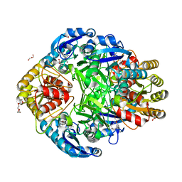
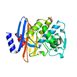
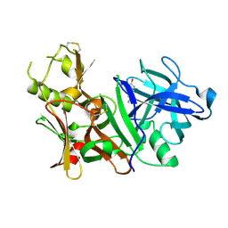
2PDI
 
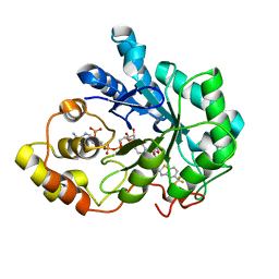 | | Human aldose reductase mutant L300A complexed with zopolrestat at 1.55 A. | | 分子名称: | 3,4-DIHYDRO-4-OXO-3-((5-TRIFLUOROMETHYL-2-BENZOTHIAZOLYL)METHYL)-1-PHTHALAZINE ACETIC ACID, Aldose reductase, NADP NICOTINAMIDE-ADENINE-DINUCLEOTIDE PHOSPHATE | | 著者 | Steuber, H, Heine, A, Klebe, G. | | 登録日 | 2007-04-01 | | 公開日 | 2008-04-01 | | 最終更新日 | 2023-08-30 | | 実験手法 | X-RAY DIFFRACTION (1.55 Å) | | 主引用文献 | Merging the binding sites of aldose and aldehyde reductase for detection of inhibitor selectivity-determining features.
J.Mol.Biol., 379, 2008
|
|
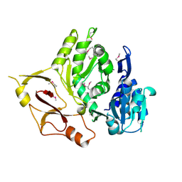
4IHZ
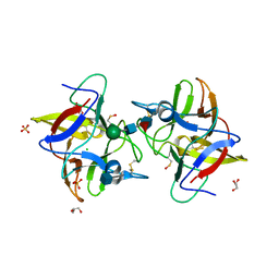 | |
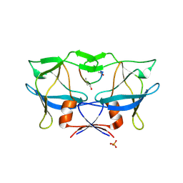
5G1B
 | | Bordetella Alcaligenes HDAH native | | 分子名称: | DI(HYDROXYETHYL)ETHER, HISTONE DEACETYLASE-LIKE AMIDOHYDROLASE, PENTAETHYLENE GLYCOL, ... | | 著者 | Kraemer, A, Meyer-Almes, F.J, Yildiz, O. | | 登録日 | 2016-03-24 | | 公開日 | 2017-04-12 | | 最終更新日 | 2024-01-10 | | 実験手法 | X-RAY DIFFRACTION (1.7 Å) | | 主引用文献 | The thermodynamic signature of ligand binding to histone deacetylase-like amidohydrolases is most sensitive to the flexibility in the L2-loop lining the active site pocket.
Biochim. Biophys. Acta, 1861, 2017
|
|
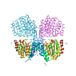
1Y0D
 | |
1JSL
 | | Crystal structure of Erwinia chrysanthemi L-asparaginase complexed with 6-HYDROXY-D-NORLEUCINE | | 分子名称: | 6-HYDROXY-D-NORLEUCINE, GLYCEROL, L-asparaginase, ... | | 著者 | Aghaiypour, K, Wlodawer, A, Lubkowski, J. | | 登録日 | 2001-08-17 | | 公開日 | 2002-01-09 | | 最終更新日 | 2023-08-16 | | 実験手法 | X-RAY DIFFRACTION (1.7 Å) | | 主引用文献 | Do bacterial L-asparaginases utilize a catalytic triad Thr-Tyr-Glu?
Biochim.Biophys.Acta, 1550, 2001
|
|
2B7F
 | | Crystal structure of human T-cell leukemia virus protease, a novel target for anti-cancer design | | 分子名称: | (ACE)APQV(STA)VMHP peptide, HTLV protease, PHOSPHATE ION | | 著者 | Li, M, Laco, G.S, Jaskolski, M, Rozycki, J, Alexandratos, J, Wlodawer, A, Gustchina, A. | | 登録日 | 2005-10-04 | | 公開日 | 2005-12-06 | | 最終更新日 | 2023-11-15 | | 実験手法 | X-RAY DIFFRACTION (2.6 Å) | | 主引用文献 | Crystal structure of human T cell leukemia virus protease, a novel target for anticancer drug design
Proc.Natl.Acad.Sci.Usa, 102, 2005
|
|
5FYR
 | | Calcium-dependent phosphoinositol-specific phospholipase C from a Gram-negative bacterium, Pseudomonas sp, apo form, myoinositol complex | | 分子名称: | 1,2,3,4,5,6-HEXAHYDROXY-CYCLOHEXANE, CALCIUM ION, PHOSPHATE ION, ... | | 著者 | Moroz, O.V, Blagova, E, Lebedev, A.A, Norgaard, A, Segura, D.R, Blicher, T.H, Wilson, K.S. | | 登録日 | 2016-03-09 | | 公開日 | 2017-01-18 | | 最終更新日 | 2024-01-10 | | 実験手法 | X-RAY DIFFRACTION (1.45 Å) | | 主引用文献 | The structure of a calcium-dependent phosphoinositide-specific phospholipase C from Pseudomonas sp. 62186, the first from a Gram-negative bacterium.
Acta Crystallogr D Struct Biol, 73, 2017
|
|
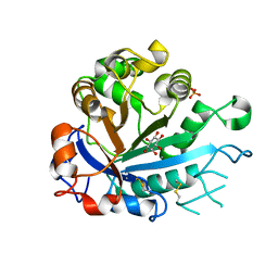
5G11
 | | Pseudomonas aeruginosa HDAH bound to PFSAHA. | | 分子名称: | 2,2,3,3,4,4,5,5,6,6,7,7-dodecakis(fluoranyl)-~{N}-oxidanyl-~{N}'-phenyl-octanediamide, HDAH, POTASSIUM ION, ... | | 著者 | Kraemer, A, Meyer-Almes, F.J, Yildiz, O. | | 登録日 | 2016-03-23 | | 公開日 | 2016-11-30 | | 最終更新日 | 2024-01-10 | | 実験手法 | X-RAY DIFFRACTION (2.48 Å) | | 主引用文献 | Crystal Structure of a Histone Deacetylase Homologue from Pseudomonas aeruginosa.
Biochemistry, 55, 2016
|
|
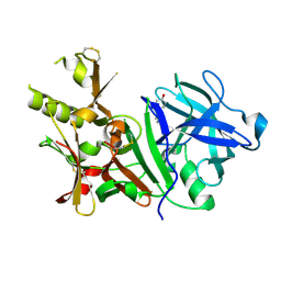
1BZA
 | | BETA-LACTAMASE TOHO-1 FROM ESCHERICHIA COLI TUH12191 | | 分子名称: | BETA-LACTAMASE, SULFATE ION | | 著者 | Ibuka, A, Taguchi, A, Ishiguro, M, Fushinobu, S, Ishii, Y, Kamitori, S, Okuyama, K, Yamaguchi, K, Konno, M, Matsuzawa, H. | | 登録日 | 1998-10-28 | | 公開日 | 1999-04-27 | | 最終更新日 | 2024-05-22 | | 実験手法 | X-RAY DIFFRACTION (1.8 Å) | | 主引用文献 | Crystal structure of the E166A mutant of extended-spectrum beta-lactamase Toho-1 at 1.8 A resolution.
J.Mol.Biol., 285, 1999
|
|
1JV0
 | | THE CRYSTAL STRUCTURE OF THE ZINC(II) ADDUCT OF THE CAI MICHIGAN 1 VARIANT | | 分子名称: | 1,2-ETHANEDIOL, CARBONIC ANHYDRASE I, CHLORIDE ION, ... | | 著者 | Briganti, F, Ferraroni, M, Chegwidden, W.R, Scozzafava, A, Supuran, C.T, Tilli, S. | | 登録日 | 2001-08-28 | | 公開日 | 2001-09-19 | | 最終更新日 | 2023-10-25 | | 実験手法 | X-RAY DIFFRACTION (2 Å) | | 主引用文献 | Crystal structure of a zinc-activated variant of human carbonic anhydrase I, CA I Michigan 1: evidence for a second zinc binding site involving arginine coordination
Biochemistry, 41, 2002
|
|
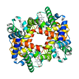
2EMN
 | |
5VF2
 | | scFv 2D10 re-refined as a complex with trehalose replacing the original alpha-1,6-mannobiose | | 分子名称: | 2-(N-MORPHOLINO)-ETHANESULFONIC ACID, MAGNESIUM ION, UNKNOWN ATOM OR ION, ... | | 著者 | Porebski, P.J, Wlodawer, A, Dauter, Z, Minor, W, Stanfield, R, Jaskolski, M, Pozharski, E, Weichenberger, C.X, Rupp, B. | | 登録日 | 2017-04-06 | | 公開日 | 2017-12-06 | | 最終更新日 | 2023-10-04 | | 実験手法 | X-RAY DIFFRACTION (1.55 Å) | | 主引用文献 | Detect, correct, retract: How to manage incorrect structural models.
FEBS J., 285, 2018
|
|
4JWK
 | | Crystal structure of the complex of peptidyl-tRNA hydrolase from Acinetobacter baumannii with cytidine at 1.87 A resolution | | 分子名称: | 4-AMINO-1-BETA-D-RIBOFURANOSYL-2(1H)-PYRIMIDINONE, Peptidyl-tRNA hydrolase | | 著者 | Kaushik, S, Singh, N, Yamini, S, Singh, A, Sinha, M, Kaur, P, Sharma, S, Singh, T.P. | | 登録日 | 2013-03-27 | | 公開日 | 2013-06-05 | | 最終更新日 | 2023-11-08 | | 実験手法 | X-RAY DIFFRACTION (1.87 Å) | | 主引用文献 | The Mode of Inhibitor Binding to Peptidyl-tRNA Hydrolase: Binding Studies and Structure Determination of Unbound and Bound Peptidyl-tRNA Hydrolase from Acinetobacter baumannii
Plos One, 8, 2013
|
|
4JHQ
 | | Crystal structure of avidin - 6-(6-biotinamidohexanamido)hexanoylferrocene complex | | 分子名称: | 2-acetamido-2-deoxy-beta-D-glucopyranose, Avidin, [(1,2,3,4,5-eta)-cyclopentadienyl][(1,2,3,4,5-eta)-{6-[(6-{[5-(2-oxohexahydro-1H-thieno[3,4-d]imidazol-4-yl)pentanoyl]amino}hexanoyl)amino]hexanoyl}cyclopentadienyl]iron | | 著者 | Strzelczyk, P, Bujacz, A, Bujacz, G. | | 登録日 | 2013-03-05 | | 公開日 | 2013-11-20 | | 最終更新日 | 2023-11-08 | | 実験手法 | X-RAY DIFFRACTION (1.99 Å) | | 主引用文献 | Ferrocene-Biotin Conjugates Targeting Cancer Cells: Synthesis, Interaction with Avidin, Cytotoxic Properties and the Crystal Structure of the Complex of Avidin with a Biotin-Linker-Ferrocene Conjugate
Organometallics, 32, 2013
|
|
1QK4
 | | TOXOPLASMA GONDII HYPOXANTHINE-GUANINE PHOSPHORIBOSYLTRANSFERASE IMP COMPLEX | | 分子名称: | HYPOXANTHINE-GUANINE PHOSPHORIBOSYLTRANSFERASE, INOSINIC ACID | | 著者 | Heroux, A, White, E.L, Ross, L.J, Borhani, D.W. | | 登録日 | 1999-07-09 | | 公開日 | 1999-10-17 | | 最終更新日 | 2023-12-13 | | 実験手法 | X-RAY DIFFRACTION (1.9 Å) | | 主引用文献 | Crystal Structures of the Toxoplasma Gondii Hypoxanthine-Guanine Phosphoribosyltransferase Gmp and -Imp Complexes: Comparison of Purine Binding Interactions with the Xmp Complex
Biochemistry, 38, 1999
|
|
5GSS
 | |
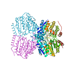
6GQX
 | | KRAS-169 Q61H GPPNHP + CH-2 | | 分子名称: | GTPase KRas, MAGNESIUM ION, PHOSPHOAMINOPHOSPHONIC ACID-GUANYLATE ESTER, ... | | 著者 | Cruz-Migoni, A, Quevedo, C.E, Carr, S.B, Phillips, S.E.V, Rabbitts, T.H. | | 登録日 | 2018-06-08 | | 公開日 | 2019-02-06 | | 最終更新日 | 2024-01-17 | | 実験手法 | X-RAY DIFFRACTION (2.2 Å) | | 主引用文献 | Structure-based development of new RAS-effector inhibitors from a combination of active and inactive RAS-binding compounds.
Proc. Natl. Acad. Sci. U.S.A., 116, 2019
|
|
3BRA
 | |
4JNV
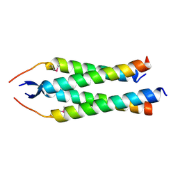 | | Crystal structure of the human Nup57CCS3* coiled-coil segment, space group C2 | | 分子名称: | Nucleoporin p54 | | 著者 | Stuwe, T, Bley, C.J, Mayo, D.J, Hoelz, A. | | 登録日 | 2013-03-15 | | 公開日 | 2014-09-17 | | 最終更新日 | 2016-02-03 | | 実験手法 | X-RAY DIFFRACTION (1.85 Å) | | 主引用文献 | Architecture of the fungal nuclear pore inner ring complex.
Science, 350, 2015
|
|
3E2U
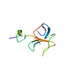 | | Crystal structure of the zink-knuckle 2 domain of human CLIP-170 in complex with CAP-Gly domain of human dynactin-1 (p150-GLUED) | | 分子名称: | CAP-Gly domain-containing linker protein 1, Dynactin subunit 1, ZINC ION | | 著者 | Weisbrich, A, Honnappa, S, Capitani, G, Steinmetz, M.O. | | 登録日 | 2008-08-06 | | 公開日 | 2008-08-19 | | 最終更新日 | 2023-11-01 | | 実験手法 | X-RAY DIFFRACTION (2.6 Å) | | 主引用文献 | Structure-function relationship of CAP-Gly domains
Nat.Struct.Mol.Biol., 14, 2007
|
|
3BUF
 | |
3E53
 | |
5VGA
 | | Alternative model for Fab 36-65 | | 分子名称: | Fab 36-65 heavy chain, Fab 36-65 light chain, TRIETHYLENE GLYCOL | | 著者 | Stanfield, R.L, Rupp, B, Wlodawer, A, Dauter, Z, Porebski, P.J, Minor, W, Jaskolski, M, Pozharski, E, Weichenberger, C.X. | | 登録日 | 2017-04-10 | | 公開日 | 2017-12-06 | | 最終更新日 | 2022-04-13 | | 実験手法 | X-RAY DIFFRACTION (2.5 Å) | | 主引用文献 | Detect, correct, retract: How to manage incorrect structural models.
FEBS J., 285, 2018
|
|
2EMD
 | |
2EMO
 | |


