6QQA
 
 | |
6QQF
 | |
3U9A
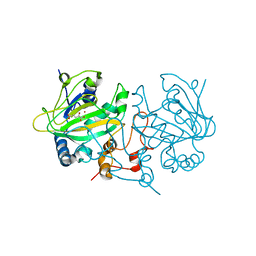
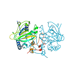
 | | Human Thrombin In Complex With MI330 | | 分子名称: | (2S)-N-[[2-(aminomethyl)-5-chloranyl-phenyl]methyl]-1-[(2S)-2-[(3-chloranyl-4-methoxy-phenyl)sulfonylamino]-4-[(4-cyanophenyl)methylamino]-4-oxidanylidene-butanoyl]pyrrolidine-2-carboxamide, 2-acetamido-2-deoxy-beta-D-glucopyranose, GLYCEROL, ... | | 著者 | Biela, A, Heine, A, Klebe, G. | | 登録日 | 2011-10-18 | | 公開日 | 2012-10-24 | | 最終更新日 | 2023-12-06 | | 実験手法 | X-RAY DIFFRACTION (1.58 Å) | | 主引用文献 | Thrombin Inhibition
To be Published
|
|
2BLE
 | | Structure of human guanosine monophosphate reductase GMPR1 in complex with GMP | | 分子名称: | GMP REDUCTASE I, GUANOSINE-5'-MONOPHOSPHATE, SODIUM ION | | 著者 | Bunkoczi, G, Haroniti, A, Ng, S, von Delft, F, Oppermann, U, Arrowsmith, C, Sundstrom, M, Edwards, A, Gileadi, O. | | 登録日 | 2005-03-03 | | 公開日 | 2005-03-09 | | 最終更新日 | 2023-12-13 | | 実験手法 | X-RAY DIFFRACTION (1.9 Å) | | 主引用文献 | Structure of Human Guanosine Monophosphate Reductase Gmpr1 in Complex with Gmp
To be Published
|
|
2HRG
 | | Crystal Structure of Blue Laccase from Trametes trogii complexed with p-methylbenzoate | | 分子名称: | 2-acetamido-2-deoxy-beta-D-glucopyranose-(1-4)-2-acetamido-2-deoxy-beta-D-glucopyranose, 4-METHYLBENZOIC ACID, CALCIUM ION, ... | | 著者 | Matera, I, Gullotto, A, Ferraroni, M, Tilli, S, Briganti, F, Scozzafava, A. | | 登録日 | 2006-07-20 | | 公開日 | 2007-09-11 | | 最終更新日 | 2024-04-03 | | 実験手法 | X-RAY DIFFRACTION (1.58 Å) | | 主引用文献 | Crystal structure of the blue multicopper oxidase from the white-rot fungus Trametes trogii complexed with p-toluate
Inorg.Chim.Acta., 361, 2008
|
|
6QQP
 | | Aplysia californica AChBP in complex with 2-Fluoro-(carbamoylpyridinyl)deschloroepibatidine analogue (2) | | 分子名称: | 1,2-ETHANEDIOL, 2-[5-[(1~{R},2~{R},4~{S})-7-azabicyclo[2.2.1]heptan-2-yl]-2-fluoranyl-pyridin-3-yl]pyridine-4-carboxamide, 2-acetamido-2-deoxy-beta-D-glucopyranose, ... | | 著者 | Bueno, R.V, Davis, S, Dawson, A, Hunter, W.N. | | 登録日 | 2019-02-18 | | 公開日 | 2020-03-18 | | 最終更新日 | 2024-01-24 | | 実験手法 | X-RAY DIFFRACTION (2.4 Å) | | 主引用文献 | Interactions between 2'-fluoro-(carbamoylpyridinyl)deschloroepibatidine analogues and acetylcholine-binding protein inform on potent antagonist activity against nicotinic receptors
Acta Crystallogr.,Sect.D, 78, 2022
|
|
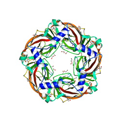
2UUL
 | | Crystal structure of C-phycocyanin from Phormidium, Lyngbya spp. (Marine) and Spirulina sp. (Fresh water) shows two different ways of energy transfer between two hexamers. | | 分子名称: | BILIVERDINE IX ALPHA, C-PHYCOCYANIN ALPHA CHAIN, C-PHYCOCYANIN BETA CHAIN, ... | | 著者 | Satyanarayana, L, Patel, A, Mishra, S, K Ghosh, P, Suresh, C.G. | | 登録日 | 2007-03-04 | | 公開日 | 2008-05-27 | | 最終更新日 | 2023-12-13 | | 実験手法 | X-RAY DIFFRACTION (3.1 Å) | | 主引用文献 | Crystal Structure of C-Phycocyanin from Phormidium, Lyngbya Spp. (Marine) and Spirulina Sp. (Fresh Water) Shows Two Different Ways of Energy Transfer between Twohexamers.
To be Published
|
|
6QSN
 | |
1QJK
 | | Metallothionein MTA from sea urchin (alpha domain) | | 分子名称: | CADMIUM ION, METALLOTHIONEIN | | 著者 | Riek, R, Precheur, B, Wang, Y, Mackay, E.A, Wider, G, Guntert, P, Liu, A, Kaegi, J.H.R, Wuthrich, K. | | 登録日 | 1999-06-24 | | 公開日 | 1999-08-31 | | 最終更新日 | 2024-05-15 | | 実験手法 | SOLUTION NMR | | 主引用文献 | NMR structure of the sea urchin (Strongylocentrotus purpuratus) metallothionein MTA.
J. Mol. Biol., 291, 1999
|
|
6FU4
 | | Ligand binding domain (LBD) of the p. aeruginosa histamine receptor TlpQ | | 分子名称: | ACETATE ION, GLYCEROL, HISTAMINE, ... | | 著者 | Gavira, J.A, Krell, T, Conejero-Muriel, M, Corral-Lugo, A, Matilla, M.A, Silva Jimenez, H, Mesa Torres, N, Martin-Mora, D. | | 登録日 | 2018-02-26 | | 公開日 | 2018-05-16 | | 最終更新日 | 2018-11-28 | | 実験手法 | X-RAY DIFFRACTION (2.45 Å) | | 主引用文献 | High-Affinity Chemotaxis to Histamine Mediated by the TlpQ Chemoreceptor of the Human Pathogen Pseudomonas aeruginosa.
MBio, 9, 2018
|
|
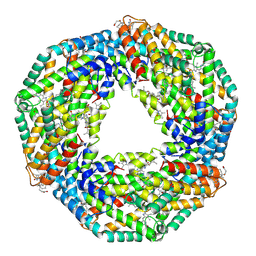
7JJJ
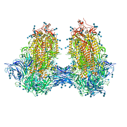 | | Structure of SARS-CoV-2 3Q-2P full-length dimers of spike trimers | | 分子名称: | 2-acetamido-2-deoxy-beta-D-glucopyranose, 2-acetamido-2-deoxy-beta-D-glucopyranose-(1-4)-2-acetamido-2-deoxy-beta-D-glucopyranose, 2-acetamido-2-deoxy-beta-D-glucopyranose-(1-4)-[alpha-L-fucopyranose-(1-6)]2-acetamido-2-deoxy-beta-D-glucopyranose, ... | | 著者 | Bangaru, S, Turner, H.L, Ozorowski, G, Antanasijevic, A, Ward, A.B. | | 登録日 | 2020-07-26 | | 公開日 | 2020-08-26 | | 最終更新日 | 2020-12-23 | | 実験手法 | ELECTRON MICROSCOPY (4.5 Å) | | 主引用文献 | Structural analysis of full-length SARS-CoV-2 spike protein from an advanced vaccine candidate.
Science, 370, 2020
|
|
5GQD
 | | Crystal structure of covalent glycosyl-enzyme intermediate of xylanase mutant (T82A, N127S, and E128H) from Streptomyces olivaceoviridis E-86 | | 分子名称: | Beta-xylanase, GLYCEROL, beta-D-xylopyranose-(1-4)-alpha-D-xylopyranose | | 著者 | Suzuki, R, Fujimoto, Z, Kaneko, S, Kuno, A. | | 登録日 | 2016-08-07 | | 公開日 | 2017-08-09 | | 最終更新日 | 2023-11-08 | | 実験手法 | X-RAY DIFFRACTION (1.8 Å) | | 主引用文献 | Azidolysis by the Formation of Stable Ser-His Catalytic Dyad in a Glycoside Hydrolase Family 10 Xylanase Mutant
J.Appl.Glyosci., 65, 2019
|
|
6QQB
 | |
6QQH
 | |
2KLS
 | | Apo-form of the second Ca2+ binding domain of NCX1.4 | | 分子名称: | Sodium/calcium exchanger 1 | | 著者 | Hilge, M, Aelen, J, Foarce, A, Perrakis, A, Vuister, G.W. | | 登録日 | 2009-07-08 | | 公開日 | 2009-08-18 | | 最終更新日 | 2024-05-22 | | 実験手法 | SOLUTION NMR | | 主引用文献 | Ca2+ regulation in the Na+/Ca2+ exchanger features a dual electrostatic switch mechanism.
Proc.Natl.Acad.Sci.USA, 106, 2009
|
|
6Y45
 | | Crystal Structure of the H33A variant of RsrR | | 分子名称: | (4S)-2-METHYL-2,4-PENTANEDIOL, 2-(N-MORPHOLINO)-ETHANESULFONIC ACID, FE2/S2 (INORGANIC) CLUSTER, ... | | 著者 | Rohac, R, Volbeda, A, Fontecilla-Camps, J.C. | | 登録日 | 2020-02-19 | | 公開日 | 2020-03-04 | | 最終更新日 | 2024-01-24 | | 実験手法 | X-RAY DIFFRACTION (1.68 Å) | | 主引用文献 | Electron and Proton Transfers Modulate DNA Binding by the Transcription Regulator RsrR.
J.Am.Chem.Soc., 142, 2020
|
|
2BL6
 | | Solution structure of the Zn complex of EIAV NCp11(22-58) peptide, including two CCHC Zn-binding motifs. | | 分子名称: | NUCLEOCAPSID PROTEIN P11, ZINC ION | | 著者 | Amodeo, P, Castiglione-Morelli, M.A, Ostuni, A, Bavoso, A. | | 登録日 | 2005-03-02 | | 公開日 | 2006-04-10 | | 最終更新日 | 2024-05-15 | | 実験手法 | SOLUTION NMR | | 主引用文献 | Structural Features in Eiav Ncp11: A Lentivirus Nucleocapsid Protein with a Short Linker
Biochemistry, 45, 2006
|
|
1CJX
 | | CRYSTAL STRUCTURE OF PSEUDOMONAS FLUORESCENS HPPD | | 分子名称: | 4-HYDROXYPHENYLPYRUVATE DIOXYGENASE, ACETATE ION, ETHYL MERCURY ION, ... | | 著者 | Serre, L, Sailland, A, Sy, D, Boudec, P, Rolland, A, Pebay-Peroulla, E, Cohen-Addad, C. | | 登録日 | 1999-04-20 | | 公開日 | 2000-04-26 | | 最終更新日 | 2023-12-27 | | 実験手法 | X-RAY DIFFRACTION (2.4 Å) | | 主引用文献 | Crystal structure of Pseudomonas fluorescens 4-hydroxyphenylpyruvate dioxygenase: an enzyme involved in the tyrosine degradation pathway.
Structure Fold.Des., 7, 1999
|
|
6G4J
 | |
5DAP
 | | Fe(II)/(alpha)ketoglutarate-dependent dioxygenase AsqJ | | 分子名称: | 2-OXOGLUTARIC ACID, NICKEL (II) ION, Phytanoyl-CoA dioxygenase family protein (AFU_orthologue AFUA_8G00230) | | 著者 | Groll, M, Braeuer, A. | | 登録日 | 2015-08-20 | | 公開日 | 2015-11-25 | | 最終更新日 | 2024-01-10 | | 実験手法 | X-RAY DIFFRACTION (1.7 Å) | | 主引用文献 | Structure of the Dioxygenase AsqJ: Mechanistic Insights into a One-Pot Multistep Quinolone Antibiotic Biosynthesis.
Angew.Chem.Int.Ed.Engl., 55, 2016
|
|
3RRL
 | | Complex structure of 3-oxoadipate coA-transferase subunit A and B from Helicobacter pylori 26695 | | 分子名称: | GLYCEROL, Succinyl-CoA:3-ketoacid-coenzyme A transferase subunit A, Succinyl-CoA:3-ketoacid-coenzyme A transferase subunit B | | 著者 | Nocek, B, Stein, A, Marshall, N, Jedrzejczak, R, Babnigg, G, Joachimiak, A, Midwest Center for Structural Genomics (MCSG) | | 登録日 | 2011-04-29 | | 公開日 | 2011-06-29 | | 最終更新日 | 2012-01-11 | | 実験手法 | X-RAY DIFFRACTION (2.29 Å) | | 主引用文献 | Complex structure of 3-oxoadipate coA-transferase subunit A and B
from Helicobacter pylori 26695
TO BE PUBLISHED
|
|
2BVD
 | | HOW FAMILY 26 GLYCOSIDE HYDROLASES ORCHESTRATE CATALYSIS ON DIFFERENT POLYSACCHARIDES. STRUCTURE AND ACTIVITY OF A CLOSTRIDIUM THERMOCELLUM LICHENASE, CtLIC26A | | 分子名称: | (3R,4R,5R)-4-hydroxy-5-(hydroxymethyl)piperidin-3-yl beta-D-glucopyranoside, ENDOGLUCANASE H | | 著者 | Taylor, E.J, Goyal, A, Guerreiro, C.I.P.D, Prates, J.A.M, Money, V.A, Ferry, N, Morland, C, Planas, A, Macdonald, J.A, Stick, R.V, Gilbert, H.J, Fontes, C.M.G.A, Davies, G.J. | | 登録日 | 2005-06-27 | | 公開日 | 2005-06-30 | | 最終更新日 | 2023-12-13 | | 実験手法 | X-RAY DIFFRACTION (1.6 Å) | | 主引用文献 | How Family 26 Glycoside Hydrolases Orchestrate Catalysis on Different Polysaccharides: Structure and Activity of a Clostridium Thermocellum Lichenase, Ctlic26A.
J.Biol.Chem., 280, 2005
|
|
5DAW
 | |
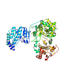
6QXK
 | | Human PIM1 bound to OX0999 | | 分子名称: | 2-[(1-methylpiperidin-4-yl)methylamino]-5-[[2-[4-(trifluoromethyloxy)phenyl]-1,3-thiazol-4-yl]methyl]-1,3-thiazol-4-one, Pimtide, Serine/threonine-protein kinase pim-1 | | 著者 | Alexander, L.T, Elkins, J.M, Russell, A, Bountra, C, Edwards, A.M, Knapp, S. | | 登録日 | 2019-03-07 | | 公開日 | 2019-04-24 | | 最終更新日 | 2024-01-24 | | 実験手法 | X-RAY DIFFRACTION (2.1 Å) | | 主引用文献 | PIM1 bound to OX0999
To Be Published
|
|
1CR7
 | | PEANUT LECTIN-LACTOSE COMPLEX MONOCLINIC FORM | | 分子名称: | CALCIUM ION, LECTIN, MANGANESE (II) ION, ... | | 著者 | Ravishankar, R, Suguna, K, Surolia, A, Vijayan, M. | | 登録日 | 1999-08-14 | | 公開日 | 2001-04-21 | | 最終更新日 | 2023-08-09 | | 実験手法 | X-RAY DIFFRACTION (2.6 Å) | | 主引用文献 | Crystal structures of the peanut lectin-lactose complex at acidic pH: retention of unusual quaternary structure, empty and carbohydrate bound combining sites, molecular mimicry and crystal packing directed by interactions at the combining site.
Proteins, 43, 2001
|
|


