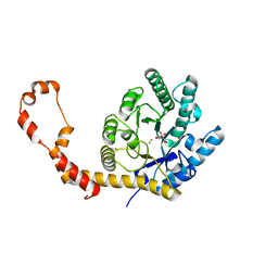
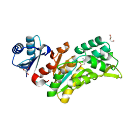
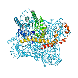
6QNJ
 
 | | Liquid Application Method for time-resolved Analyses (LAMA) by serial synchrotron crystallography, Xylose Isomerase 4.5 s timepoint | | 分子名称: | COBALT (II) ION, MAGNESIUM ION, Xylose isomerase, ... | | 著者 | Mehrabi, P, Schulz, E.C, Miller, R.J.D. | | 登録日 | 2019-02-11 | | 公開日 | 2019-10-02 | | 最終更新日 | 2024-05-15 | | 実験手法 | X-RAY DIFFRACTION (1.851 Å) | | 主引用文献 | Liquid application method for time-resolved analyses by serial synchrotron crystallography.
Nat.Methods, 16, 2019
|
|
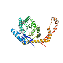
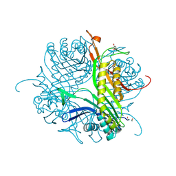
6QND
 | | Liquid Application Method for time-resolved Analyses (LAMA) by serial synchrotron crystallography, Xylose Isomerase 60 s timepoint | | 分子名称: | COBALT (II) ION, D-glucose, MAGNESIUM ION, ... | | 著者 | Mehrabi, P, Schulz, E.C, Miller, R.J.D. | | 登録日 | 2019-02-11 | | 公開日 | 2019-10-02 | | 最終更新日 | 2024-05-15 | | 実験手法 | X-RAY DIFFRACTION (2.001 Å) | | 主引用文献 | Liquid application method for time-resolved analyses by serial synchrotron crystallography.
Nat.Methods, 16, 2019
|
|
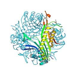
6QNI
 | | Liquid Application Method for time-resolved Analyses (LAMA) by serial synchrotron crystallography, Xylose Isomerase 1.0 s timepoint | | 分子名称: | COBALT (II) ION, MAGNESIUM ION, Xylose isomerase, ... | | 著者 | Mehrabi, P, Schulz, E.C, Miller, R.J.D. | | 登録日 | 2019-02-11 | | 公開日 | 2019-10-02 | | 最終更新日 | 2024-05-15 | | 実験手法 | X-RAY DIFFRACTION (1.846 Å) | | 主引用文献 | Liquid application method for time-resolved analyses by serial synchrotron crystallography.
Nat.Methods, 16, 2019
|
|
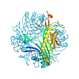
6QNH
 | | Liquid Application Method for time-resolved Analyses (LAMA) by serial synchrotron crystallography, Xylose Isomerase 0ms timepoint | | 分子名称: | COBALT (II) ION, MAGNESIUM ION, Xylose isomerase | | 著者 | Mehrabi, P, Schulz, E.C, Miller, R.J.D. | | 登録日 | 2019-02-11 | | 公開日 | 2019-10-02 | | 最終更新日 | 2024-05-15 | | 実験手法 | X-RAY DIFFRACTION (1.849 Å) | | 主引用文献 | Liquid application method for time-resolved analyses by serial synchrotron crystallography.
Nat.Methods, 16, 2019
|
|
7N36
 | |
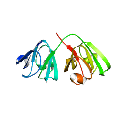
7N37
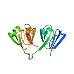
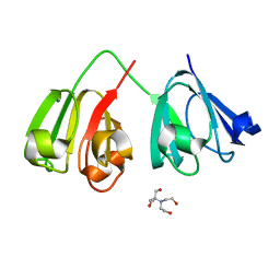 | | Crystal structure of 3-site deamidated variant of human gamma(S)-crystallin | | 分子名称: | 2-[BIS-(2-HYDROXY-ETHYL)-AMINO]-2-HYDROXYMETHYL-PROPANE-1,3-DIOL, Gamma-crystallin S, MAGNESIUM ION | | 著者 | Norton-Baker, B, Mehrabi, P, Martin, R.W. | | 登録日 | 2021-05-31 | | 公開日 | 2022-03-23 | | 最終更新日 | 2023-10-18 | | 実験手法 | X-RAY DIFFRACTION (1.3 Å) | | 主引用文献 | Deamidation of the human eye lens protein gamma S-crystallin accelerates oxidative aging.
Structure, 30, 2022
|
|
7N38
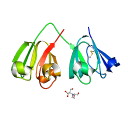 | | Crystal structure of 5-site deamidated variant of human gamma(S)-crystallin | | 分子名称: | 2-[BIS-(2-HYDROXY-ETHYL)-AMINO]-2-HYDROXYMETHYL-PROPANE-1,3-DIOL, Gamma-crystallin S, MAGNESIUM ION | | 著者 | Norton-Baker, B, Mehrabi, P, Martin, R.W. | | 登録日 | 2021-05-31 | | 公開日 | 2022-03-23 | | 最終更新日 | 2023-10-18 | | 実験手法 | X-RAY DIFFRACTION (1.22 Å) | | 主引用文献 | Deamidation of the human eye lens protein gamma S-crystallin accelerates oxidative aging.
Structure, 30, 2022
|
|
7N39
 | |
7N3A
 | |
7N3B
 | |
8Q4S
 | |
8AMS
 | | Complex of human TRIM2 RING domain, UBCH5C, and Ubiquitin | | 分子名称: | 3,6,9,12,15,18,21-HEPTAOXATRICOSANE-1,23-DIOL, GLYCEROL, Polyubiquitin-C, ... | | 著者 | Perez-Borrajero, C, Kotova, I, Murciano, B, Hennig, J. | | 登録日 | 2022-08-04 | | 公開日 | 2023-11-15 | | 実験手法 | X-RAY DIFFRACTION (2.4 Å) | | 主引用文献 | Structural and biophysical studies of TRIM2 and TRIM3
To Be Published
|
|
8Q3L
 | |
6RNC
 | | Liquid Application Method for time-resolved Analyses (LAMA) by serial synchrotron crystallography, Lysozyme with GlcNAc3 - 100ms diffusion time. | | 分子名称: | 2-acetamido-2-deoxy-beta-D-glucopyranose-(1-4)-2-acetamido-2-deoxy-beta-D-glucopyranose-(1-4)-2-acetamido-2-deoxy-beta-D-glucopyranose, CHLORIDE ION, Lysozyme C, ... | | 著者 | Mehrabi, P, Schulz, E.C, Miller, R.J.D. | | 登録日 | 2019-05-08 | | 公開日 | 2019-10-02 | | 最終更新日 | 2024-01-24 | | 実験手法 | X-RAY DIFFRACTION (1.799 Å) | | 主引用文献 | Liquid application method for time-resolved analyses by serial synchrotron crystallography.
Nat.Methods, 16, 2019
|
|
6RNB
 | | Liquid Application Method for time-resolved Analyses (LAMA) by serial synchrotron crystallography, Lysozyme with GlcNAc3 50ms diffusion time | | 分子名称: | 2-acetamido-2-deoxy-beta-D-glucopyranose-(1-4)-2-acetamido-2-deoxy-beta-D-glucopyranose-(1-4)-2-acetamido-2-deoxy-beta-D-glucopyranose, CHLORIDE ION, Lysozyme C, ... | | 著者 | Mehrabi, P, Schulz, E.C, Miller, R.J.D. | | 登録日 | 2019-05-08 | | 公開日 | 2019-10-02 | | 最終更新日 | 2024-01-24 | | 実験手法 | X-RAY DIFFRACTION (1.699 Å) | | 主引用文献 | Liquid application method for time-resolved analyses by serial synchrotron crystallography.
Nat.Methods, 16, 2019
|
|
6RNF
 | | Liquid Application Method for time-resolved Analyses (LAMA) by serial synchrotron crystallography, Xylose Isomerase 30 ms timepoint | | 分子名称: | MAGNESIUM ION, Xylose isomerase, alpha-D-glucopyranose | | 著者 | Mehrabi, P, Schulz, E.C, Miller, R.J.D. | | 登録日 | 2019-05-08 | | 公開日 | 2019-10-02 | | 最終更新日 | 2024-01-24 | | 実験手法 | X-RAY DIFFRACTION (1.7 Å) | | 主引用文献 | Liquid application method for time-resolved analyses by serial synchrotron crystallography.
Nat.Methods, 16, 2019
|
|
6RND
 | | Liquid Application Method for time-resolved Analyses (LAMA) by serial synchrotron crystallography, Xylose Isomerase 15 ms timepoint | | 分子名称: | MAGNESIUM ION, Xylose isomerase, alpha-D-glucopyranose | | 著者 | Mehrabi, P, Schulz, E.C, Miller, R.J.D. | | 登録日 | 2019-05-08 | | 公開日 | 2019-10-02 | | 最終更新日 | 2024-01-24 | | 実験手法 | X-RAY DIFFRACTION (1.7 Å) | | 主引用文献 | Liquid application method for time-resolved analyses by serial synchrotron crystallography.
Nat.Methods, 16, 2019
|
|
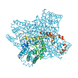
7B83
 | | Structure of SARS-CoV-2 Main Protease bound to pyrithione zinc | | 分子名称: | 3C-like proteinase, 9-oxa-7-thia-1-azonia-8$l^{2}-zincabicyclo[4.3.0]nona-1,3,5-triene, CHLORIDE ION, ... | | 著者 | Guenther, S, Reinke, P, Oberthuer, D, Yefanov, O, Gelisio, L, Ginn, H, Lieske, J, Domaracky, M, Brehm, W, Rahmani Mashour, A, White, T.A, Knoska, J, Pena Esperanza, G, Koua, F, Tolstikova, A, Groessler, M, Fischer, P, Hennicke, V, Fleckenstein, H, Trost, F, Galchenkova, M, Gevorkov, Y, Li, C, Awel, S, Paulraj, L.X, Ullah, N, Falke, S, Alves Franca, B, Schwinzer, M, Brognaro, H, Werner, N, Perbandt, M, Tidow, H, Seychell, B, Beck, T, Meier, S, Doyle, J.J, Giseler, H, Melo, D, Dunkel, I, Lane, T.J, Peck, A, Saouane, S, Hakanpaeae, J, Meyer, J, Noei, H, Gribbon, P, Ellinger, B, Kuzikov, M, Wolf, M, Zhang, L, Ehrt, C, Pletzer-Zelgert, J, Wollenhaupt, J, Feiler, C, Weiss, M, Schulz, E.C, Mehrabi, P, Norton-Baker, B, Schmidt, C, Lorenzen, K, Schubert, R, Han, H, Chari, A, Fernandez Garcia, Y, Turk, D, Hilgenfeld, R, Rarey, M, Zaliani, A, Chapman, H.N, Pearson, A, Betzel, C, Meents, A. | | 登録日 | 2020-12-12 | | 公開日 | 2021-01-13 | | 最終更新日 | 2024-01-31 | | 実験手法 | X-RAY DIFFRACTION (1.8 Å) | | 主引用文献 | X-ray screening identifies active site and allosteric inhibitors of SARS-CoV-2 main protease.
Science, 372, 2021
|
|
7AQE
 | | Structure of SARS-CoV-2 Main Protease bound to UNC-2327 | | 分子名称: | 3C-like proteinase, CHLORIDE ION, DIMETHYL SULFOXIDE, ... | | 著者 | Guenther, S, Reinke, P, Meents, A. | | 登録日 | 2020-10-21 | | 公開日 | 2021-03-03 | | 最終更新日 | 2024-01-31 | | 実験手法 | X-RAY DIFFRACTION (1.39 Å) | | 主引用文献 | X-ray screening identifies active site and allosteric inhibitors of SARS-CoV-2 main protease.
Science, 372, 2021
|
|
4CW3
 | | Crystal structure of cofactor-free urate oxidase in complex with the 5-peroxo derivative of 9-metyl uric acid (X-ray dose, 665 kGy) | | 分子名称: | (4S)-2-METHYL-2,4-PENTANEDIOL, (5S)-5-(dioxidanyl)-9-methyl-7H-purine-2,6,8-trione, 9-METHYL URIC ACID, ... | | 著者 | Bui, S, Steiner, R.A. | | 登録日 | 2014-04-01 | | 公開日 | 2014-10-29 | | 最終更新日 | 2018-02-21 | | 実験手法 | X-RAY DIFFRACTION (1.34 Å) | | 主引用文献 | Direct evidence for a peroxide intermediate and a reactive enzyme-substrate-dioxygen configuration in a cofactor-free oxidase.
Angew. Chem. Int. Ed. Engl., 53, 2014
|
|
4CW6
 | | Crystal structure of cofactor-free urate oxidase in complex with the 5-peroxo derivative of 9-metyl uric acid (X-ray dose, 92 kGy) | | 分子名称: | (4S)-2-METHYL-2,4-PENTANEDIOL, (5S)-5-(dioxidanyl)-9-methyl-7H-purine-2,6,8-trione, 9-METHYL URIC ACID, ... | | 著者 | Bui, S, Steiner, R.A. | | 登録日 | 2014-04-01 | | 公開日 | 2014-10-29 | | 最終更新日 | 2018-02-21 | | 実験手法 | X-RAY DIFFRACTION (1.28 Å) | | 主引用文献 | Direct evidence for a peroxide intermediate and a reactive enzyme-substrate-dioxygen configuration in a cofactor-free oxidase.
Angew. Chem. Int. Ed. Engl., 53, 2014
|
|
4CW0
 | |
4CW2
 | | Crystal structure of cofactor-free urate oxidase in complex with the 5-peroxo derivative of 9-metyl uric acid (X-ray dose, 2.5 kGy) | | 分子名称: | (4S)-2-METHYL-2,4-PENTANEDIOL, (5S)-5-(dioxidanyl)-9-methyl-7H-purine-2,6,8-trione, URICASE | | 著者 | Bui, S, Steiner, R.A. | | 登録日 | 2014-04-01 | | 公開日 | 2014-10-29 | | 最終更新日 | 2018-02-21 | | 実験手法 | X-RAY DIFFRACTION (1.32 Å) | | 主引用文献 | Direct evidence for a peroxide intermediate and a reactive enzyme-substrate-dioxygen configuration in a cofactor-free oxidase.
Angew. Chem. Int. Ed. Engl., 53, 2014
|
|
4D19
 | | Crystal structure of cofactor-free urate oxidase in complex with its 5-peroxoisourate intermediate (X-ray dose, 1.75 MGy) | | 分子名称: | (4S)-2-METHYL-2,4-PENTANEDIOL, 5-(HYDRO)PEROXOISOURATE, OXYGEN MOLECULE, ... | | 著者 | Bui, S, Steiner, R.A. | | 登録日 | 2014-05-01 | | 公開日 | 2014-10-29 | | 最終更新日 | 2024-05-08 | | 実験手法 | X-RAY DIFFRACTION (1.35 Å) | | 主引用文献 | Direct evidence for a peroxide intermediate and a reactive enzyme-substrate-dioxygen configuration in a cofactor-free oxidase.
Angew. Chem. Int. Ed. Engl., 53, 2014
|
|
4D13
 | |


