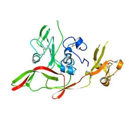
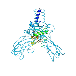
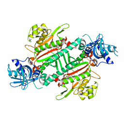
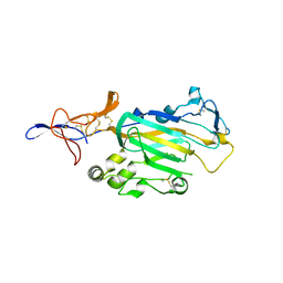
1UZK
 
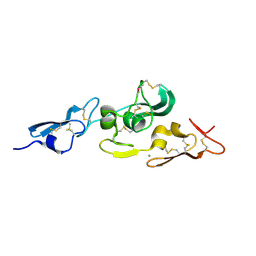 | | Integrin binding cbEGF22-TB4-cbEGF33 fragment of human fibrillin-1, Ca bound to cbEGF23 domain only | | 分子名称: | CALCIUM ION, FIBRILLIN-1 | | 著者 | Lee, S.S.J, Knott, V, Harlos, K, Handford, P.A, Stuart, D.I. | | 登録日 | 2004-03-13 | | 公開日 | 2006-05-24 | | 最終更新日 | 2024-05-01 | | 実験手法 | X-RAY DIFFRACTION (1.35 Å) | | 主引用文献 | Structure of the Integrin Binding Fragment from Fibrillin-1 Gives New Insights Into Microfibril Organization
Structure, 12, 2004
|
|
4BXT
 | |
4CNC
 | | Crystal structure of human 5T4 (Wnt-activated inhibitory factor 1, Trophoblast glycoprotein) | | 分子名称: | 2-acetamido-2-deoxy-beta-D-glucopyranose, 2-acetamido-2-deoxy-beta-D-glucopyranose-(1-4)-2-acetamido-2-deoxy-beta-D-glucopyranose, DI(HYDROXYETHYL)ETHER, ... | | 著者 | Zhao, Y, Malinauskas, T, Harlos, K, Jones, E.Y. | | 登録日 | 2014-01-21 | | 公開日 | 2014-02-26 | | 最終更新日 | 2024-05-01 | | 実験手法 | X-RAY DIFFRACTION (1.77 Å) | | 主引用文献 | Structural Insights Into the Inhibition of Wnt Signaling by Cancer Antigen 5T4/Wnt-Activated Inhibitory Factor 1.
Structure, 22, 2014
|
|
4CNM
 | | Crystal structure of human 5T4 (Wnt-activated inhibitory factor 1, Trophoblast glycoprotein) | | 分子名称: | 2-acetamido-2-deoxy-beta-D-glucopyranose, 2-acetamido-2-deoxy-beta-D-glucopyranose-(1-4)-2-acetamido-2-deoxy-beta-D-glucopyranose, CITRIC ACID, ... | | 著者 | Zhao, Y, Malinauskas, T, Harlos, K, Jones, E.Y. | | 登録日 | 2014-01-23 | | 公開日 | 2014-02-26 | | 最終更新日 | 2023-12-20 | | 実験手法 | X-RAY DIFFRACTION (1.75 Å) | | 主引用文献 | Structural Insights Into the Inhibition of Wnt Signaling by Cancer Antigen 5T4/Wnt-Activated Inhibitory Factor 1.
Structure, 22, 2014
|
|
4CS8
 | | Crystal structure of the asymmetric human metapneumovirus M2-1 tetramer, form 2 | | 分子名称: | M2-1, ZINC ION | | 著者 | Leyrat, C, Renner, M, Harlos, K, Grimes, J.M. | | 登録日 | 2014-03-05 | | 公開日 | 2014-05-28 | | 最終更新日 | 2023-12-20 | | 実験手法 | X-RAY DIFFRACTION (2.1 Å) | | 主引用文献 | Drastic Changes in Conformational Dynamics of the Antiterminator M2-1 Regulate Transcription Efficiency in Pneumovirinae.
Elife, 3, 2014
|
|
6IPA
 | |
6JRE
 | | Structure of N-terminal domain of Plasmodium vivax p43 (PfNTD) solved by Co-SAD phasing | | 分子名称: | Aminoacyl-tRNA synthetase-interacting multifunctional protein p43, COBALT (II) ION | | 著者 | Manickam, Y, Harlos, K, Sharma, M, Gupta, S, Sharma, A. | | 登録日 | 2019-04-03 | | 公開日 | 2020-03-11 | | 実験手法 | X-RAY DIFFRACTION (2.59 Å) | | 主引用文献 | Crystal structures of the two domains that constitute the Plasmodium vivax p43 protein.
Acta Crystallogr D Struct Biol, 76, 2020
|
|
6FKM
 | |
6F8P
 | |
6FKN
 | |
6FKK
 | | Drosophila Semaphorin 1b, extracellular domains 1-2 | | 分子名称: | 2-acetamido-2-deoxy-beta-D-glucopyranose, MIP07328p, alpha-D-mannopyranose-(1-2)-alpha-D-mannopyranose-(1-2)-alpha-D-mannopyranose-(1-3)-[alpha-D-mannopyranose-(1-3)-[alpha-D-mannopyranose-(1-6)]alpha-D-mannopyranose-(1-6)]beta-D-mannopyranose-(1-4)-2-acetamido-2-deoxy-beta-D-glucopyranose-(1-4)-2-acetamido-2-deoxy-beta-D-glucopyranose, ... | | 著者 | Rozbesky, D, Harlos, K, Jones, E.Y. | | 登録日 | 2018-01-24 | | 公開日 | 2019-02-06 | | 最終更新日 | 2024-01-17 | | 実験手法 | X-RAY DIFFRACTION (2.78 Å) | | 主引用文献 | Diversity of oligomerization in Drosophila semaphorins suggests a mechanism of functional fine-tuning.
Nat Commun, 10, 2019
|
|
1H15
 | | X-ray crystal structure of HLA-DRA1*0101/DRB5*0101 complexed with a peptide from Epstein Barr Virus DNA polymerase | | 分子名称: | 2-acetamido-2-deoxy-beta-D-glucopyranose, 2-acetamido-2-deoxy-beta-D-glucopyranose-(1-4)-2-acetamido-2-deoxy-beta-D-glucopyranose, DNA POLYMERASE, ... | | 著者 | Lang, H, Jacobsen, H, Ikemizu, S, Andersson, C, Harlos, K, Madsen, L, Hjorth, P, Sondergaard, L, Svejgaard, A, Wucherpfennig, K, Stuart, D.I, Bell, J.I, Jones, E.Y, Fugger, L. | | 登録日 | 2002-07-02 | | 公開日 | 2002-10-03 | | 最終更新日 | 2023-12-13 | | 実験手法 | X-RAY DIFFRACTION (3.1 Å) | | 主引用文献 | A Functional and Structural Basis for Tcr Cross-Reactivity in Multiple Sclerosis
Nat.Immunol., 3, 2002
|
|
4AE2
 | | Crystal structure of Human fibrillar procollagen type III C- propeptide trimer | | 分子名称: | CALCIUM ION, COLLAGEN ALPHA-1(III) CHAIN, NITRATE ION | | 著者 | Bourhis, J.M, Mariano, N, Zhao, Y, Harlos, K, Jones, E.Y, Moali, C, Aghajari, N, Hulmes, D.J. | | 登録日 | 2012-01-05 | | 公開日 | 2012-09-12 | | 最終更新日 | 2019-04-03 | | 実験手法 | X-RAY DIFFRACTION (1.68 Å) | | 主引用文献 | Structural Basis of Fibrillar Collagen Trimerization and Related Genetic Disorders.
Nat.Struct.Mol.Biol., 19, 2012
|
|
4AK3
 | | Crystal structure of Human fibrillar procollagen type III C- propeptide trimer | | 分子名称: | CALCIUM ION, COLLAGEN ALPHA-1(III) CHAIN | | 著者 | Bourhis, J.M, Mariano, N, Zhao, Y, Harlos, K, Jones, E.Y, Moali, C, Aghajari, N, Hulmes, D.J.S. | | 登録日 | 2012-02-21 | | 公開日 | 2012-09-12 | | 最終更新日 | 2023-12-20 | | 実験手法 | X-RAY DIFFRACTION (3.5 Å) | | 主引用文献 | Structural Basis of Fibrillar Collagen Trimerization and Related Genetic Disorders.
Nat.Struct.Mol.Biol., 19, 2012
|
|
4AEJ
 | | Crystal structure of Human fibrillar procollagen type III C- propeptide trimer | | 分子名称: | CALCIUM ION, COLLAGEN ALPHA-1(III) CHAIN, GLYCEROL | | 著者 | Bourhis, J.M, Mariano, N, Zhao, Y, Harlos, K, Jones, E.Y, Moali, C, Aghajari, N, Hulmes, D.J.S. | | 登録日 | 2012-01-11 | | 公開日 | 2012-09-12 | | 最終更新日 | 2024-10-09 | | 実験手法 | X-RAY DIFFRACTION (2.21 Å) | | 主引用文献 | Structural Basis of Fibrillar Collagen Trimerization and Related Genetic Disorders.
Nat.Struct.Mol.Biol., 19, 2012
|
|
5ZH4
 | | CRYSTAL STRUCTURE OF PfKRS WITH INHIBITOR CLADO-7 | | 分子名称: | (3R)-6,8-dihydroxy-3-{[(2S,6R)-6-methyloxan-2-yl]methyl}-3,4-dihydro-1H-2-benzopyran-1-one, CHLORIDE ION, LYSINE, ... | | 著者 | Babbar, P, Malhotra, N, Sharma, M, Harlos, K, Reddy, D.S, Manickam, Y, Sharma, A. | | 登録日 | 2018-03-11 | | 公開日 | 2018-06-27 | | 最終更新日 | 2023-11-22 | | 実験手法 | X-RAY DIFFRACTION (2.6 Å) | | 主引用文献 | Specific Stereoisomeric Conformations Determine the Drug Potency of Cladosporin Scaffold against Malarial Parasite
J. Med. Chem., 61, 2018
|
|
5ZH3
 | | CRYSTAL STRUCTURE OF PfKRS WITH INHIBITOR CLADO-6 | | 分子名称: | (3S)-6,8-dihydroxy-3-{[(2R,6R)-6-methyloxan-2-yl]methyl}-3,4-dihydro-1H-2-benzopyran-1-one, LYSINE, Lysine-tRNA ligase | | 著者 | Babbar, P, Malhotra, N, Sharma, M, Harlos, K, Reddy, D.S, Manickam, Y, Sharma, A. | | 登録日 | 2018-03-11 | | 公開日 | 2018-06-27 | | 最終更新日 | 2023-11-22 | | 実験手法 | X-RAY DIFFRACTION (2.86 Å) | | 主引用文献 | Specific Stereoisomeric Conformations Determine the Drug Potency of Cladosporin Scaffold against Malarial Parasite
J. Med. Chem., 61, 2018
|
|
5ZH5
 | | CRYSTAL STRUCTURE OF PfKRS WITH INHIBITOR CLADO-2 | | 分子名称: | (3S)-6,8-dihydroxy-3-{[(2R,6S)-6-methyloxan-2-yl]methyl}-3,4-dihydro-1H-2-benzopyran-1-one, CHLORIDE ION, LYSINE, ... | | 著者 | Babbar, P, Malhotra, N, Sharma, M, Harlos, K, Reddy, D.S, Manickam, Y, Sharma, A. | | 登録日 | 2018-03-11 | | 公開日 | 2018-06-27 | | 最終更新日 | 2023-11-22 | | 実験手法 | X-RAY DIFFRACTION (3.08 Å) | | 主引用文献 | Specific Stereoisomeric Conformations Determine the Drug Potency of Cladosporin Scaffold against Malarial Parasite
J. Med. Chem., 61, 2018
|
|
3ZN4
 | | VP16, a capsid protein of bacteriophage P23-77 (VP16-type-2) | | 分子名称: | CHLORIDE ION, CITRIC ACID, VP16 | | 著者 | Rissanen, I, Grimes, J.M, Pawlowski, A, Mantynen, S, Harlos, K, Bamford, J.K.H, Stuart, D.I. | | 登録日 | 2013-02-13 | | 公開日 | 2013-05-15 | | 最終更新日 | 2023-12-20 | | 実験手法 | X-RAY DIFFRACTION (1.26 Å) | | 主引用文献 | Bacteriophage P23-77 Capsid Protein Structures Reveal the Archetype of an Ancient Branch from a Major Virus Lineage.
Structure, 21, 2013
|
|
3ZMN
 | | VP17, a capsid protein of bacteriophage P23-77 | | 分子名称: | VP17 | | 著者 | Rissanen, I, Grimes, J.M, Pawlowski, A, Mantynen, S, Harlos, K, Bamford, J.K.H, Stuart, D.I. | | 登録日 | 2013-02-11 | | 公開日 | 2013-05-15 | | 最終更新日 | 2024-05-08 | | 実験手法 | X-RAY DIFFRACTION (2.26 Å) | | 主引用文献 | Bacteriophage P23-77 Capsid Protein Structures Reveal the Archetype of an Ancient Branch from a Major Virus Lineage.
Structure, 21, 2013
|
|
3ZYI
 | | NetrinG2 in complex with NGL2 | | 分子名称: | 2-acetamido-2-deoxy-beta-D-glucopyranose, CALCIUM ION, LEUCINE-RICH REPEAT-CONTAINING PROTEIN 4, ... | | 著者 | Seiradake, E, Coles, C.H, Perestenko, P.V, Harlos, K, McIlhinney, R.A.J, Aricescu, A.R, Jones, E.Y. | | 登録日 | 2011-08-23 | | 公開日 | 2011-10-05 | | 最終更新日 | 2024-10-16 | | 実験手法 | X-RAY DIFFRACTION (2.6 Å) | | 主引用文献 | Structural Basis for Cell Surface Patterning Through Netring-Ngl Interactions
Embo J., 30, 2011
|
|
3ZYG
 | | NETRING2 LAM AND EGF1 DOMAINS | | 分子名称: | 2-acetamido-2-deoxy-beta-D-glucopyranose, CALCIUM ION, NETRIN-G2 | | 著者 | Seiradake, E, Coles, C.H, Perestenko, P.V, Harlos, K, Mcilhinney, R.A.J, Aricescu, A.R, Jones, E.Y. | | 登録日 | 2011-08-22 | | 公開日 | 2011-10-05 | | 最終更新日 | 2023-12-20 | | 実験手法 | X-RAY DIFFRACTION (2.2 Å) | | 主引用文献 | Structural Basis for Cell Surface Patterning Through Netring-Ngl Interactions.
Embo J., 30, 2011
|
|
3ZYO
 | | Crystal structure of the N-terminal leucine rich repeats and immunoglobulin domain of netrin-G ligand-3 | | 分子名称: | 2-acetamido-2-deoxy-beta-D-glucopyranose, LEUCINE-RICH REPEAT-CONTAINING PROTEIN 4B, ZINC ION | | 著者 | Seiradake, E, Coles, C.H, Perestenko, P.V, Harlos, K, McIlhinney, R.A.J, Aricescu, A.R, Jones, E.Y. | | 登録日 | 2011-08-24 | | 公開日 | 2011-10-05 | | 最終更新日 | 2023-12-20 | | 実験手法 | X-RAY DIFFRACTION (3.1 Å) | | 主引用文献 | Structural Basis for Cell Surface Patterning Through Netring-Ngl Interactions.
Embo J., 30, 2011
|
|
3ZYN
 | | Crystal structure of the N-terminal leucine rich repeats of Netrin-G Ligand-3 | | 分子名称: | 2-acetamido-2-deoxy-beta-D-glucopyranose, LEUCINE-RICH REPEAT-CONTAINING PROTEIN 4B | | 著者 | Seiradake, E, Coles, C.H, Perestenko, P.V, Harlos, K, McIlhinney, R.A.J, Aricescu, A.R, Jones, E.Y. | | 登録日 | 2011-08-23 | | 公開日 | 2011-10-05 | | 最終更新日 | 2023-12-20 | | 実験手法 | X-RAY DIFFRACTION (3.2 Å) | | 主引用文献 | Structural Basis for Cell Surface Patterning Through Netring-Ngl Interactions.
Embo J., 30, 2011
|
|
3ZYJ
 | | NetrinG1 in complex with NGL1 | | 分子名称: | 2-acetamido-2-deoxy-beta-D-glucopyranose, CALCIUM ION, LEUCINE-RICH REPEAT-CONTAINING PROTEIN 4C, ... | | 著者 | Seiradake, E, Coles, C.H, Perestenko, P.V, Harlos, K, McIlhinney, R.A.J, Aricescu, A.R, Jones, E.Y. | | 登録日 | 2011-08-23 | | 公開日 | 2011-10-05 | | 最終更新日 | 2023-12-20 | | 実験手法 | X-RAY DIFFRACTION (3.25 Å) | | 主引用文献 | Structural Basis for Cell Surface Patterning Through Netring-Ngl Interactions.
Embo J., 30, 2011
|
|


