2M97
 
 | |
6O3Y
 | |
6O3W
 | |
6O3X
 | |
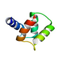
6NNW
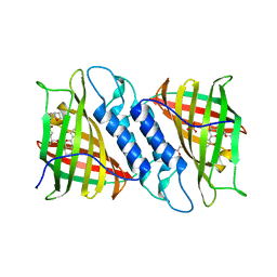 | | Tsn15 in complex with substrate intermediate | | 分子名称: | (3E)-3-{(1S,4S,4aS,5R,8aS)-1-[(2E,4R,7S,8E,10S)-1,7-dihydroxy-10-{(2R,3S,5R)-5-[(1S)-1-methoxyethyl]-3-methyloxolan-2-yl}-4-methylundeca-2,8-dien-2-yl]-4,5-dimethyloctahydro-3H-2-benzopyran-3-ylidene}oxolane-2,4-dione, PHOSPHATE ION, PROPANOIC ACID, ... | | 著者 | Paiva, F.C.R, Little, R, Leadlay, P, Dias, M.V.B. | | 登録日 | 2019-01-15 | | 公開日 | 2019-10-23 | | 最終更新日 | 2023-10-11 | | 実験手法 | X-RAY DIFFRACTION (1.7 Å) | | 主引用文献 | Unexpected enzyme-catalysed [4+2] cycloaddition and rearrangement in polyether antibiotic biosynthesis
Nat Catal, 2019
|
|
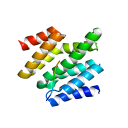
6NOI
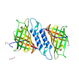 | | Crystal structure of Tsn15 in apo form | | 分子名称: | DI(HYDROXYETHYL)ETHER, PHOSPHATE ION, TRIETHYLENE GLYCOL, ... | | 著者 | Little, R, Paiva, F.C.R, Dias, M.V.B, Leadlay, P. | | 登録日 | 2019-01-16 | | 公開日 | 2019-10-23 | | 実験手法 | X-RAY DIFFRACTION (1.8 Å) | | 主引用文献 | Unexpected enzyme-catalysed [4+2] cycloaddition and rearrangement in polyether antibiotic biosynthesis
Nat Catal, 2019
|
|
2D6F
 | |
7QO8
 | |
1MEA
 | |
1MED
 | |
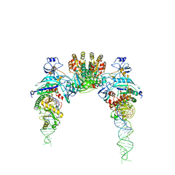
1EG0
 | | FITTING OF COMPONENTS WITH KNOWN STRUCTURE INTO AN 11.5 A CRYO-EM MAP OF THE E.COLI 70S RIBOSOME | | 分子名称: | FORMYL-METHIONYL-TRNA, FRAGMENT OF 16S RRNA HELIX 23, FRAGMENT OF 23S RRNA, ... | | 著者 | Gabashvili, I.S, Agrawal, R.K, Spahn, C.M.T, Grassucci, R.A, Svergun, D.I, Frank, J, Penczek, P. | | 登録日 | 2000-02-11 | | 公開日 | 2000-03-06 | | 最終更新日 | 2024-02-07 | | 実験手法 | ELECTRON MICROSCOPY (11.5 Å) | | 主引用文献 | Solution structure of the E. coli 70S ribosome at 11.5 A resolution.
Cell(Cambridge,Mass.), 100, 2000
|
|
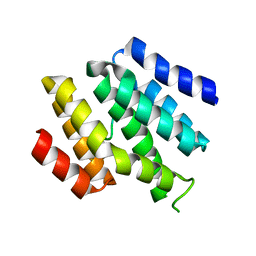
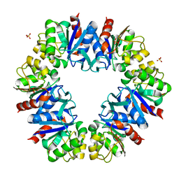
3B2R
 | | Crystal Structure of PDE5A1 catalytic domain in complex with Vardenafil | | 分子名称: | 2-{2-ETHOXY-5-[(4-ETHYLPIPERAZIN-1-YL)SULFONYL]PHENYL}-5-METHYL-7-PROPYLIMIDAZO[5,1-F][1,2,4]TRIAZIN-4(1H)-ONE, cGMP-specific 3',5'-cyclic phosphodiesterase | | 著者 | Huanchen, W, Mengchun, Y, Howard, R, Sharron, H.F, Hengming, K. | | 登録日 | 2007-10-19 | | 公開日 | 2008-05-20 | | 最終更新日 | 2024-04-03 | | 実験手法 | X-RAY DIFFRACTION (2.07 Å) | | 主引用文献 | Conformational variations of both phosphodiesterase-5 and inhibitors provide the structural basis for the physiological effects of vardenafil and sildenafil.
Mol.Pharmacol., 73, 2008
|
|


