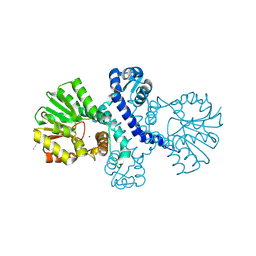
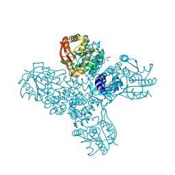
5O25
 
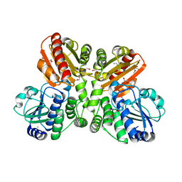 | | Structure of wildtype T.maritima PDE (TM1595) in ligand-free state | | 分子名称: | CALCIUM ION, DI(HYDROXYETHYL)ETHER, MANGANESE (II) ION, ... | | 著者 | Witte, G, Drexler, D, Mueller, M. | | 登録日 | 2017-05-19 | | 公開日 | 2017-10-25 | | 最終更新日 | 2024-01-17 | | 実験手法 | X-RAY DIFFRACTION (1.75 Å) | | 主引用文献 | Structural and Biophysical Analysis of the Soluble DHH/DHHA1-Type Phosphodiesterase TM1595 from Thermotoga maritima.
Structure, 25, 2017
|
|
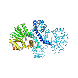
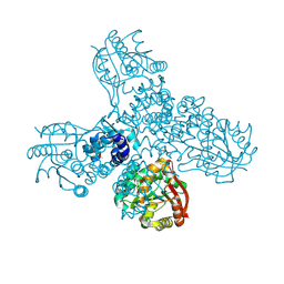
5O4Z
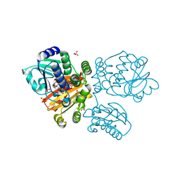 | | Structure of the inactive T.maritima PDE (TM1595) D80N D154N mutant with substrate 5'-pApA | | 分子名称: | ADENOSINE-5'-MONOPHOSPHATE, CHLORIDE ION, DHH/DHHA1-type phosphodiesterase TM1595, ... | | 著者 | Witte, G, Drexler, D, Mueller, M. | | 登録日 | 2017-05-31 | | 公開日 | 2017-10-25 | | 最終更新日 | 2024-01-17 | | 実験手法 | X-RAY DIFFRACTION (1.7 Å) | | 主引用文献 | Structural and Biophysical Analysis of the Soluble DHH/DHHA1-Type Phosphodiesterase TM1595 from Thermotoga maritima.
Structure, 25, 2017
|
|
5QU8
 | |
5QU3
 | |
5QUA
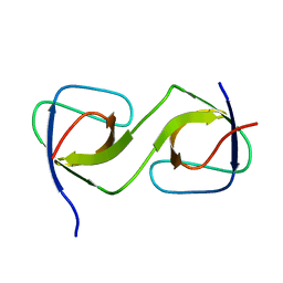 | |
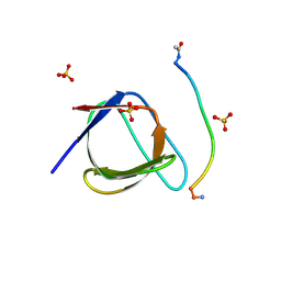
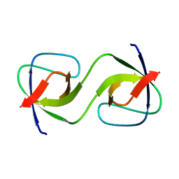
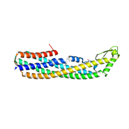
5QU1
 | | Crystal Structure of the monomeric human Nck SH3.1 domain, triclinic, 1.08A | | 分子名称: | Cytoplasmic protein NCK1, SULFATE ION | | 著者 | Burger, D, Ruf, A, Benz, J, Schlatter, D, Rudolph, M.G. | | 登録日 | 2019-12-13 | | 公開日 | 2020-02-12 | | 最終更新日 | 2024-04-03 | | 実験手法 | X-RAY DIFFRACTION (1.08 Å) | | 主引用文献 | Small molecule AX-024 reduces T cell proliferation independently of CD3ε/Nck1 interaction, which is governed by a domain swap in the Nck1-SH3.1 domain.
J.Biol.Chem., 295, 2020
|
|
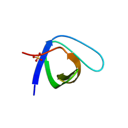
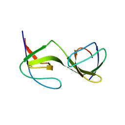
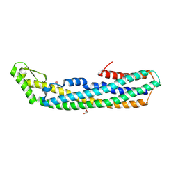
5QU5
 | | Domain Swap in the first SH3 domain of human Nck1 | | 分子名称: | Cytoplasmic protein NCK1 | | 著者 | Burger, D, Ruf, A, Benz, J, Schlatter, D, Rudolph, M.G. | | 登録日 | 2019-12-13 | | 公開日 | 2020-02-12 | | 最終更新日 | 2024-04-03 | | 実験手法 | X-RAY DIFFRACTION (1.11 Å) | | 主引用文献 | Small molecule AX-024 reduces T cell proliferation independently of CD3ε/Nck1 interaction, which is governed by a domain swap in the Nck1-SH3.1 domain.
J.Biol.Chem., 295, 2020
|
|
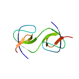
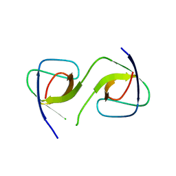
5QU2
 | | Crystal Structure of human Nck SH3.1 in complex with peptide PPPVPNPDY | | 分子名称: | ACE-PRO-PRO-PRO-VAL-PRO-ASN-PRO-ASP-TYR-NH2, Cytoplasmic protein NCK1, SULFATE ION | | 著者 | Rudolph, M.G. | | 登録日 | 2019-12-13 | | 公開日 | 2020-02-12 | | 最終更新日 | 2024-04-03 | | 実験手法 | X-RAY DIFFRACTION (1.04 Å) | | 主引用文献 | Small molecule AX-024 reduces T cell proliferation independently of CD3ε/Nck1 interaction, which is governed by a domain swap in the Nck1-SH3.1 domain.
J.Biol.Chem., 295, 2020
|
|
5QU7
 | |
5QU4
 | |
4A7H
 | | Structure of the Actin-Tropomyosin-Myosin Complex (rigor ATM 2) | | 分子名称: | ACTIN, ALPHA SKELETAL MUSCLE, ADENOSINE-5'-DIPHOSPHATE, ... | | 著者 | Behrmann, E, Mueller, M, Penczek, P.A, Mannherz, H.G, Manstein, D.J, Raunser, S. | | 登録日 | 2011-11-14 | | 公開日 | 2012-08-01 | | 最終更新日 | 2017-08-30 | | 実験手法 | ELECTRON MICROSCOPY (7.8 Å) | | 主引用文献 | Structure of the Rigor Actin-Tropomyosin-Myosin Complex.
Cell(Cambridge,Mass.), 150, 2012
|
|
5QU6
 | |
5O70
 | |
4A7F
 | | Structure of the Actin-Tropomyosin-Myosin Complex (rigor ATM 3) | | 分子名称: | ACTIN, ALPHA SKELETAL MUSCLE, ADENOSINE-5'-DIPHOSPHATE, ... | | 著者 | Behrmann, E, Mueller, M, Penczek, P.A, Mannherz, H.G, Manstein, D.J, Raunser, S. | | 登録日 | 2011-11-14 | | 公開日 | 2012-08-01 | | 最終更新日 | 2017-08-30 | | 実験手法 | ELECTRON MICROSCOPY (7.7 Å) | | 主引用文献 | Structure of the Rigor Actin-Tropomyosin-Myosin Complex.
Cell(Cambridge,Mass.), 150, 2012
|
|
4A7L
 | | Structure of the Actin-Tropomyosin-Myosin Complex (rigor ATM 1) | | 分子名称: | ACTIN, ALPHA SKELETON MUSCLE, ADENOSINE-5'-DIPHOSPHATE, ... | | 著者 | Behrmann, E, Mueller, M, Penczek, P.A, Mannherz, H.G, Manstein, D.J, Raunser, S. | | 登録日 | 2011-11-14 | | 公開日 | 2012-08-01 | | 最終更新日 | 2019-10-23 | | 実験手法 | ELECTRON MICROSCOPY (8.1 Å) | | 主引用文献 | Structure of the Rigor Actin-Tropomyosin-Myosin Complex.
Cell(Cambridge,Mass.), 150, 2012
|
|
4A7N
 | | Structure of bare F-actin filaments obtained from the same sample as the Actin-Tropomyosin-Myosin Complex | | 分子名称: | ADENOSINE-5'-DIPHOSPHATE, CALCIUM ION, F-ACTIN | | 著者 | Behrmann, E, Mueller, M, Penczek, P.A, Mannherz, H.G, Manstein, D.J, Raunser, S. | | 登録日 | 2011-11-14 | | 公開日 | 2012-08-01 | | 最終更新日 | 2017-08-30 | | 実験手法 | ELECTRON MICROSCOPY (8.9 Å) | | 主引用文献 | Structure of the Rigor Actin-Tropomyosin-Myosin Complex.
Cell(Cambridge,Mass.), 150, 2012
|
|
8R4Z
 | |
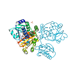
8RVC
 | | Crystal structure of alpha keto acid C-methyl-transferases MrsA bound to ketoarginine | | 分子名称: | 1,2-ETHANEDIOL, 2-ketoarginine methyltransferase, 5-[(diaminomethylidene)amino]-2-oxopentanoic acid, ... | | 著者 | Gerhardt, S, Kemper, F, Andexer, J.N. | | 登録日 | 2024-02-01 | | 公開日 | 2024-07-03 | | 最終更新日 | 2024-07-24 | | 実験手法 | X-RAY DIFFRACTION (1.969 Å) | | 主引用文献 | Structures and Protein Engineering of the alpha-Keto Acid C-Methyltransferases SgvM and MrsA for Rational Substrate Transfer.
Chembiochem, 2024
|
|
8RWW
 | |
8RXF
 | |
8RVS
 | |
8RWM
 | |
8RXG
 | |
4PHO
 | | ClyA CC6/264 ox (2-303) | | 分子名称: | DI(HYDROXYETHYL)ETHER, GLYCEROL, Hemolysin E, ... | | 著者 | Roderer, D.J.A, Glockshuber, R, Ban, N. | | 登録日 | 2014-05-06 | | 公開日 | 2014-09-24 | | 最終更新日 | 2023-12-20 | | 実験手法 | X-RAY DIFFRACTION (2.123 Å) | | 主引用文献 | Characterization of Variants of the Pore-Forming Toxin ClyA from Escherichia coli Controlled by a Redox Switch.
Biochemistry, 53, 2014
|
|
4PHQ
 | | ClyA CC6/264 ox (6-303) | | 分子名称: | ACETATE ION, GLYCEROL, Hemolysin E, ... | | 著者 | Roderer, D.J.A, Glockshuber, R, Ban, N. | | 登録日 | 2014-05-06 | | 公開日 | 2014-09-24 | | 最終更新日 | 2023-12-20 | | 実験手法 | X-RAY DIFFRACTION (1.94 Å) | | 主引用文献 | Characterization of Variants of the Pore-Forming Toxin ClyA from Escherichia coli Controlled by a Redox Switch.
Biochemistry, 53, 2014
|
|


