5OXL
 
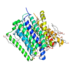 | | PepTSt in complex with dipeptide Ala-Leu | | 分子名称: | (2R)-2,3-DIHYDROXYPROPYL(7Z)-PENTADEC-7-ENOATE, (2S)-2,3-DIHYDROXYPROPYL(7Z)-PENTADEC-7-ENOATE, 4-(2-HYDROXYETHYL)-1-PIPERAZINE ETHANESULFONIC ACID, ... | | 著者 | Martinez Molledo, M, Quistgaard, E.M, Loew, C. | | 登録日 | 2017-09-07 | | 公開日 | 2018-02-21 | | 最終更新日 | 2024-01-17 | | 実験手法 | X-RAY DIFFRACTION (2.66 Å) | | 主引用文献 | Multispecific Substrate Recognition in a Proton-Dependent Oligopeptide Transporter.
Structure, 26, 2018
|
|
2F52
 | |
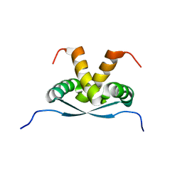
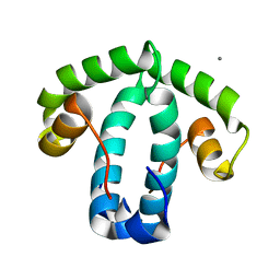
3FT7
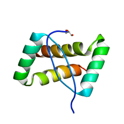 | | Crystal structure of an extremely stable dimeric protein from sulfolobus islandicus | | 分子名称: | GLYCEROL, Uncharacterized protein ORF56 | | 著者 | Neumann, P, Loew, C, Weininger, U, Stubbs, M.T. | | 登録日 | 2009-01-12 | | 公開日 | 2009-10-20 | | 最終更新日 | 2023-11-01 | | 実験手法 | X-RAY DIFFRACTION (2 Å) | | 主引用文献 | Structure-Based Stability Analysis of an Extremely Stable Dimeric DNA Binding Protein from Sulfolobus islandicus
Biochemistry, 48, 2009
|
|
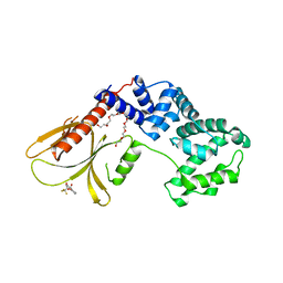
1Z1L
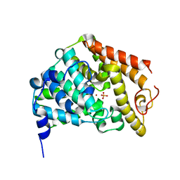 | | The Crystal Structure of the Phosphodiesterase 2A Catalytic Domain | | 分子名称: | MAGNESIUM ION, PHOSPHATE ION, ZINC ION, ... | | 著者 | Ding, Y.H, Kohls, D, Low, C. | | 登録日 | 2005-03-04 | | 公開日 | 2005-06-21 | | 最終更新日 | 2023-08-23 | | 実験手法 | X-RAY DIFFRACTION (1.7 Å) | | 主引用文献 | Structural Determinants for Inhibitor Specificity and Selectivity in PDE2A Using the Wheat Germ in Vitro Translation System.
Biochemistry, 44, 2005
|
|
2K9I
 | |
6FNE
 | | Structure of human Brag2 (Sec7-PH domains) with the inhibitor Bragsin bound to the PH domain | | 分子名称: | (2~{S})-6-methyl-5-nitro-2-(trifluoromethyl)-2,3-dihydrochromen-4-one, IQ motif and SEC7 domain-containing protein 1, NONAETHYLENE GLYCOL | | 著者 | Nawrotek, A, Zeghouf, M, Cherfils, J. | | 登録日 | 2018-02-03 | | 公開日 | 2019-03-13 | | 最終更新日 | 2024-01-17 | | 実験手法 | X-RAY DIFFRACTION (2.501 Å) | | 主引用文献 | PH-domain-binding inhibitors of nucleotide exchange factor BRAG2 disrupt Arf GTPase signaling.
Nat.Chem.Biol., 15, 2019
|
|
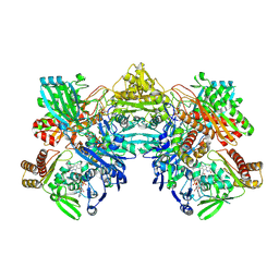
7Z1H
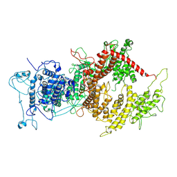 | | VAR2CSA APO | | 分子名称: | VAR2CSA APO | | 著者 | Raghavan, S.S.R, Wang, K.T. | | 登録日 | 2022-02-24 | | 公開日 | 2022-11-02 | | 最終更新日 | 2024-07-17 | | 実験手法 | ELECTRON MICROSCOPY (3.12 Å) | | 主引用文献 | Cryo-EM reveals the conformational epitope of human monoclonal antibody PAM1.4 broadly reacting with polymorphic malarial protein VAR2CSA.
Plos Pathog., 18, 2022
|
|
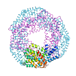
7Z12
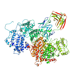 | | VAR2 complex with PAM1.4 | | 分子名称: | PAM1.4, Heavy Chain, light Chain, ... | | 著者 | Raghavan, S.S.R, Wang, K.T. | | 登録日 | 2022-02-24 | | 公開日 | 2022-11-02 | | 最終更新日 | 2022-11-30 | | 実験手法 | ELECTRON MICROSCOPY (3 Å) | | 主引用文献 | Cryo-EM reveals the conformational epitope of human monoclonal antibody PAM1.4 broadly reacting with polymorphic malarial protein VAR2CSA.
Plos Pathog., 18, 2022
|
|
8C8E
 | |
8UEM
 | |
3BRP
 | |
2P33
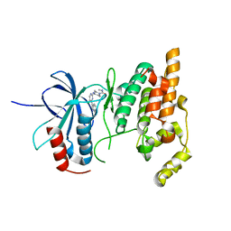 | | Synthesis and SAR of Aminopyrimidines as Novel c-Jun N-Terminal Kinase (JNK) Inhibitors | | 分子名称: | 4-{[5-chloro-4-(1H-indol-3-yl)pyrimidin-2-yl]amino}-N-ethylpiperidine-1-carboxamide, c-Jun N-terminal kinase 3 | | 著者 | Ceska, T.A, Platt, A, Fortunato, M, Dickson, K.M, Sharpe, A. | | 登録日 | 2007-03-08 | | 公開日 | 2007-06-19 | | 最終更新日 | 2023-08-30 | | 実験手法 | X-RAY DIFFRACTION (2.4 Å) | | 主引用文献 | Synthesis and SAR of aminopyrimidines as novel c-Jun N-terminal kinase (JNK) inhibitors
Bioorg.Med.Chem.Lett., 17, 2007
|
|


