4FIQ
 
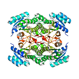
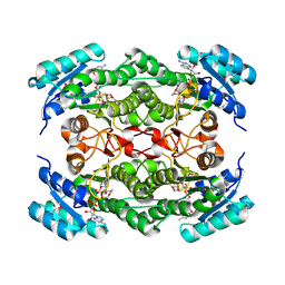
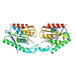
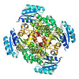
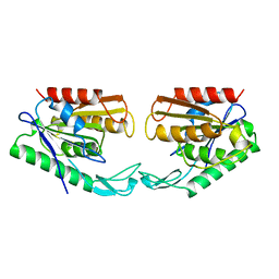
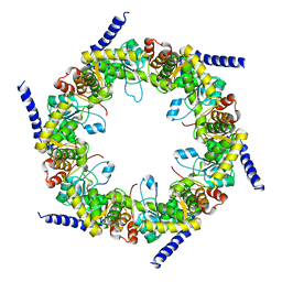 | | Crystal structure of pyridoxal biosynthesis lyase PdxS from Pyrococcus horikoshii | | 分子名称: | Pyridoxal biosynthesis lyase pdxS | | 著者 | Matsuura, A, Yoon, J.Y, Yoon, H.J, Lee, H.H, Suh, S.W. | | 登録日 | 2012-06-11 | | 公開日 | 2012-11-14 | | 最終更新日 | 2023-09-13 | | 実験手法 | X-RAY DIFFRACTION (2.7 Å) | | 主引用文献 | Crystal structure of pyridoxal biosynthesis lyase PdxS from Pyrococcus horikoshii.
Mol.Cells, 34, 2012
|
|
4FIR
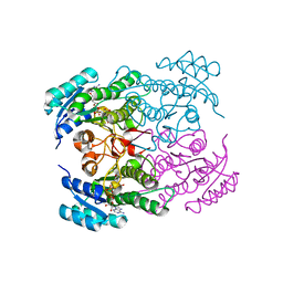
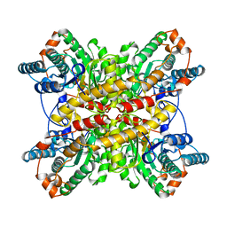
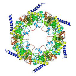 | | Crystal structure of pyridoxal biosynthesis lyase PdxS from Pyrococcus | | 分子名称: | Pyridoxal biosynthesis lyase pdxS, RIBOSE-5-PHOSPHATE | | 著者 | Matsuura, A, Yoon, J.Y, Yoon, H.J, Lee, H.H, Suh, S.W. | | 登録日 | 2012-06-11 | | 公開日 | 2012-11-14 | | 最終更新日 | 2023-09-13 | | 実験手法 | X-RAY DIFFRACTION (3.1 Å) | | 主引用文献 | Crystal structure of pyridoxal biosynthesis lyase PdxS from Pyrococcus horikoshii.
Mol.Cells, 34, 2012
|
|
4FB9
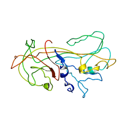
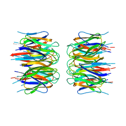
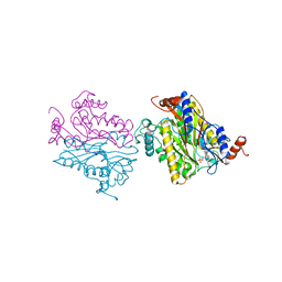
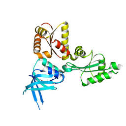
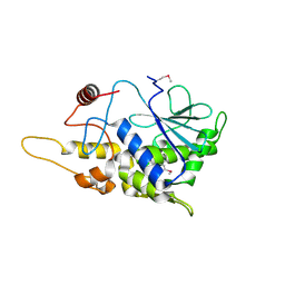 | | Structure of mutant RIP from barley seeds | | 分子名称: | Protein synthesis inhibitor I | | 著者 | Lee, B.-G, Kim, M.K, Suh, S.W, Song, H.K. | | 登録日 | 2012-05-22 | | 公開日 | 2012-10-31 | | 最終更新日 | 2013-01-23 | | 実験手法 | X-RAY DIFFRACTION (1.75 Å) | | 主引用文献 | Structures of the ribosome-inactivating protein from barley seeds reveal a unique activation mechanism.
Acta Crystallogr.,Sect.D, 68, 2012
|
|
1JUV
 | |
1NFQ
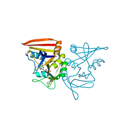
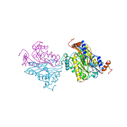
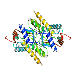
 | | Rv2002 gene product from Mycobacterium tuberculosis | | 分子名称: | 1,4-DIHYDRONICOTINAMIDE ADENINE DINUCLEOTIDE, Androsterone, Putative oxidoreductase Rv2002 | | 著者 | Yang, J.K, Park, M.S, Waldo, G.S, Suh, S.W, TB Structural Genomics Consortium (TBSGC) | | 登録日 | 2002-12-15 | | 公開日 | 2002-12-30 | | 最終更新日 | 2024-05-29 | | 実験手法 | X-RAY DIFFRACTION (2.4 Å) | | 主引用文献 | Directed evolution approach to a structural genomics project: Rv2002 from Mycobacterium tuberculosis
Proc.Natl.Acad.Sci.USA, 100, 2003
|
|
1KCT
 | | ALPHA1-ANTITRYPSIN | | 分子名称: | ALPHA1-ANTITRYPSIN | | 著者 | Song, H.K, Suh, S.W. | | 登録日 | 1996-08-06 | | 公開日 | 1997-01-11 | | 最終更新日 | 2024-02-07 | | 実験手法 | X-RAY DIFFRACTION (3.46 Å) | | 主引用文献 | Crystal structure of an uncleaved alpha 1-antitrypsin reveals the conformation of its inhibitory reactive loop.
FEBS Lett., 377, 1995
|
|
1NFF
 | | Crystal structure of Rv2002 gene product from Mycobacterium tuberculosis | | 分子名称: | NICOTINAMIDE-ADENINE-DINUCLEOTIDE, Putative oxidoreductase Rv2002 | | 著者 | Yang, J.K, Park, M.S, Waldo, G.S, Suh, S.W, TB Structural Genomics Consortium (TBSGC) | | 登録日 | 2002-12-14 | | 公開日 | 2002-12-30 | | 最終更新日 | 2024-05-29 | | 実験手法 | X-RAY DIFFRACTION (1.8 Å) | | 主引用文献 | Directed evolution approach to a structural genomics project: Rv2002 from Mycobacterium tuberculosis
Proc.Natl.Acad.Sci.USA, 100, 2003
|
|
1NFR
 | | Rv2002 gene product from Mycobacterium tuberculosis | | 分子名称: | NICOTINAMIDE-ADENINE-DINUCLEOTIDE, Putative oxidoreductase Rv2002 | | 著者 | Yang, J.K, Park, M.S, Waldo, G.S, Suh, S.W, TB Structural Genomics Consortium (TBSGC) | | 登録日 | 2002-12-16 | | 公開日 | 2002-12-30 | | 最終更新日 | 2024-10-16 | | 実験手法 | X-RAY DIFFRACTION (2.1 Å) | | 主引用文献 | Directed evolution approach to a structural genomics project: Rv2002 from Mycobacterium tuberculosis
Proc.Natl.Acad.Sci.USA, 100, 2003
|
|
4XZZ
 | | Structure of Helicobacter pylori Csd6 in the ligand-free state | | 分子名称: | Conserved hypothetical secreted protein, GLYCEROL | | 著者 | Kim, H.S, Im, H.N, Yoon, H.J, Suh, S.W. | | 登録日 | 2015-02-05 | | 公開日 | 2015-09-02 | | 最終更新日 | 2024-05-29 | | 実験手法 | X-RAY DIFFRACTION (2.03 Å) | | 主引用文献 | The Cell Shape-determining Csd6 Protein from Helicobacter pylori Constitutes a New Family of l,d-Carboxypeptidase
J.Biol.Chem., 290, 2015
|
|
4Y4V
 | | Structure of Helicobacter pylori Csd6 in the D-Ala-bound state | | 分子名称: | Conserved hypothetical secreted protein, D-ALANINE, GLYCEROL | | 著者 | Kim, H.S, Im, H.N, Yoon, H.J, Suh, S.W. | | 登録日 | 2015-02-11 | | 公開日 | 2015-09-02 | | 最終更新日 | 2022-03-23 | | 実験手法 | X-RAY DIFFRACTION (2.04 Å) | | 主引用文献 | The Cell Shape-determining Csd6 Protein from Helicobacter pylori Constitutes a New Family of l,d-Carboxypeptidase
J.Biol.Chem., 290, 2015
|
|
2P1B
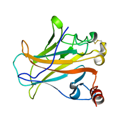
 | | Crystal structure of human nucleophosmin-core | | 分子名称: | Nucleophosmin | | 著者 | Lee, H.H, Kim, H.S, Kang, J.Y, Lee, B.I, Ha, J.Y, Yoon, H.J, Lim, S.O, Jung, G, Suh, S.W. | | 登録日 | 2007-03-03 | | 公開日 | 2007-03-27 | | 最終更新日 | 2023-10-25 | | 実験手法 | X-RAY DIFFRACTION (2.75 Å) | | 主引用文献 | Crystal structure of human nucleophosmin-core reveals plasticity of the pentamer-pentamer interface
Proteins, 69, 2007
|
|
2O4C
 | | Crystal Structure of D-Erythronate-4-phosphate Dehydrogenase Complexed with NAD | | 分子名称: | Erythronate-4-phosphate dehydrogenase, GLYCEROL, L(+)-TARTARIC ACID, ... | | 著者 | Ha, J.Y, Lee, J.H, Kim, K.H, Kim, D.J, Lee, H.H, Kim, H.K, Yoon, H.J, Suh, S.W. | | 登録日 | 2006-12-04 | | 公開日 | 2007-02-20 | | 最終更新日 | 2023-12-27 | | 実験手法 | X-RAY DIFFRACTION (2.3 Å) | | 主引用文献 | Crystal Structure of d-Erythronate-4-phosphate Dehydrogenase Complexed with NAD
J.Mol.Biol., 366, 2007
|
|
2P52
 | |
1AUR
 | |
1B5D
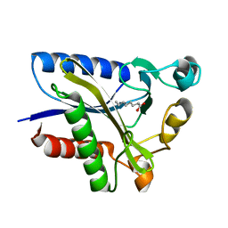
 | | DCMP Hydroxymethylase from T4 (Intact) | | 分子名称: | 2'-DEOXYCYTIDINE-5'-MONOPHOSPHATE, PROTEIN (DEOXYCYTIDYLATE HYDROXYMETHYLASE) | | 著者 | Song, H.K, Sohn, S.H, Suh, S.W. | | 登録日 | 1999-01-06 | | 公開日 | 1999-01-13 | | 最終更新日 | 2024-02-07 | | 実験手法 | X-RAY DIFFRACTION (2.2 Å) | | 主引用文献 | Crystal structure of deoxycytidylate hydroxymethylase from bacteriophage T4, a component of the deoxyribonucleoside triphosphate-synthesizing complex.
EMBO J., 18, 1999
|
|
2QHU
 | | Structural Basis of Octanoic Acid Recognition by Lipoate-Protein Ligase B | | 分子名称: | Lipoyltransferase, OCTANAL | | 著者 | Kim, D.J, Lee, S.J, Kim, H.S, Kim, K.H, Lee, H.H, Yoon, H.J, Suh, S.W. | | 登録日 | 2007-07-02 | | 公開日 | 2008-02-26 | | 最終更新日 | 2017-10-18 | | 実験手法 | X-RAY DIFFRACTION (1.9 Å) | | 主引用文献 | Structural basis of octanoic acid recognition by lipoate-protein ligase B
Proteins, 70, 2008
|
|
2PD4
 | | Crystal Structure of the Helicobacter pylori Enoyl-Acyl Carrier Protein Reductase in Complex with Hydroxydiphenyl Ether Compounds, Triclosan and Diclosan | | 分子名称: | DICLOSAN, Enoyl-[acyl-carrier-protein] reductase [NADH], NICOTINAMIDE-ADENINE-DINUCLEOTIDE | | 著者 | Lee, H.H, Moon, J.H, Suh, S.W. | | 登録日 | 2007-03-31 | | 公開日 | 2007-04-17 | | 最終更新日 | 2023-10-25 | | 実験手法 | X-RAY DIFFRACTION (2.3 Å) | | 主引用文献 | Crystal structure of the Helicobacter pylori enoyl-acyl carrier protein reductase in complex with hydroxydiphenyl ether compounds, triclosan and diclosan
Proteins, 69, 2007
|
|
1B5E
 | | DCMP HYDROXYMETHYLASE FROM T4 | | 分子名称: | 2'-DEOXYCYTIDINE-5'-MONOPHOSPHATE, PROTEIN (DEOXYCYTIDYLATE HYDROXYMETHYLASE) | | 著者 | Song, H.K, Sohn, S.H, Suh, S.W. | | 登録日 | 1999-01-06 | | 公開日 | 1999-01-13 | | 最終更新日 | 2024-02-07 | | 実験手法 | X-RAY DIFFRACTION (1.6 Å) | | 主引用文献 | Crystal structure of deoxycytidylate hydroxymethylase from bacteriophage T4, a component of the deoxyribonucleoside triphosphate-synthesizing complex.
EMBO J., 18, 1999
|
|
1B49
 | | DCMP HYDROXYMETHYLASE FROM T4 (PHOSPHATE-BOUND) | | 分子名称: | PHOSPHATE ION, PROTEIN (DEOXYCYTIDYLATE HYDROXYMETHYLASE) | | 著者 | Song, H.K, Sohn, S.H, Suh, S.W. | | 登録日 | 1999-01-06 | | 公開日 | 1999-01-13 | | 最終更新日 | 2024-02-07 | | 実験手法 | X-RAY DIFFRACTION (2.3 Å) | | 主引用文献 | Crystal structure of deoxycytidylate hydroxymethylase from bacteriophage T4, a component of the deoxyribonucleoside triphosphate-synthesizing complex.
EMBO J., 18, 1999
|
|
2QHS
 | | Structural Basis of Octanoic Acid Recognition by Lipoate-Protein Ligase B | | 分子名称: | Lipoyltransferase, OCTANOIC ACID (CAPRYLIC ACID) | | 著者 | Kim, D.J, Lee, S.J, Kim, H.S, Kim, K.H, Lee, H.H, Yoon, H.J, Suh, S.W. | | 登録日 | 2007-07-02 | | 公開日 | 2008-02-26 | | 最終更新日 | 2011-07-13 | | 実験手法 | X-RAY DIFFRACTION (1.5 Å) | | 主引用文献 | Structural basis of octanoic acid recognition by lipoate-protein ligase B
Proteins, 70, 2008
|
|
2QHV
 | | Structural Basis of Octanoic Acid Recognition by Lipoate-Protein Ligase B | | 分子名称: | Lipoyltransferase, OCTAN-1-OL | | 著者 | Kim, D.J, Lee, S.J, Kim, H.S, Kim, K.H, Lee, H.H, Yoon, H.J, Suh, S.W. | | 登録日 | 2007-07-03 | | 公開日 | 2008-02-26 | | 最終更新日 | 2024-03-13 | | 実験手法 | X-RAY DIFFRACTION (1.6 Å) | | 主引用文献 | Structural basis of octanoic acid recognition by lipoate-protein ligase B
Proteins, 70, 2008
|
|
2QHT
 | | Structural Basis of Octanoic Acid Recognition by Lipoate-Protein Ligase B | | 分子名称: | Lipoyltransferase | | 著者 | Kim, D.J, Lee, S.J, Kim, H.S, Kim, K.H, Lee, H.H, Yoon, H.J, Suh, S.W. | | 登録日 | 2007-07-02 | | 公開日 | 2008-02-26 | | 最終更新日 | 2024-03-13 | | 実験手法 | X-RAY DIFFRACTION (1.5 Å) | | 主引用文献 | Structural basis of octanoic acid recognition by lipoate-protein ligase B
Proteins, 70, 2008
|
|
1AUO
 | |
1BXC
 | |
2QI2
 | | Crystal structure of the Thermoplasma acidophilum Pelota protein | | 分子名称: | Cell division protein pelota related protein | | 著者 | Lee, H.H, Kim, Y.S, Kim, K.H, Heo, I.H, Kim, S.K, Kim, O, Suh, S.W. | | 登録日 | 2007-07-03 | | 公開日 | 2007-10-09 | | 最終更新日 | 2024-03-13 | | 実験手法 | X-RAY DIFFRACTION (2.9 Å) | | 主引用文献 | Structural and functional insights into dom34, a key component of no-go mRNA decay
Mol.Cell, 27, 2007
|
|


