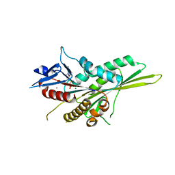
4HWI
 
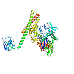 | | Crystal structure of ATBAG1 in complex with HSP70 | | 分子名称: | BAG family molecular chaperone regulator 1, Heat shock cognate 71 kDa protein | | 著者 | Shen, Y, Fang, S. | | 登録日 | 2012-11-07 | | 公開日 | 2013-05-29 | | 最終更新日 | 2023-09-20 | | 実験手法 | X-RAY DIFFRACTION (2.273 Å) | | 主引用文献 | Structural insight into plant programmed cell death mediated by BAG proteins in Arabidopsis thaliana.
Acta Crystallogr.,Sect.D, 69, 2013
|
|
5WW0
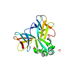 | | Crystal structure of Set7, a novel histone methyltransferase in Schizossacharomyces pombe | | 分子名称: | SET domain-containing protein 7, SULFATE ION | | 著者 | Mevius, D.E.H.F, Shen, Y, Morishita, M, Carrozzini, B, Caliandro, R, di Luccio, E. | | 登録日 | 2016-12-30 | | 公開日 | 2017-12-06 | | 最終更新日 | 2023-11-29 | | 実験手法 | X-RAY DIFFRACTION (2.1 Å) | | 主引用文献 | Set7 Is a H3K37 Methyltransferase in Schizosaccharomyces pombe and Is Required for Proper Gametogenesis.
Structure, 27, 2019
|
|
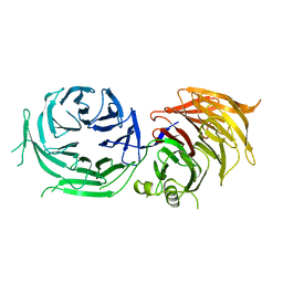
6HX0
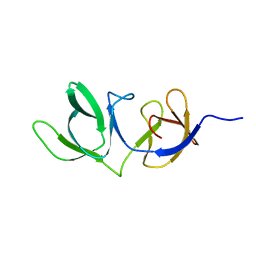 | | Cell wall binding domain of endolysin from Listeria phage A500. | | 分子名称: | L-alanyl-D-glutamate peptidase | | 著者 | Dunne, M, Taylor, N.M.I, Shen, Y, Loessner, M.J, Leiman, P.G. | | 登録日 | 2018-10-15 | | 公開日 | 2019-10-30 | | 最終更新日 | 2024-01-24 | | 実験手法 | X-RAY DIFFRACTION (1.59 Å) | | 主引用文献 | Structural basis for recognition of bacterial cell wall teichoic acid by pseudo-symmetric SH3b-like repeats of a viral peptidoglycan hydrolase
Chem Sci, 2020
|
|
2FOJ
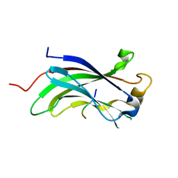 | | The Crystal Structure of the N-terminal domain of HAUSP/USP7 complexed with p53 peptide 364-367 | | 分子名称: | Ubiquitin carboxyl-terminal hydrolase 7, p53 peptide | | 著者 | Saridakis, V, Sheng, Y, Sarkari, F, Duan, S, Wu, T, Arrowsmith, C.H, Frappier, L. | | 登録日 | 2006-01-13 | | 公開日 | 2006-02-14 | | 最終更新日 | 2023-08-30 | | 実験手法 | X-RAY DIFFRACTION (1.6 Å) | | 主引用文献 | Molecular recognition of p53 and MDM2 by USP7/HAUSP
Nat.Struct.Mol.Biol., 13, 2006
|
|
2FOP
 | | The Crystal Structure of the N-terminal domain of HAUSP/USP7 complexed with mdm2 peptide 147-150 | | 分子名称: | Ubiquitin carboxyl-terminal hydrolase 7, mdm2 peptide | | 著者 | Saridakis, V, Sheng, Y, Sarkari, F, Duan, S, Wu, T, Arrowsmith, C.H, Frappier, L. | | 登録日 | 2006-01-13 | | 公開日 | 2006-02-14 | | 最終更新日 | 2023-08-30 | | 実験手法 | X-RAY DIFFRACTION (2.1 Å) | | 主引用文献 | Molecular recognition of p53 and MDM2 by USP7/HAUSP
Nat.Struct.Mol.Biol., 13, 2006
|
|
2FOO
 | | The Crystal Structure of the N-terminal domain of HAUSP/USP7 complexed with p53 peptide 359-362 | | 分子名称: | Ubiquitin carboxyl-terminal hydrolase 7, p53 peptide | | 著者 | Saridakis, V, Sheng, Y, Sarkari, F, Duan, S, Wu, T, Arrowsmith, C.H, Frappier, L. | | 登録日 | 2006-01-13 | | 公開日 | 2006-02-14 | | 最終更新日 | 2023-08-30 | | 実験手法 | X-RAY DIFFRACTION (2.2 Å) | | 主引用文献 | Molecular recognition of p53 and MDM2 by USP7/HAUSP
Nat.Struct.Mol.Biol., 13, 2006
|
|
2K2D
 | | Solution NMR structure of C-terminal domain of human pirh2. Northeast Structural Genomics Consortium (NESG) target HT2C | | 分子名称: | RING finger and CHY zinc finger domain-containing protein 1, ZINC ION | | 著者 | Lemak, A, Sheng, Y, Karra, M, Srisailam, S, Laister, R.C, Duan, S, Arrowsmith, C.H, Northeast Structural Genomics Consortium (NESG) | | 登録日 | 2008-03-31 | | 公開日 | 2008-04-15 | | 最終更新日 | 2024-05-01 | | 実験手法 | SOLUTION NMR | | 主引用文献 | Molecular basis of Pirh2-mediated p53 ubiquitylation.
Nat.Struct.Mol.Biol., 15, 2008
|
|
2K2C
 | | Solution NMR structure of N-terminal domain of human pirh2. Northeast Structural Genomics Consortium (NESG) target HT2A | | 分子名称: | RING finger and CHY zinc finger domain-containing protein 1, ZINC ION | | 著者 | Wu, B, Lemak, A, Sheng, Y, Karra, M, Srisailam, S, Sunnerhagen, M, Arrowsmith, C.H, Northeast Structural Genomics Consortium (NESG) | | 登録日 | 2008-03-31 | | 公開日 | 2008-04-15 | | 最終更新日 | 2024-05-08 | | 実験手法 | SOLUTION NMR | | 主引用文献 | Molecular basis of Pirh2-mediated p53 ubiquitylation.
Nat.Struct.Mol.Biol., 15, 2008
|
|
1MI1
 | | Crystal Structure of the PH-BEACH Domain of Human Neurobeachin | | 分子名称: | Neurobeachin | | 著者 | Jogl, G, Shen, Y, Gebauer, D, Li, J, Wiegmann, K, Kashkar, H, Kroenke, M, Tong, L, Northeast Structural Genomics Consortium (NESG) | | 登録日 | 2002-08-21 | | 公開日 | 2002-09-27 | | 最終更新日 | 2011-07-13 | | 実験手法 | X-RAY DIFFRACTION (2.9 Å) | | 主引用文献 | Crystal structure of the BEACH domain reveals an unusual fold and extensive association with a novel PH domain.
EMBO J., 21, 2002
|
|
3V70
 | | Crystal Structure of Human GTPase IMAP family member 1 | | 分子名称: | GTPase IMAP family member 1, GUANOSINE-5'-DIPHOSPHATE, MAGNESIUM ION | | 著者 | Nedyalkova, L, Shen, Y, Tong, Y, Tempel, W, Mackenzie, F, Arrowsmith, C.H, Edwards, A.M, Bountra, C, Weigelt, J, Bochkarev, A, Andrews, D.W, Park, H, Structural Genomics Consortium (SGC) | | 登録日 | 2011-12-20 | | 公開日 | 2012-01-11 | | 最終更新日 | 2023-09-13 | | 実験手法 | X-RAY DIFFRACTION (2.206 Å) | | 主引用文献 | Crystal Structure of Human GTPase IMAP family member 1
to be published
|
|
3CBQ
 | | Crystal structure of the human REM2 GTPase with bound GDP | | 分子名称: | 1,2-ETHANEDIOL, GTP-binding protein REM 2, GUANOSINE-5'-DIPHOSPHATE, ... | | 著者 | Nedyalkova, L, Shen, Y, Tong, Y, Tempel, W, MacKenzie, F, Arrowsmith, C.H, Edwards, A.M, Bountra, C, Weigelt, J, Bochkarev, A, Park, H, Structural Genomics Consortium (SGC) | | 登録日 | 2008-02-22 | | 公開日 | 2008-03-04 | | 最終更新日 | 2023-08-30 | | 実験手法 | X-RAY DIFFRACTION (1.82 Å) | | 主引用文献 | Crystal structure of the human REM2 GTPase with bound GDP.
To be Published
|
|
3UIT
 | | Overall structure of Patj/Pals1/Mals complex | | 分子名称: | ACETATE ION, InaD-like protein, MAGUK p55 subfamily member 5, ... | | 著者 | Zhang, J, Yang, X, Long, J, Shen, Y. | | 登録日 | 2011-11-06 | | 公開日 | 2012-02-22 | | 最終更新日 | 2024-03-20 | | 実験手法 | X-RAY DIFFRACTION (2.05 Å) | | 主引用文献 | Structure of an L27 domain heterotrimer from cell polarity complex Patj/Pals1/Mals2 reveals mutually independent L27 domain assembly mode
J.Biol.Chem., 287, 2012
|
|
3UIU
 | | Crystal structure of Apo-PKR kinase domain | | 分子名称: | Interferon-induced, double-stranded RNA-activated protein kinase | | 著者 | Li, F, Li, S, Yang, X, Shen, Y, Zhang, T. | | 登録日 | 2011-11-06 | | 公開日 | 2012-11-07 | | 最終更新日 | 2024-03-20 | | 実験手法 | X-RAY DIFFRACTION (2.903 Å) | | 主引用文献 | Crystal structure of Apo-PKR kinase domain
TO BE PUBLISHED
|
|
7Y53
 | | The cryo-EM structure of human ERAD retro-translocation complex | | 分子名称: | ADENOSINE-5'-DIPHOSPHATE, Derlin-1, Transitional endoplasmic reticulum ATPase | | 著者 | Cao, Y, Rao, B, Wang, Q, Yao, D, Xia, Y, Li, W, Li, S, Shen, Y. | | 登録日 | 2022-06-16 | | 公開日 | 2023-10-18 | | 最終更新日 | 2024-04-03 | | 実験手法 | ELECTRON MICROSCOPY (3.61 Å) | | 主引用文献 | The cryo-EM structure of the human ERAD retrotranslocation complex.
Sci Adv, 9, 2023
|
|
7Y59
 | | The cryo-EM structure of human ERAD retro-translocation complex | | 分子名称: | ADENOSINE-5'-DIPHOSPHATE, ADENOSINE-5'-TRIPHOSPHATE, Derlin-1, ... | | 著者 | Cao, Y, Rao, B, Wang, Q, Yao, D, Xia, Y, Li, W, Li, S, Shen, Y. | | 登録日 | 2022-06-16 | | 公開日 | 2023-10-18 | | 最終更新日 | 2024-04-03 | | 実験手法 | ELECTRON MICROSCOPY (4.51 Å) | | 主引用文献 | The cryo-EM structure of the human ERAD retrotranslocation complex.
Sci Adv, 9, 2023
|
|
7Y4W
 | | The cryo-EM structure of human ERAD retro-translocation complex | | 分子名称: | ADENOSINE-5'-DIPHOSPHATE, Derlin-1, Transitional endoplasmic reticulum ATPase | | 著者 | Cao, Y, Rao, B, Wang, Q, Yao, D, Xia, Y, Li, W, Li, S, Shen, Y. | | 登録日 | 2022-06-16 | | 公開日 | 2023-10-18 | | 最終更新日 | 2024-04-03 | | 実験手法 | ELECTRON MICROSCOPY (3.67 Å) | | 主引用文献 | The cryo-EM structure of the human ERAD retrotranslocation complex.
Sci Adv, 9, 2023
|
|
3CEQ
 | | The TPR domain of Human Kinesin Light Chain 2 (hKLC2) | | 分子名称: | Kinesin light chain 2 | | 著者 | Zhu, H, Shen, Y, MacKenzie, F, Arrowsmith, C.H, Edwards, A.M, Bountra, C, Weigelt, J, Bochkarev, A, Park, H, Structural Genomics Consortium (SGC) | | 登録日 | 2008-02-29 | | 公開日 | 2008-08-12 | | 最終更新日 | 2017-10-25 | | 実験手法 | X-RAY DIFFRACTION (2.75 Å) | | 主引用文献 | The TPR domain of Human Kinesin Light Chain 2 (hKLC2)
To be Published
|
|
7CFM
 | | Cryo-EM structure of the P395-bound GPBAR-Gs complex | | 分子名称: | 2-(ethylamino)-6-[3-(4-propan-2-ylphenyl)propanoyl]-7,8-dihydro-5H-pyrido[4,3-d]pyrimidine-4-carboxamide, CHOLESTEROL, G-protein coupled bile acid receptor 1, ... | | 著者 | Yang, F, Mao, C, Guo, L, Lin, J, Ming, Q, Xiao, P, Wu, X, Shen, Q, Guo, S, Shen, D, Lu, R, Zhang, L, Huang, S, Ping, Y, Zhang, C, Ma, C, Zhang, K, Liang, X, Shen, Y, Nan, F, Yi, F, Luca, V, Zhou, J, Jiang, C, Sun, J, Xie, X, Yu, X, Zhang, Y. | | 登録日 | 2020-06-27 | | 公開日 | 2020-09-09 | | 最終更新日 | 2020-12-02 | | 実験手法 | ELECTRON MICROSCOPY (3 Å) | | 主引用文献 | Structural basis of GPBAR activation and bile acid recognition.
Nature, 587, 2020
|
|
7CFN
 | | Cryo-EM structure of the INT-777-bound GPBAR-Gs complex | | 分子名称: | (2S,4R)-4-[(3R,5S,6R,7R,8R,9S,10S,12S,13R,14S,17R)-6-ethyl-10,13-dimethyl-3,7,12-tris(oxidanyl)-2,3,4,5,6,7,8,9,11,12,14,15,16,17-tetradecahydro-1H-cyclopenta[a]phenanthren-17-yl]-2-methyl-pentanoic acid, CHOLESTEROL, G-protein coupled bile acid receptor 1, ... | | 著者 | Yang, F, Mao, C, Guo, L, Lin, J, Ming, Q, Xiao, P, Wu, X, Shen, Q, Guo, S, Shen, D, Lu, R, Zhang, L, Huang, S, Ping, Y, Zhang, C, Ma, C, Zhang, K, Liang, X, Shen, Y, Nan, F, Yi, F, Luca, V, Zhou, J, Jiang, C, Sun, J, Xie, X, Yu, X, Zhang, Y. | | 登録日 | 2020-06-27 | | 公開日 | 2020-09-09 | | 最終更新日 | 2021-04-07 | | 実験手法 | ELECTRON MICROSCOPY (3 Å) | | 主引用文献 | Structural basis of GPBAR activation and bile acid recognition.
Nature, 587, 2020
|
|
2L15
 | | Solution Structure of Cold Shock Protein CspA Using Combined NMR and CS-Rosetta method | | 分子名称: | Cold shock protein CspA | | 著者 | Tang, Y, Schneider, W.M, Shen, Y, Raman, S, Inouye, M, Baker, D, Roth, M.J, Montelione, G.T. | | 登録日 | 2010-07-22 | | 公開日 | 2010-09-15 | | 最終更新日 | 2024-05-01 | | 実験手法 | SOLUTION NMR | | 主引用文献 | Fully automated high-quality NMR structure determination of small (2)H-enriched proteins.
J Struct Funct Genomics, 11, 2010
|
|
3GBJ
 | | Crystal structure of the motor domain of kinesin KIF13B bound with ADP | | 分子名称: | ADENOSINE-5'-DIPHOSPHATE, KIF13B protein, MAGNESIUM ION, ... | | 著者 | Tong, Y, Shen, L, Shen, Y, Tempel, W, Arrowsmith, C.H, Edwards, A.M, Bountra, C, Weigelt, J, Bochkarev, A, Park, H, Structural Genomics Consortium (SGC) | | 登録日 | 2009-02-19 | | 公開日 | 2009-03-03 | | 最終更新日 | 2023-09-06 | | 実験手法 | X-RAY DIFFRACTION (2.102 Å) | | 主引用文献 | Crystal structure of the motor domain of kinesin KIF13B bound with ADP
To be Published
|
|
3NF1
 | | Crystal structure of the TPR domain of kinesin light chain 1 | | 分子名称: | Kinesin light chain 1 | | 著者 | Tong, Y, Tempel, W, Shen, L, Shen, Y, Nedyalkova, L, Arrowsmith, C.H, Edwards, A.M, Bountra, C, Weigelt, J, Bochkarev, A, Park, H, Structural Genomics Consortium (SGC) | | 登録日 | 2010-06-09 | | 公開日 | 2011-05-25 | | 最終更新日 | 2023-09-06 | | 実験手法 | X-RAY DIFFRACTION (2.8 Å) | | 主引用文献 | Crystal structure of the TPR domain of kinesin light chain 1
to be published
|
|
1O8T
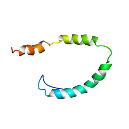 | | Global Structure and Dynamics of Human Apolipoprotein CII in Complex with Micelles: Evidence for increased mobility of the helix involved in the activation of lipoprotein lipase | | 分子名称: | APOLIPOPROTEIN C-II | | 著者 | Zdunek, J, Martinez, G.V, Schleucher, J, Lycksell, P.O, Yin, Y, Nilsson, S, Shen, Y, Olivecrona, G, Wijmenga, S. | | 登録日 | 2002-11-29 | | 公開日 | 2003-02-27 | | 最終更新日 | 2024-05-15 | | 実験手法 | SOLUTION NMR | | 主引用文献 | Global Structure and Dynamics of Human Apolipoprotein Cii in Complex with Micelles: Evidence for Increased Mobility of the Helix Involved in the Activation of Lipoprotein Lipase
Biochemistry, 42, 2003
|
|
6OBI
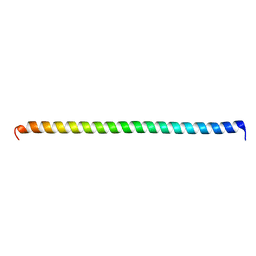 | | Remarkable rigidity of the single alpha-helical domain of myosin-VI revealed by NMR spectroscopy | | 分子名称: | Myosin-VI | | 著者 | Barnes, A, Shen, Y, Ying, J, Takagi, Y, Torchia, D.A, Sellers, J, Bax, A. | | 登録日 | 2019-03-20 | | 公開日 | 2019-06-12 | | 最終更新日 | 2024-05-15 | | 実験手法 | SOLUTION NMR | | 主引用文献 | Remarkable Rigidity of the Single alpha-Helical Domain of Myosin-VI As Revealed by NMR Spectroscopy.
J.Am.Chem.Soc., 141, 2019
|
|
4XFV
 | | Crystal Structure of Elp2 | | 分子名称: | Elongator complex protein 2 | | 著者 | Lin, Z, Dong, C, Long, J, Shen, Y. | | 登録日 | 2014-12-29 | | 公開日 | 2015-05-20 | | 最終更新日 | 2023-11-08 | | 実験手法 | X-RAY DIFFRACTION (3.2 Å) | | 主引用文献 | The elp2 subunit is essential for elongator complex assembly and functional regulation
Structure, 23, 2015
|
|


