2FDC
 
 | |
6ON5
 | |
6ON8
 | |
6ON7
 | |
6WP0
 | |
6WP2
 | |
6WNF
 | |
8W00
 | |
8VZX
 | |
6WP1
 | |
6C7Z
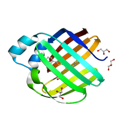
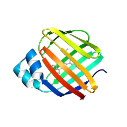 | | Crystal structure of the Q108K:K40L:T51V:R58F mutant of human Cellular Retinol Binding Protein II in complex with synthetic Ligand Julolidine | | 分子名称: | (2E,4E)-3-methyl-5-(2,3,6,7-tetrahydro-1H,5H-pyrido[3,2,1-ij]quinolin-9-yl)penta-2,4-dienal, ACETATE ION, Retinol-binding protein 2 | | 著者 | Nosrati, M, Geiger, J.H. | | 登録日 | 2018-01-23 | | 公開日 | 2018-04-25 | | 最終更新日 | 2023-10-04 | | 実験手法 | X-RAY DIFFRACTION (1.42 Å) | | 主引用文献 | A Genetically Encoded Ratiometric pH Probe: Wavelength Regulation-Inspired Design of pH Indicators.
Chembiochem, 19, 2018
|
|
6WNJ
 | |
6VIT
 | |
7MFZ
 | |
7MFX
 | |
7MFY
 | |
6VIS
 | |
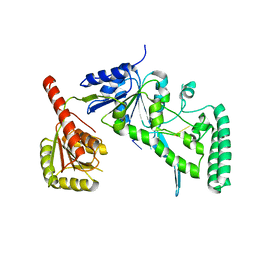
2HD5
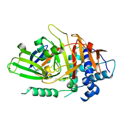 | | USP2 in complex with ubiquitin | | 分子名称: | Polyubiquitin, Ubiquitin carboxyl-terminal hydrolase 2, ZINC ION | | 著者 | Renatus, M, Kroemer, M. | | 登録日 | 2006-06-20 | | 公開日 | 2006-08-15 | | 最終更新日 | 2023-08-30 | | 実験手法 | X-RAY DIFFRACTION (1.85 Å) | | 主引用文献 | Structural Basis of Ubiquitin Recognition by the Deubiquitinating Protease USP2.
Structure, 14, 2006
|
|
3I17
 | |
6MCU
 | |
6MCV
 | |
6MQJ
 | |
6MOQ
 | |
6MOP
 | |
6MPK
 | |


