6SXV
 
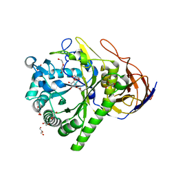 | | GH51 a-l-arabinofuranosidase soaked with aziridine inhibitor | | 分子名称: | 2-{2-[2-2-(METHOXY-ETHOXY)-ETHOXY]-ETHOXY}-ETHANOL, DI(HYDROXYETHYL)ETHER, GH51 a-l-arabinofuranosidase, ... | | 著者 | McGregor, N.G.S, Davies, G.J. | | 登録日 | 2019-09-26 | | 公開日 | 2020-02-26 | | 最終更新日 | 2024-01-24 | | 実験手法 | X-RAY DIFFRACTION (1.402 Å) | | 主引用文献 | Rational Design of Mechanism-Based Inhibitors and Activity-Based Probes for the Identification of Retaining alpha-l-Arabinofuranosidases.
J.Am.Chem.Soc., 142, 2020
|
|
6SXS
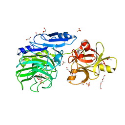 | | GH54 a-l-arabinofuranosidase soaked with cyclic sulfate inhibitor | | 分子名称: | 1,2-ETHANEDIOL, 2-acetamido-2-deoxy-beta-D-glucopyranose, 2-acetamido-2-deoxy-beta-D-glucopyranose-(1-4)-2-acetamido-2-deoxy-beta-D-glucopyranose, ... | | 著者 | McGregor, N.G.S, Davies, G.J, Nin-Hill, A, Rovira, C. | | 登録日 | 2019-09-26 | | 公開日 | 2020-02-26 | | 最終更新日 | 2024-01-24 | | 実験手法 | X-RAY DIFFRACTION (1.859 Å) | | 主引用文献 | Rational Design of Mechanism-Based Inhibitors and Activity-Based Probes for the Identification of Retaining alpha-l-Arabinofuranosidases.
J.Am.Chem.Soc., 142, 2020
|
|
6SXT
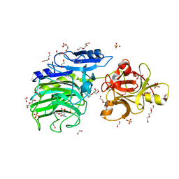 | | GH54 a-l-arabinofuranosidase soaked with aziridine inhibitor | | 分子名称: | 1,2-ETHANEDIOL, 2-acetamido-2-deoxy-beta-D-glucopyranose, 2-acetamido-2-deoxy-beta-D-glucopyranose-(1-4)-2-acetamido-2-deoxy-beta-D-glucopyranose, ... | | 著者 | McGregor, N.G.S, Davies, G.J, Nin-Hill, A, Rovira, C. | | 登録日 | 2019-09-26 | | 公開日 | 2020-02-26 | | 最終更新日 | 2024-01-24 | | 実験手法 | X-RAY DIFFRACTION (1.466 Å) | | 主引用文献 | Rational Design of Mechanism-Based Inhibitors and Activity-Based Probes for the Identification of Retaining alpha-l-Arabinofuranosidases.
J.Am.Chem.Soc., 142, 2020
|
|
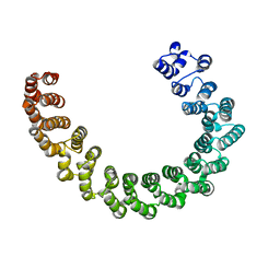
6SXU
 | |
6EF4
 | | Crystal structure of mouse PP2A Aalpha P179R mutant | | 分子名称: | Serine/threonine-protein phosphatase 2A 65 kDa regulatory subunit A alpha isoform | | 著者 | Wang, Z, Shen, G, Xu, W. | | 登録日 | 2018-08-16 | | 公開日 | 2019-06-26 | | 最終更新日 | 2023-10-11 | | 実験手法 | X-RAY DIFFRACTION (3.4 Å) | | 主引用文献 | The Highly Recurrent PP2A A alpha-Subunit Mutation P179R Alters Protein Structure and Impairs PP2A Enzyme Function to Promote Endometrial Tumorigenesis.
Cancer Res., 79, 2019
|
|
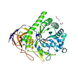
6SXR
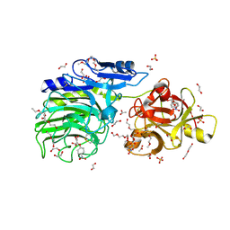 | | E221Q mutant of GH54 a-l-arabinofuranosidase soaked with 4-nitrophenyl a-l-arabinofuranoside | | 分子名称: | 1,2-ETHANEDIOL, 2-acetamido-2-deoxy-beta-D-glucopyranose, 2-acetamido-2-deoxy-beta-D-glucopyranose-(1-4)-2-acetamido-2-deoxy-beta-D-glucopyranose, ... | | 著者 | McGregor, N.G.S, Davies, G.J, Nin-Hill, A, Rovira, C. | | 登録日 | 2019-09-26 | | 公開日 | 2020-02-26 | | 最終更新日 | 2024-01-24 | | 実験手法 | X-RAY DIFFRACTION (1.64 Å) | | 主引用文献 | Rational Design of Mechanism-Based Inhibitors and Activity-Based Probes for the Identification of Retaining alpha-l-Arabinofuranosidases.
J.Am.Chem.Soc., 142, 2020
|
|
3EWI
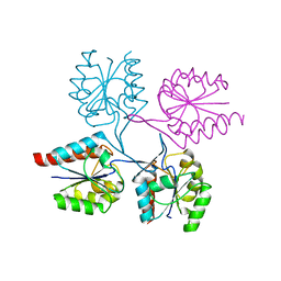 | | Structural analysis of the C-terminal domain of murine CMP-Sialic acid Synthetase | | 分子名称: | N-acylneuraminate cytidylyltransferase | | 著者 | Oschlies, M, Dickmanns, A, Stummeyer, K, Gerardy-Schahn, R, Ficner, R, Muenster-Kuehnel, A.K. | | 登録日 | 2008-10-15 | | 公開日 | 2009-08-18 | | 最終更新日 | 2023-12-27 | | 実験手法 | X-RAY DIFFRACTION (1.9 Å) | | 主引用文献 | A C-terminal phosphatase module conserved in vertebrate CMP-sialic acid synthetases provides a tetramerization interface for the physiologically active enzyme.
J.Mol.Biol., 393, 2009
|
|
4O5J
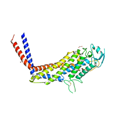 | | Crystal structure of SabA from Helicobacter pylori | | 分子名称: | 1,2-ETHANEDIOL, GLYCEROL, Uncharacterized protein | | 著者 | Pang, S.S, Nguyen, S.T.S, Whisstock, J.C. | | 登録日 | 2013-12-19 | | 公開日 | 2014-01-01 | | 最終更新日 | 2014-03-26 | | 実験手法 | X-RAY DIFFRACTION (2.2 Å) | | 主引用文献 | The three-dimensional structure of the extracellular adhesion domain of the sialic acid-binding adhesin SabA from Helicobacter pylori
J.Biol.Chem., 289, 2013
|
|


