Loading
PDBj
メニューPDBj@FacebookPDBj@TwitterPDBj@YouTubewwPDB FoundationwwPDB
RCSB PDBPDBeBMRBAdv. SearchSearch help
Search by PDB author
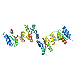
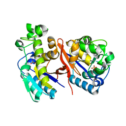
3HDO
DownloadVisualize
BU of 3hdo by Molmil
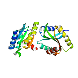
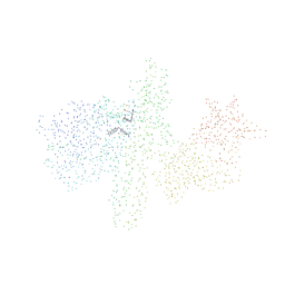
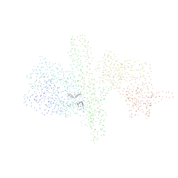
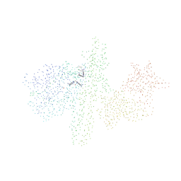
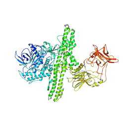
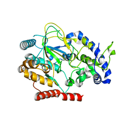
Crystal Structure of a Histidinol-phosphate aminotransferase from Geobacter metallireducens
分子名称: GLYCEROL, Histidinol-phosphate aminotransferase, MAGNESIUM ION
著者Damodharan, L, Burley, S.K, Swaminathan, S, New York SGX Research Center for Structural Genomics (NYSGXRC)
登録日2009-05-07
公開日2009-05-19
最終更新日2021-02-10
実験手法X-RAY DIFFRACTION (1.61 Å)
主引用文献Crystal Structure of an Histidinol-phosphate aminotransferase from Geobacter metallireducens
To be Published
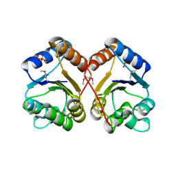
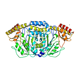
3HD5
DownloadVisualize
BU of 3hd5 by Molmil
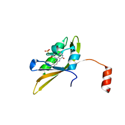
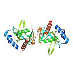
Crystal structure of a thiol:disulfide interchange protein dsbA from Bordetella parapertussis
分子名称: Thiol:disulfide interchange protein dsbA
著者Palani, K, Burley, S.K, Swaminathan, S, New York SGX Research Center for Structural Genomics (NYSGXRC)
登録日2009-05-06
公開日2009-05-19
最終更新日2021-02-10
実験手法X-RAY DIFFRACTION (2.35 Å)
主引用文献Crystal structure of a thiol:disulfide interchange protein dsbA from Bordetella parapertussis
To be Published
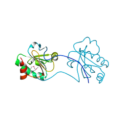
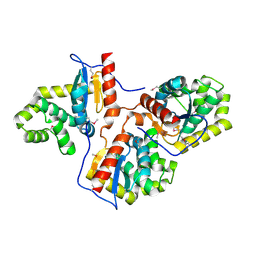
3HDV
DownloadVisualize
BU of 3hdv by Molmil
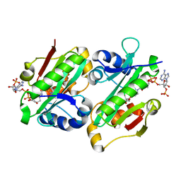
Crystal structure of response regulator receiver protein from Pseudomonas putida
分子名称: Response regulator
著者Bagaria, A, Burley, S.K, Swaminathan, S, New York SGX Research Center for Structural Genomics (NYSGXRC)
登録日2009-05-07
公開日2009-05-26
最終更新日2021-02-10
実験手法X-RAY DIFFRACTION (2.09 Å)
主引用文献Crystal structure of response regulator receiver protein from Pseudomonas putida
To be Published
3HEB
DownloadVisualize
BU of 3heb by Molmil
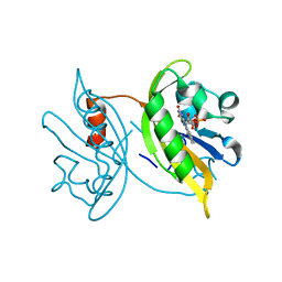
Crystal Structure of Response regulator receiver domain from Rhodospirillum rubrum
分子名称: PHOSPHATE ION, Response regulator receiver domain protein (CheY)
著者Syed Ibrahim, B, Burley, S.K, Swaminathan, S, New York SGX Research Center for Structural Genomics (NYSGXRC)
登録日2009-05-08
公開日2009-05-19
最終更新日2021-02-10
実験手法X-RAY DIFFRACTION (2.4 Å)
主引用文献Crystal Structure of Response regulator receiver domain from Rhodospirillum rubrum
To be Published
3HDC
DownloadVisualize
BU of 3hdc by Molmil
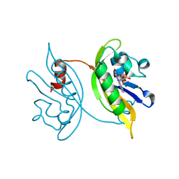
The crystal structure of thioredoxin protein from Geobacter metallireducens
分子名称: Thioredoxin family protein
著者Zhang, Z, Burley, S.K, Swaminathan, S, New York SGX Research Center for Structural Genomics (NYSGXRC)
登録日2009-05-07
公開日2009-05-26
最終更新日2021-02-10
実験手法X-RAY DIFFRACTION (1.771 Å)
主引用文献The crystal structure of thioredoxin protein from Geobacter metallireducens
To be Published
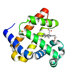
8EKO
DownloadVisualize
BU of 8eko by Molmil
Sperm whale myoglobin mutant L29H F33W F43H (F33W CuBMb)
分子名称: Myoglobin, PROTOPORPHYRIN IX CONTAINING FE
著者Ledray, A.P, Dwaraknath, S, Lu, Y.
登録日2022-09-21
公開日2022-10-26
最終更新日2023-10-25
実験手法X-RAY DIFFRACTION (1.34 Å)
主引用文献Tryptophan Can Promote Oxygen Reduction to Water in a Biosynthetic Model of Heme Copper Oxidases.
Biochemistry, 62, 2023
6K2F
DownloadVisualize
BU of 6k2f by Molmil
Structure of Eh TWINFILIN
分子名称: Actin binding protein family protein
著者Kumar, N, Gourinath, S, Rath, P.P.
登録日2019-05-14
公開日2020-06-03
最終更新日2023-11-22
実験手法X-RAY DIFFRACTION (1.55 Å)
主引用文献Structure of N-terminal Twinfilin domain from Entamoeba histolytica
To Be Published
1CCD
DownloadVisualize
BU of 1ccd by Molmil
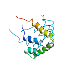
REFINED STRUCTURE OF RAT CLARA CELL 17 KDA PROTEIN AT 3.0 ANGSTROMS RESOLUTION
分子名称: CLARA CELL 17 kD PROTEIN, SULFATE ION
著者Umland, T.C, Swaminathan, S, Furey, W, Singh, G, Pletcher, J, Sax, M.
登録日1991-09-17
公開日1994-01-31
最終更新日2017-11-29
実験手法X-RAY DIFFRACTION (3 Å)
主引用文献Refined structure of rat Clara cell 17 kDa protein at 3.0 A resolution.
J.Mol.Biol., 224, 1992
1AF9
DownloadVisualize
BU of 1af9 by Molmil
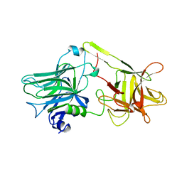
TETANUS NEUROTOXIN C FRAGMENT
分子名称: TETANUS NEUROTOXIN
著者Umland, T.C, Wingert, L, Swaminathan, S, Furey, W.F, Schmidt, J.J, Sax, M.
登録日1997-03-24
公開日1998-04-29
最終更新日2024-02-07
実験手法X-RAY DIFFRACTION (2.7 Å)
主引用文献Structure of the receptor binding fragment HC of tetanus neurotoxin.
Nat.Struct.Biol., 4, 1997
1G2X
DownloadVisualize
BU of 1g2x by Molmil
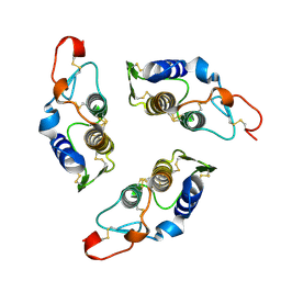
Sequence induced trimerization of krait PLA2: crystal structure of the trimeric form of krait PLA2
分子名称: PHOSPHOLIPASE A2
著者Singh, G, Gourinath, S, Sharma, S, Bhanumathi, S, Paramsivam, M, Singh, T.P.
登録日2000-10-22
公開日2003-06-17
最終更新日2023-08-09
実験手法X-RAY DIFFRACTION (2.5 Å)
主引用文献Sequence-induced trimerization of phospholipase A2: structure of a trimeric isoform of PLA2 from common krait (Bungarus caeruleus) at 2.5 A resolution.
Acta Crystallogr.,Sect.F, 61, 2005
1G9C
DownloadVisualize
BU of 1g9c by Molmil
CRYSTAL STRUCTURE OF CLOSTRIDIUM BOTULINUM NEUROTOXIN B COMPLEXED WITH AN INHIBITOR (EXPERIMENT 4)
分子名称: BIS(5-AMIDINO-BENZIMIDAZOLYL)METHANE, BOTULINUM NEUROTOXIN TYPE B, ZINC ION
著者Eswaramoorthy, S, Swaminathan, S.
登録日2000-11-22
公開日2002-11-13
最終更新日2023-08-09
実験手法X-RAY DIFFRACTION (2.35 Å)
主引用文献A Novel Mechanism for Clostridium botulinum Neurotoxin Inhibition
BIOCHEMISTRY, 41, 2002
1G9B
DownloadVisualize
BU of 1g9b by Molmil
CRYSTAL STRUCTURE OF CLOSTRIDIUM BOTULINUM NEUROTOXIN B COMPLEXED WITH AN INHIBITOR (EXPERIMENT 1)
分子名称: BIS(5-AMIDINO-BENZIMIDAZOLYL)METHANE, BOTULINUM NEUROTOXIN TYPE B, ZINC ION
著者Eswaramoorthy, S, Swaminathan, S.
登録日2000-11-22
公開日2002-11-13
最終更新日2023-08-09
実験手法X-RAY DIFFRACTION (2 Å)
主引用文献A Novel Mechanism for Clostridium botulinum Neurotoxin Inhibition
BIOCHEMISTRY, 41, 2002
1G9A
DownloadVisualize
BU of 1g9a by Molmil
CRYSTAL STRUCTURE OF CLOSTRIDIUM BOTULINUM NEUROTOXIN B COMPLEXED WITH AN INHIBITOR (EXPERIMENT 3)
分子名称: BIS(5-AMIDINO-BENZIMIDAZOLYL)METHANE, BOTULINUM NEUROTOXIN TYPE B, ZINC ION
著者Eswaramoorthy, S, Swaminathan, S.
登録日2000-11-22
公開日2002-11-13
最終更新日2023-08-09
実験手法X-RAY DIFFRACTION (2.1 Å)
主引用文献A Novel Mechanism for Clostridium botulinum Neurotoxin Inhibition
BIOCHEMISTRY, 41, 2002
1G9D
DownloadVisualize
BU of 1g9d by Molmil
CRYSTAL STRUCTURE OF CLOSTRIDIUM BOTULINUM NEUROTOXIN B COMPLEXED WITH AN INHIBITOR (EXPERIMENT 2)
分子名称: BIS(5-AMIDINO-BENZIMIDAZOLYL)METHANE, BOTULINUM NEUROTOXIN TYPE B, ZINC ION
著者Eswaramoorthy, S, Swaminathan, S.
登録日2000-11-22
公開日2002-11-13
最終更新日2023-08-09
実験手法X-RAY DIFFRACTION (2.2 Å)
主引用文献A Novel Mechanism for Clostridium botulinum Neurotoxin Inhibition
BIOCHEMISTRY, 41, 2002
8I11
DownloadVisualize
BU of 8i11 by Molmil
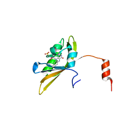
Crystal structure of LOV1 domain of phototropin from Klebsormidium nitens
分子名称: FLAVIN MONONUCLEOTIDE, Phototropin
著者Gautam, A.K, Sharma, S, Gourinath, S, Kateriya, S.
登録日2023-01-12
公開日2024-01-24
実験手法X-RAY DIFFRACTION (1.855 Å)
主引用文献Crystal structure of LOV1 domain of phototropin from klebsormidium nitens
To Be Published
8IL9
DownloadVisualize
BU of 8il9 by Molmil
Crystal structure of the LOV1 Q122N mutant of Klebsormidium nitens phototropin
分子名称: FLAVIN MONONUCLEOTIDE, Phototropin
著者Gautam, A.K, Sharma, S, Gourinath, S, Kateriya, S.
登録日2023-03-03
公開日2024-03-06
実験手法X-RAY DIFFRACTION (2.16 Å)
主引用文献Crystal structure of LOV1 Q122N mutant of phototropin from klebsormidium nitens
To Be Published
8IYN
DownloadVisualize
BU of 8iyn by Molmil
Crystal structure of LOV1 D33N mutant of phototropin from Klebsormidium nitens
分子名称: FLAVIN MONONUCLEOTIDE, Phototropin
著者Gautam, A.K, Sharma, S, Gourinath, S, Kateriya, S.
登録日2023-04-05
公開日2024-04-10
実験手法X-RAY DIFFRACTION (2.081 Å)
主引用文献Crystal structure of LOV1 D33N mutant of phototropin from Klebsormidium nitens
To Be Published
8J68
DownloadVisualize
BU of 8j68 by Molmil
Crystal structure of the LOV1 R60K mutant of Klebsormidium nitens phototropin
分子名称: FLAVIN MONONUCLEOTIDE, Phototropin
著者Gautam, A.K, Sharma, S, Gourinath, S, Kateriya, S.
登録日2023-04-25
公開日2024-05-01
実験手法X-RAY DIFFRACTION (1.845 Å)
主引用文献Crystal structure of LOV1 domain of phototropin from klebsormidium nitens
To Be Published
1I1E
DownloadVisualize
BU of 1i1e by Molmil
CRYSTAL STRUCTURE OF CLOSTRIDIUM BOTULINUM NEUROTOXIN B COMPLEXED WITH DOXORUBICIN
分子名称: BOTULINUM NEUROTOXIN TYPE B, DOXORUBICIN, SULFATE ION, ...
著者Eswaramoorthy, S, Kumaran, D, Swaminathan, S.
登録日2001-02-01
公開日2001-11-21
最終更新日2023-08-09
実験手法X-RAY DIFFRACTION (2.5 Å)
主引用文献Crystallographic evidence for doxorubicin binding to the receptor-binding site in Clostridium botulinum neurotoxin B.
Acta Crystallogr.,Sect.D, 57, 2001
6AKZ
DownloadVisualize
BU of 6akz by Molmil
Crystal structure of GlcNAc Inducible Gene 2, GIG2 (DUF1479) from Candida albicans
分子名称: FE (III) ION, GlcNAc Inducible Gene 2, GIG2
著者Gautam, G, Rani, P, Dutta, A, Gourinath, S.
登録日2018-09-05
公開日2019-09-11
最終更新日2023-11-22
実験手法X-RAY DIFFRACTION (1.69 Å)
主引用文献Crystal structure of Gig2 protein from Candida albicans provides a structural insight into DUF1479 family oxygenases.
Int.J.Biol.Macromol., 150, 2020
1I4P
DownloadVisualize
BU of 1i4p by Molmil
CRYSTAL STRUCTURE OF STAPHYLOCOCCAL ENTEROTOXIN C2 AT 100K CRYSTALLIZED AT PH 5.5
分子名称: ENTEROTOXIN TYPE C-2, ZINC ION
著者Kumaran, D, Swaminathan, S.
登録日2001-02-22
公開日2001-03-14
最終更新日2023-08-09
実験手法X-RAY DIFFRACTION (2 Å)
主引用文献Structure of staphylococcal enterotoxin C2 at various pH levels.
Acta Crystallogr.,Sect.D, 57, 2001
3FCM
DownloadVisualize
BU of 3fcm by Molmil
Crystal structure of a NUDIX hydrolase from Clostridium perfringens
分子名称: Hydrolase, NUDIX family, MANGANESE (II) ION
著者Palani, K, Burley, S.K, Swaninathan, S, New York SGX Research Center for Structural Genomics (NYSGXRC)
登録日2008-11-21
公開日2008-12-09
最終更新日2023-12-27
実験手法X-RAY DIFFRACTION (2.2 Å)
主引用文献Crystal structure of a NUDIX hydrolase from Clostridium perfringens
To be Published
6ADD
DownloadVisualize
BU of 6add by Molmil
The crystal structure of Rv2747 from Mycobacterium tuberculosis in complex with CoA and NLQ
分子名称: Amino-acid acetyltransferase, COENZYME A, N~2~-ACETYL-L-GLUTAMINE
著者Das, U, Singh, E, Pal, R.K, Tiruttani Subhramanyam, U.K, Gourinath, S, Srinivasan, A.
登録日2018-07-31
公開日2018-12-26
最終更新日2023-11-22
実験手法X-RAY DIFFRACTION (2.301 Å)
主引用文献Structural insights into the substrate binding mechanism of novel ArgA from Mycobacterium tuberculosis
Int. J. Biol. Macromol., 125, 2019
6M1X
DownloadVisualize
BU of 6m1x by Molmil
Crystal structure of Phosphoserine Phosphatase in complex with 3-Phosphoglyceric Acid from Entamoeba histolytica
分子名称: 3-PHOSPHOGLYCERIC ACID, PHOSPHATE ION, Phosphoglycerate mutase family protein
著者Kumari, P, Gourinath, S.
登録日2020-02-26
公開日2021-03-03
最終更新日2023-11-29
実験手法X-RAY DIFFRACTION (2.48 Å)
主引用文献Structural analysis of EhPSP in complex with 3-phosphoglyceric acid from Entamoeba histolytica reveals a basis for its lack of phosphoglycerate mutase activity.
Int.J.Biol.Macromol., 178, 2021
1I4Q
DownloadVisualize
BU of 1i4q by Molmil
CRYSTAL STRUCTURE OF STAPHYLOCOCCAL ENTEROTOXIN C2 AT 100K CRYSTALLIZED AT PH 6.0
分子名称: ENTEROTOXIN TYPE C-2, ZINC ION
著者Kumaran, D, Swaminathan, S.
登録日2001-02-22
公開日2001-03-14
最終更新日2023-08-09
実験手法X-RAY DIFFRACTION (2.2 Å)
主引用文献Structure of staphylococcal enterotoxin C2 at various pH levels.
Acta Crystallogr.,Sect.D, 57, 2001

222415

件を2024-07-10に公開中

PDB statisticsPDBj update infoContact PDBjnumon