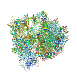
5J91
 
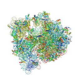 | | Structure of the Wild-type 70S E coli ribosome bound to Tigecycline | | 分子名称: | (4S)-2-METHYL-2,4-PENTANEDIOL, 1,2-ETHANEDIOL, 1,4-DIAMINOBUTANE, ... | | 著者 | Cocozaki, A, Ferguson, A. | | 登録日 | 2016-04-08 | | 公開日 | 2016-07-06 | | 最終更新日 | 2016-08-03 | | 実験手法 | X-RAY DIFFRACTION (2.96 Å) | | 主引用文献 | Resistance mutations generate divergent antibiotic susceptibility profiles against translation inhibitors.
Proc.Natl.Acad.Sci.USA, 113, 2016
|
|
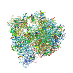
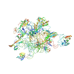
5J5B
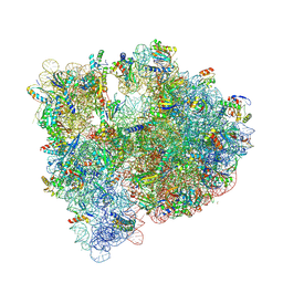 | | Structure of the WT E coli ribosome bound to tetracycline | | 分子名称: | (4S)-2-METHYL-2,4-PENTANEDIOL, 1,2-ETHANEDIOL, 1,4-DIAMINOBUTANE, ... | | 著者 | Cocozaki, A, Ferguson, A. | | 登録日 | 2016-04-01 | | 公開日 | 2016-07-27 | | 最終更新日 | 2018-08-15 | | 実験手法 | X-RAY DIFFRACTION (2.8 Å) | | 主引用文献 | Resistance mutations generate divergent antibiotic susceptibility profiles against translation inhibitors.
Proc.Natl.Acad.Sci.USA, 113, 2016
|
|
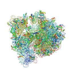
5J8A
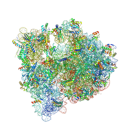 | | Structure of the E coli 70S ribosome with the U1052G mutation in 16S rRNA bound to tigecycline | | 分子名称: | (4S)-2-METHYL-2,4-PENTANEDIOL, 1,2-ETHANEDIOL, 1,4-DIAMINOBUTANE, ... | | 著者 | Cocozaki, A, Ferguson, A. | | 登録日 | 2016-04-07 | | 公開日 | 2016-07-06 | | 最終更新日 | 2016-08-03 | | 実験手法 | X-RAY DIFFRACTION (3.1 Å) | | 主引用文献 | Resistance mutations generate divergent antibiotic susceptibility profiles against translation inhibitors.
Proc.Natl.Acad.Sci.USA, 113, 2016
|
|
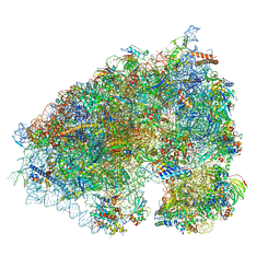
5JC9
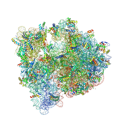 | | Structure of the Escherichia coli ribosome with the U1052G mutation in the 16S rRNA | | 分子名称: | (4S)-2-METHYL-2,4-PENTANEDIOL, 1,2-ETHANEDIOL, 1,4-DIAMINOBUTANE, ... | | 著者 | Cocozaki, A, Ferguson, A. | | 登録日 | 2016-04-14 | | 公開日 | 2016-07-06 | | 最終更新日 | 2016-08-03 | | 実験手法 | X-RAY DIFFRACTION (3.03 Å) | | 主引用文献 | Resistance mutations generate divergent antibiotic susceptibility profiles against translation inhibitors.
Proc.Natl.Acad.Sci.USA, 113, 2016
|
|
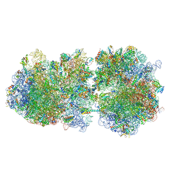
5IT8
 | | High-resolution structure of the Escherichia coli ribosome | | 分子名称: | (4S)-2-METHYL-2,4-PENTANEDIOL, 1,2-ETHANEDIOL, 1,4-DIAMINOBUTANE, ... | | 著者 | Cocozaki, A, Ferguson, A. | | 登録日 | 2016-03-16 | | 公開日 | 2016-07-27 | | 最終更新日 | 2023-11-15 | | 実験手法 | X-RAY DIFFRACTION (3.12 Å) | | 主引用文献 | Resistance mutations generate divergent antibiotic susceptibility profiles against translation inhibitors.
Proc.Natl.Acad.Sci.USA, 113, 2016
|
|
5J7L
 | | Structure of the 70S E coli ribosome with the U1052G mutation in the 16S rRNA bound to tetracycline | | 分子名称: | (4S)-2-METHYL-2,4-PENTANEDIOL, 1,2-ETHANEDIOL, 1,4-DIAMINOBUTANE, ... | | 著者 | Cocozaki, A, Ferguson, A. | | 登録日 | 2016-04-06 | | 公開日 | 2016-07-27 | | 最終更新日 | 2018-08-15 | | 実験手法 | X-RAY DIFFRACTION (3 Å) | | 主引用文献 | Resistance mutations generate divergent antibiotic susceptibility profiles against translation inhibitors.
Proc.Natl.Acad.Sci.USA, 113, 2016
|
|
5J88
 | | Structure of the E coli 70S ribosome with the U1060A mutation in 16S rRNA | | 分子名称: | (4S)-2-METHYL-2,4-PENTANEDIOL, 1,2-ETHANEDIOL, 1,4-DIAMINOBUTANE, ... | | 著者 | Cocozaki, A, Ferguson, A. | | 登録日 | 2016-04-07 | | 公開日 | 2016-07-06 | | 最終更新日 | 2016-12-07 | | 実験手法 | X-RAY DIFFRACTION (3.32 Å) | | 主引用文献 | Resistance mutations generate divergent antibiotic susceptibility profiles against translation inhibitors.
Proc.Natl.Acad.Sci.USA, 113, 2016
|
|
6XA1
 | |
6I7V
 | | Ribosomal protein paralogs bL31 and bL36 | | 分子名称: | (4S)-2-METHYL-2,4-PENTANEDIOL, 1,2-ETHANEDIOL, 1,4-DIAMINOBUTANE, ... | | 著者 | Pulk, A, Cate, J.H.D, Remme, J, Lilleorg, S, Reier, K, Peil, L, Liiv, A, Tammsalu, T. | | 登録日 | 2018-11-19 | | 公開日 | 2018-12-05 | | 最終更新日 | 2024-04-24 | | 実験手法 | X-RAY DIFFRACTION (2.9 Å) | | 主引用文献 | Bacterial ribosome heterogeneity: Changes in ribosomal protein composition during transition into stationary growth phase.
Biochimie, 156, 2018
|
|
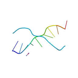
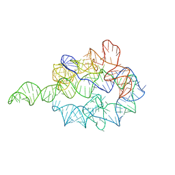
1HR2
 | | CRYSTAL STRUCTURE ANALYSIS OF A MUTANT P4-P6 DOMAIN (DELC209) OF TETRAHYMENA THEMOPHILA GROUP I INTRON. | | 分子名称: | MAGNESIUM ION, P4-P6 DELC209 MUTANT RNA RIBOZYME DOMAIN | | 著者 | Juneau, K, Podell, E.R, Harrington, D.J, Cech, T.R. | | 登録日 | 2000-12-20 | | 公開日 | 2001-04-12 | | 最終更新日 | 2023-08-09 | | 実験手法 | X-RAY DIFFRACTION (2.25 Å) | | 主引用文献 | Structural basis of the enhanced stability of a mutant ribozyme domain and a detailed view of RNA--solvent interactions.
Structure, 9, 2001
|
|
376D
 | | A ZIPPER-LIKE DNA DUPLEX D(GCGAAAGCT) | | 分子名称: | COBALT HEXAMMINE(III), DNA (5'-D(*GP*(CBR)P*GP*AP*AP*AP*GP*CP*T)-3') | | 著者 | Cruse, W.B.T, Shepard, W, Prange, T, delalFortelle, E, Fourme, R. | | 登録日 | 1998-01-22 | | 公開日 | 1999-10-26 | | 最終更新日 | 2024-04-03 | | 実験手法 | X-RAY DIFFRACTION (2.1 Å) | | 主引用文献 | A zipper-like duplex in DNA: the crystal structure of d(GCGAAAGCT) at 2.1 A resolution.
Structure, 6, 1998
|
|
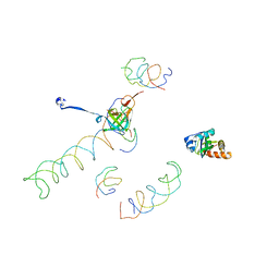
3IY9
 | | Leishmania Tarentolae Mitochondrial Large Ribosomal Subunit Model | | 分子名称: | 39S ribosomal protein L11, mitochondrial, 39S ribosomal protein L16, ... | | 著者 | Sharma, M.R, Booth, T.M, Simpson, L, Maslov, D.A, Agrawal, R.K. | | 登録日 | 2009-04-20 | | 公開日 | 2009-07-07 | | 最終更新日 | 2024-02-21 | | 実験手法 | ELECTRON MICROSCOPY (14.1 Å) | | 主引用文献 | Structure of a mitochondrial ribosome with minimal RNA
Proc.Natl.Acad.Sci.USA, 106, 2009
|
|
1GRZ
 | |
1MVR
 | | Decoding Center & Peptidyl transferase center from the X-ray structure of the Thermus thermophilus 70S ribosome, aligned to the low resolution Cryo-EM map of E.coli 70S Ribosome | | 分子名称: | 30S RIBOSOMAL PROTEIN S12, 50S ribosomal protein L11, Helix 34 of 16S rRNA, ... | | 著者 | Rawat, U.B, Zavialov, A.V, Sengupta, J, Valle, M, Grassucci, R.A, Linde, J, Vestergaard, B, Ehrenberg, M, Frank, J. | | 登録日 | 2002-09-26 | | 公開日 | 2003-04-01 | | 最終更新日 | 2024-02-14 | | 実験手法 | ELECTRON MICROSCOPY (12.8 Å) | | 主引用文献 | A cryo-electron microscopic study of ribosome-bound termination factor RF2
Nature, 421, 2003
|
|
2RDO
 | | 50S subunit with EF-G(GDPNP) and RRF bound | | 分子名称: | 23S RIBOSOMAL RNA, 50S ribosomal protein L1, 50S ribosomal protein L11, ... | | 著者 | Gao, N, Zavialov, A.V, Ehrenberg, M, Frank, J. | | 登録日 | 2007-09-24 | | 公開日 | 2008-03-04 | | 最終更新日 | 2024-02-21 | | 実験手法 | ELECTRON MICROSCOPY (9.1 Å) | | 主引用文献 | Specific interaction between EF-G and RRF and its implication for GTP-dependent ribosome splitting into subunits.
J.Mol.Biol., 374, 2007
|
|


