Loading
PDBj
メニューPDBj@FacebookPDBj@TwitterPDBj@YouTubewwPDB FoundationwwPDB
RCSB PDBPDBeBMRBAdv. SearchSearch help
Search by PDB author
3DNM
DownloadVisualize
BU of 3dnm by Molmil
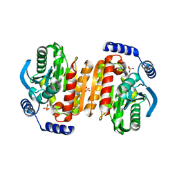
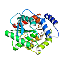
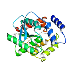
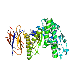
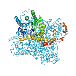
Crystal Structure Hormone-Sensitive Lipase from a Metagenome Library
分子名称: BETA-MERCAPTOETHANOL, Esterase/lipase, SULFATE ION
著者Hwang, K.Y, Nam, K.H.
登録日2008-07-02
公開日2009-01-13
最終更新日2023-11-01
実験手法X-RAY DIFFRACTION (2.8 Å)
主引用文献Structural and functional analysis of a novel hormone-sensitive lipase from a metagenome library
Proteins, 74, 2008
8IH0
DownloadVisualize
BU of 8ih0 by Molmil
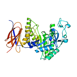
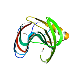
Crystal structure of GH11 from Thermoanaerobacterium saccharolyticum
分子名称: ACETATE ION, Endo-1,4-beta-xylanase
著者Nam, K.H.
登録日2023-02-22
公開日2023-11-22
実験手法X-RAY DIFFRACTION (1.5 Å)
主引用文献Characterization and structural analysis of the endo-1,4-beta-xylanase GH11 from the hemicellulose-degrading Thermoanaerobacterium saccharolyticum useful for lignocellulose saccharification.
Sci Rep, 13, 2023
8IH1
DownloadVisualize
BU of 8ih1 by Molmil
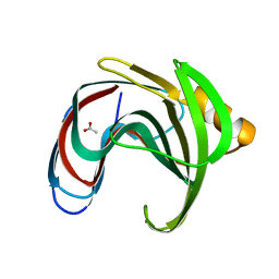
Room temperature structure of GH11 from Thermoanaerobacterium saccharolyticum by serial crystallography
分子名称: ACETATE ION, Endo-1,4-beta-xylanase
著者Nam, K.H.
登録日2023-02-22
公開日2023-11-22
実験手法X-RAY DIFFRACTION (1.75 Å)
主引用文献Characterization and structural analysis of the endo-1,4-beta-xylanase GH11 from the hemicellulose-degrading Thermoanaerobacterium saccharolyticum useful for lignocellulose saccharification.
Sci Rep, 13, 2023
6IRJ
DownloadVisualize
BU of 6irj by Molmil
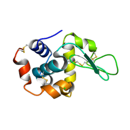
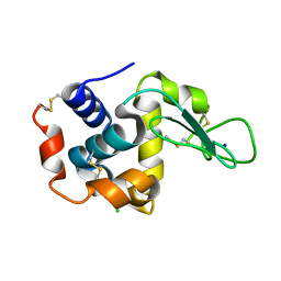
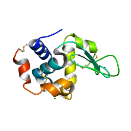
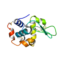
Crystal structure of lysozyme by fixed-target serial femtosecond crystallography
分子名称: CHLORIDE ION, Lysozyme C, SODIUM ION
著者Nam, K.H.
登録日2018-11-13
公開日2019-05-22
最終更新日2023-11-22
実験手法X-RAY DIFFRACTION (1.65 Å)
主引用文献Nylon mesh-based sample holder for fixed-target serial femtosecond crystallography.
Sci Rep, 9, 2019
6IG7
DownloadVisualize
BU of 6ig7 by Molmil
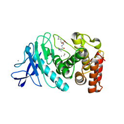
Crystal structure of thermolysin delivered in polyacrylamide using x-ray free electron laser
分子名称: CALCIUM ION, LEUCINE, LYSINE, ...
著者Nam, K.H.
登録日2018-09-25
公開日2018-11-07
最終更新日2023-11-22
実験手法X-RAY DIFFRACTION (1.8 Å)
主引用文献Polyacrylamide injection matrix for serial femtosecond crystallography.
Sci Rep, 9, 2019
3II1
DownloadVisualize
BU of 3ii1 by Molmil
Structural characterization of difunctional glucanase-xylanse CelM2
分子名称: Cellulase, ZINC ION, beta-D-glucopyranose
著者Hwang, K.Y, Nam, K.H.
登録日2009-07-31
公開日2009-08-25
最終更新日2023-11-01
実験手法X-RAY DIFFRACTION (2.25 Å)
主引用文献Structural characterization of the bifunctional glucanase-xylanase CelM2 reveals the metal effect and substrate-binding moiety
Biochem.Biophys.Res.Commun., 391, 2010
3K6K
DownloadVisualize
BU of 3k6k by Molmil
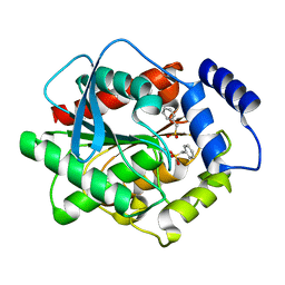
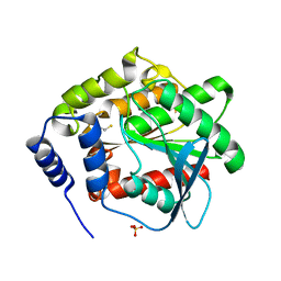
Crystal structure at 2.2 angstrom of HSL-homolog EstE7 from a metagenome library
分子名称: BETA-MERCAPTOETHANOL, Esterase/lipase, SULFATE ION
著者Hwang, K.Y, Nam, K.H.
登録日2009-10-09
公開日2009-10-27
最終更新日2023-11-01
実験手法X-RAY DIFFRACTION (2.2 Å)
主引用文献Biochemical and structural analysis of hormone-sensitive lipase homolog EstE7; Insight into the stabilized dimerization of HSL-homolog proteins
to be published
8WDG
DownloadVisualize
BU of 8wdg by Molmil
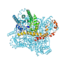
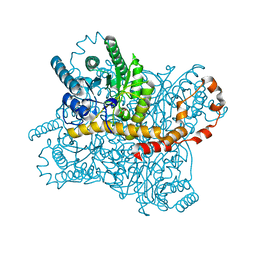
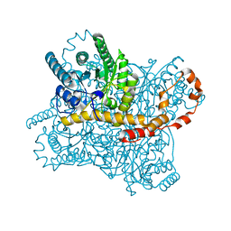
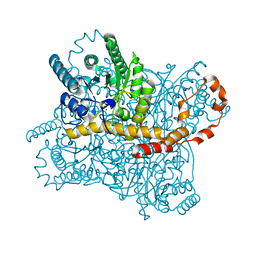
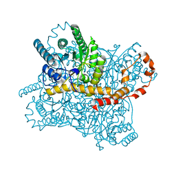
Subatomic crystal structure of glucose isomerase from Streptomyces rubiginosus
分子名称: MAGNESIUM ION, Xylitol, Xylose isomerase
著者Nam, K.H.
登録日2023-09-15
公開日2023-10-04
最終更新日2024-04-17
実験手法X-RAY DIFFRACTION (0.99 Å)
主引用文献Xylitol binding to the M1 site of glucose isomerase induces a conformational change in the substrate binding channel.
Biochem.Biophys.Res.Commun., 682, 2023
3G6N
DownloadVisualize
BU of 3g6n by Molmil
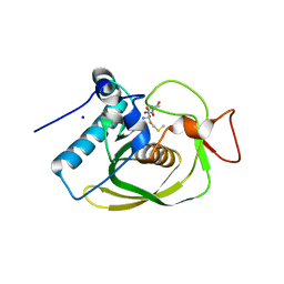
Crystal structure of an EfPDF complex with Met-Ala-Ser
分子名称: FE (III) ION, Peptide deformylase, SODIUM ION, ...
著者Hwang, K.Y, Nam, K.H.
登録日2009-02-07
公開日2009-03-03
最終更新日2023-11-01
実験手法X-RAY DIFFRACTION (2.5 Å)
主引用文献Crystal structure of an EfPDF complex with Met-Ala-Ser based on crystallographic packing.
Biochem.Biophys.Res.Commun., 381, 2009
3H17
DownloadVisualize
BU of 3h17 by Molmil
Crystal structure of EstE5-PMSF (I)
分子名称: Esterase/lipase, phenylmethanesulfonic acid
著者Hwang, K.Y, Nam, K.H.
登録日2009-04-11
公開日2009-04-28
最終更新日2023-11-01
実験手法X-RAY DIFFRACTION (2.5 Å)
主引用文献The crystal structure of an HSL-homolog EstE5 complex with PMSF reveals a unique configuration that inhibits the nucleophile Ser144 in catalytic triads.
Biochem.Biophys.Res.Commun., 389, 2009
6IRK
DownloadVisualize
BU of 6irk by Molmil
Crystal structure of glucose isomerase by fixed-target serial femtosecond crystallography
分子名称: MAGNESIUM ION, Xylose isomerase
著者Nam, K.H.
登録日2018-11-13
公開日2019-05-22
最終更新日2023-11-22
実験手法X-RAY DIFFRACTION (1.75 Å)
主引用文献Nylon mesh-based sample holder for fixed-target serial femtosecond crystallography.
Sci Rep, 9, 2019
6IG6
DownloadVisualize
BU of 6ig6 by Molmil
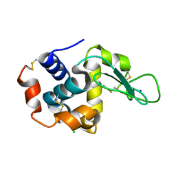
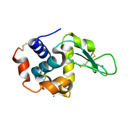
Crystal structure of lysozyme delivered in polyacrylamide using x-ray free electron laser
分子名称: CHLORIDE ION, Lysozyme C, SODIUM ION
著者Nam, K.H.
登録日2018-09-25
公開日2018-11-07
最終更新日2023-11-22
実験手法X-RAY DIFFRACTION (1.7 Å)
主引用文献Polyacrylamide injection matrix for serial femtosecond crystallography.
Sci Rep, 9, 2019
3H18
DownloadVisualize
BU of 3h18 by Molmil
Crystal structure of EstE5-PMSF (II)
分子名称: Esterase/lipase, phenylmethanesulfonic acid
著者Hwang, K.Y, Nam, K.H.
登録日2009-04-11
公開日2009-04-28
最終更新日2023-11-01
実験手法X-RAY DIFFRACTION (2.4 Å)
主引用文献The crystal structure of an HSL-homolog EstE5 complex with PMSF reveals a unique configuration that inhibits the nucleophile Ser144 in catalytic triads.
Biochem.Biophys.Res.Commun., 389, 2009
3FAK
DownloadVisualize
BU of 3fak by Molmil
Structural and Functional Analysis of a Hormone-Sensitive Lipase like EstE5 from a Metagenome Library
分子名称: CHLORIDE ION, Esterase/lipase, SODIUM ION
著者Hwang, K.Y, Nam, K.H.
登録日2008-11-17
公開日2009-02-10
最終更新日2023-11-01
実験手法X-RAY DIFFRACTION (1.9 Å)
主引用文献Structural and functional analysis of a novel EstE5 belonging to the subfamily of hormone-sensitive lipase
Biochem.Biophys.Res.Commun., 379, 2009
3GVY
DownloadVisualize
BU of 3gvy by Molmil
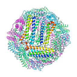
Crystal structure of bacterioferritin from R.sphaeroides
分子名称: Bacterioferritin, FE (III) ION, PROTOPORPHYRIN IX CONTAINING FE
著者Hwang, K.Y, Nam, K.H.
登録日2009-03-31
公開日2009-12-22
最終更新日2023-11-01
実験手法X-RAY DIFFRACTION (2.8 Å)
主引用文献Crystal structure of bacterioferritin from Rhodobacter sphaeroides
Biochem.Biophys.Res.Commun., 391, 2010
3FW6
DownloadVisualize
BU of 3fw6 by Molmil
Crystal structure of CelM2, a bifunctional glucanase-xylanase protein from a metagenome library
分子名称: Cellulase, ZINC ION
著者Hwang, K.Y, Nam, K.H.
登録日2009-01-17
公開日2009-03-03
最終更新日2023-11-01
実験手法X-RAY DIFFRACTION (2.3 Å)
主引用文献Crystal structure of CelM2, a bifunctional glucanase-xylanase protein from a metagenome library
Biochem.Biophys.Res.Commun., 383, 2009
7WKR
DownloadVisualize
BU of 7wkr by Molmil
Room temperature structure of lysozyme solved by serial synchrotron crystallography
分子名称: CHLORIDE ION, Lysozyme C
著者Nam, K.H.
登録日2022-01-11
公開日2022-02-02
最終更新日2023-11-29
実験手法X-RAY DIFFRACTION (1.6 Å)
主引用文献Combination of an inject-and-transfer system for serial femtosecond crystallography.
J.Appl.Crystallogr., 55, 2022
7WUC
DownloadVisualize
BU of 7wuc by Molmil
Room-temperature structure of lysozyme by serial femtosecond crystallography (BITS)
分子名称: Lysozyme C
著者Nam, K.H.
登録日2022-02-08
公開日2022-03-02
最終更新日2023-11-29
実験手法X-RAY DIFFRACTION (2.1 Å)
主引用文献Combination of an inject-and-transfer system for serial femtosecond crystallography.
J.Appl.Crystallogr., 55, 2022
6JXQ
DownloadVisualize
BU of 6jxq by Molmil
Room temperature structure of lysozyme delivered in polyacrylamide by serial millisecond crystallography
分子名称: CHLORIDE ION, Lysozyme C
著者Nam, K.H.
登録日2019-04-24
公開日2019-05-15
最終更新日2023-11-22
実験手法X-RAY DIFFRACTION (1.76 Å)
主引用文献Sample delivery using viscous media, a syringe and a syringe pump for serial crystallography.
J.Synchrotron Radiat., 26, 2019
6JXP
DownloadVisualize
BU of 6jxp by Molmil
Room temperature structure of lysozyme delivered in LCP by serial millisecond crystallography
分子名称: CHLORIDE ION, Lysozyme C
著者Nam, K.H.
登録日2019-04-24
公開日2019-05-15
最終更新日2023-11-22
実験手法X-RAY DIFFRACTION (1.56 Å)
主引用文献Sample delivery using viscous media, a syringe and a syringe pump for serial crystallography.
J.Synchrotron Radiat., 26, 2019
5ZYC
DownloadVisualize
BU of 5zyc by Molmil
Crystal Structure of Glucose Isomerase Soaked with Mn2+
分子名称: 1,2-ETHANEDIOL, ACETATE ION, MANGANESE (II) ION, ...
著者Nam, K.H.
登録日2018-05-24
公開日2018-11-28
最終更新日2023-11-22
実験手法X-RAY DIFFRACTION (1.75 Å)
主引用文献Structural analysis of substrate recognition by glucose isomerase in Mn2+binding mode at M2 site in S. rubiginosus
Biochem. Biophys. Res. Commun., 503, 2018
5ZYD
DownloadVisualize
BU of 5zyd by Molmil
Crystal Structure of Glucose Isomerase Soaked with Glucose
分子名称: ACETATE ION, MAGNESIUM ION, Xylose isomerase
著者Nam, K.H.
登録日2018-05-24
公開日2018-11-28
最終更新日2023-11-22
実験手法X-RAY DIFFRACTION (1.4 Å)
主引用文献Structural analysis of substrate recognition by glucose isomerase in Mn2+binding mode at M2 site in S. rubiginosus
Biochem. Biophys. Res. Commun., 503, 2018
5ZYE
DownloadVisualize
BU of 5zye by Molmil
Crystal Structure of Glucose Isomerase Soaked with Mn2+ and Glucose
分子名称: MANGANESE (II) ION, Xylose isomerase, alpha-D-glucopyranose
著者Nam, K.H.
登録日2018-05-24
公開日2018-11-28
最終更新日2023-11-22
実験手法X-RAY DIFFRACTION (1.4 Å)
主引用文献Structural analysis of substrate recognition by glucose isomerase in Mn2+binding mode at M2 site in S. rubiginosus
Biochem. Biophys. Res. Commun., 503, 2018
6LOF
DownloadVisualize
BU of 6lof by Molmil
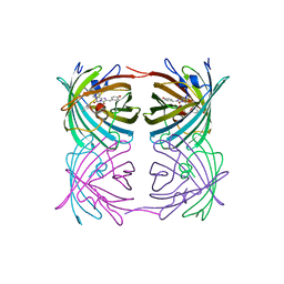
Crystal structure of ZsYellow soaked by Cu2+
分子名称: GFP-like fluorescent chromoprotein FP538
著者Nam, K.H.
登録日2020-01-05
公開日2020-01-22
最終更新日2023-11-29
実験手法X-RAY DIFFRACTION (2.6 Å)
主引用文献Spectroscopic and Structural Analysis of Cu 2+ -Induced Fluorescence Quenching of ZsYellow.
Biosensors (Basel), 10, 2020
7CVM
DownloadVisualize
BU of 7cvm by Molmil
Crystal structure of glucose isomerase by fixed-target serial synchrotron crystallography (500 ms)
分子名称: MAGNESIUM ION, Xylose isomerase
著者Nam, K.H.
登録日2020-08-26
公開日2020-09-09
最終更新日2023-11-29
実験手法X-RAY DIFFRACTION (2 Å)
主引用文献Fixed-Target Serial Synchrotron Crystallography Using Nylon Mesh and Enclosed Film-Based Sample Holder
Crystals, 10, 2020

221371

件を2024-06-19に公開中

PDB statisticsPDBj update infoContact PDBjnumon