Loading
PDBj
メニューPDBj@FacebookPDBj@TwitterPDBj@YouTubewwPDB FoundationwwPDB
RCSB PDBPDBeBMRBAdv. SearchSearch help
Search by PDB author
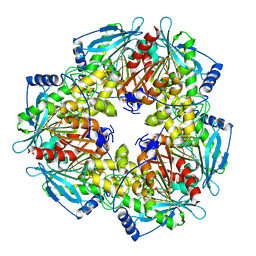
2F46
DownloadVisualize
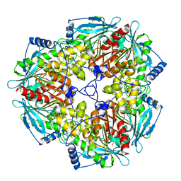
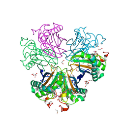
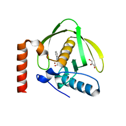
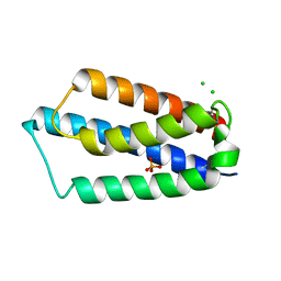
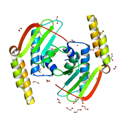
BU of 2f46 by Molmil
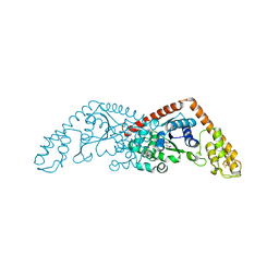
Crystal structure of a putative phosphatase (nma1982) from neisseria meningitidis z2491 at 1.41 A resolution
分子名称: CHLORIDE ION, UNKNOWN LIGAND, hypothetical protein
著者Joint Center for Structural Genomics (JCSG)
登録日2005-11-22
公開日2006-02-07
最終更新日2023-01-25
実験手法X-RAY DIFFRACTION (1.41 Å)
主引用文献Crystal structure of NMA1982 from Neisseria meningitidis at 1.5 A resolution provides a structural scaffold for nonclassical, eukaryotic-like phosphatases.
Proteins, 69, 2007
2FG0
DownloadVisualize
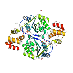
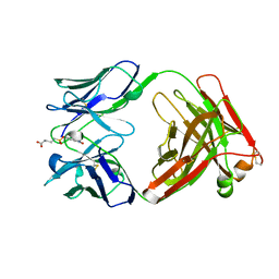
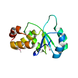
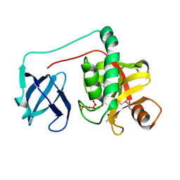
BU of 2fg0 by Molmil
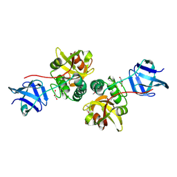
Crystal structure of a putative gamma-d-glutamyl-l-diamino acid endopeptidase (npun_r0659) from nostoc punctiforme pcc 73102 at 1.79 A resolution
分子名称: COG0791: Cell wall-associated hydrolases (invasion-associated proteins), GLYCEROL
著者Joint Center for Structural Genomics (JCSG)
登録日2005-12-20
公開日2006-01-10
最終更新日2023-01-25
実験手法X-RAY DIFFRACTION (1.79 Å)
主引用文献Structural Basis of Murein Peptide Specificity of a gamma-D-Glutamyl-L-Diamino Acid Endopeptidase.
Structure, 17, 2009
2FEA
DownloadVisualize
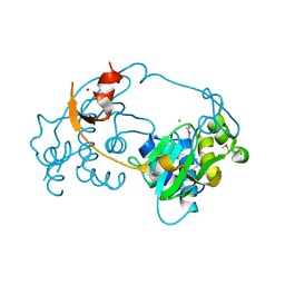
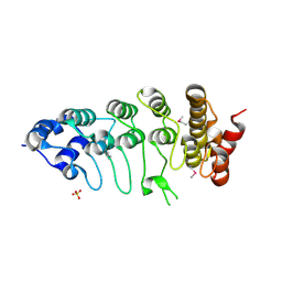
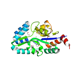
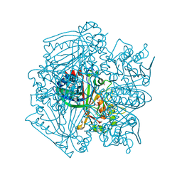
BU of 2fea by Molmil
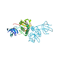
Crystal structure of MtnX phosphatase from Bacillus Subtilis at 2.00 A resolution
分子名称: 1,2-ETHANEDIOL, 2-hydroxy-3-keto-5-methylthiopentenyl-1-phosphate phosphatase, MAGNESIUM ION, ...
著者Joint Center for Structural Genomics (JCSG)
登録日2005-12-15
公開日2005-12-27
最終更新日2023-01-25
実験手法X-RAY DIFFRACTION (2 Å)
主引用文献Crystal structure of MtnX phosphatase from Bacillus subtilis at 2.0 A resolution provides a structural basis for bipartite phosphomonoester hydrolysis of 2-hydroxy-3-keto-5-methylthiopentenyl-1-phosphate.
Proteins, 69, 2007
2EVR
DownloadVisualize
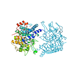
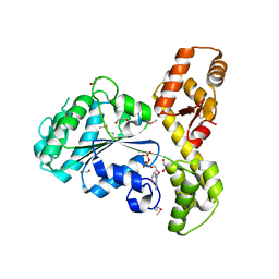
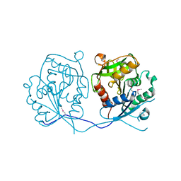
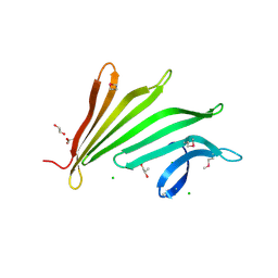
BU of 2evr by Molmil
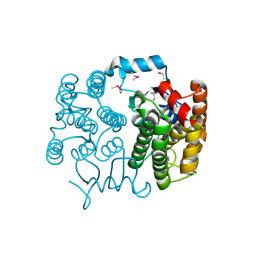
CRYSTAL STRUCTURE OF A PUTATIVE GAMMA-D-GLUTAMYL-L-DIAMINO ACID ENDOPEPTIDASE (NPUN_R0659) FROM NOSTOC PUNCTIFORME PCC 73102 AT 1.60 A RESOLUTION
分子名称: 1,2-ETHANEDIOL, CHLORIDE ION, COG0791: Cell wall-associated hydrolases (invasion-associated proteins), ...
著者Joint Center for Structural Genomics (JCSG)
登録日2005-10-31
公開日2005-11-22
最終更新日2023-01-25
実験手法X-RAY DIFFRACTION (1.6 Å)
主引用文献Structural Basis of Murein Peptide Specificity of a gamma-D-Glutamyl-L-Diamino Acid Endopeptidase.
Structure, 17, 2009
3ROX
DownloadVisualize
BU of 3rox by Molmil
Crystal Structure of Mouse Apolipoprotein A-I Binding Protein in Complex with Theophylline
分子名称: Apolipoprotein A-I-binding protein, SULFATE ION, THEOPHYLLINE
著者Shumilin, I.A, Jha, K.N, Cymborowski, M, Herr, J.C, Minor, W.
登録日2011-04-26
公開日2012-07-18
最終更新日2023-12-06
実験手法X-RAY DIFFRACTION (2.4 Å)
主引用文献Identification of unknown protein function using metabolite cocktail screening.
Structure, 20, 2012
3RQ5
DownloadVisualize
BU of 3rq5 by Molmil
Crystal Structure of ADP/ATP-dependent NAD(P)H-hydrate dehydratase from Bacillus subtilis co-crystallized with ATP/Mg2+ and soaked with CoA
分子名称: ADP/ATP-dependent NAD(P)H-hydrate dehydratase, COENZYME A, GLYCEROL
著者Shumilin, I.A, Cymborowski, M, Joachimiak, A, Minor, W, Midwest Center for Structural Genomics (MCSG)
登録日2011-04-27
公開日2011-07-27
最終更新日2023-09-13
実験手法X-RAY DIFFRACTION (1.7 Å)
主引用文献Identification of unknown protein function using metabolite cocktail screening.
Structure, 20, 2012
3RQH
DownloadVisualize
BU of 3rqh by Molmil
Crystal Structure of ADP/ATP-dependent NAD(P)H-hydrate dehydratase from Bacillus subtilis in complex with P1,P6-Di(adenosine-5') hexaphosphate
分子名称: ADP/ATP-DEPENDENT NAD(P)H-HYDRATE DEHYDRATASE, MAGNESIUM ION, P1,P6-Di(adenosine-5') hexaphosphate
著者Shumilin, I.A, Cymborowski, M, Joachimiak, A, Minor, W, Midwest Center for Structural Genomics (MCSG)
登録日2011-04-28
公開日2011-07-27
最終更新日2023-09-13
実験手法X-RAY DIFFRACTION (1.75 Å)
主引用文献Identification of unknown protein function using metabolite cocktail screening.
Structure, 20, 2012
3ROG
DownloadVisualize
BU of 3rog by Molmil
Crystal Structure of Mouse Apolipoprotein A-I Binding Protein in Complex with Thymidine 3'-monophosphate
分子名称: Apolipoprotein A-I-binding protein, SULFATE ION, THYMIDINE-3'-PHOSPHATE
著者Shumilin, I.A, Jha, K.N, Cymborowski, M, Herr, J.C, Minor, W.
登録日2011-04-25
公開日2012-07-18
最終更新日2023-12-06
実験手法X-RAY DIFFRACTION (2.05 Å)
主引用文献Identification of unknown protein function using metabolite cocktail screening.
Structure, 20, 2012
3RQQ
DownloadVisualize
BU of 3rqq by Molmil
Crystal Structure of ADP/ATP-dependent NAD(P)H-hydrate dehydratase from Bacillus subtilis in complex with P1,P3-Di(adenosine-5') triphosphate
分子名称: ADP/ATP-DEPENDENT NAD(P)H-HYDRATE DEHYDRATASE, BIS(ADENOSINE)-5'-TRIPHOSPHATE, MAGNESIUM ION
著者Shumilin, I.A, Cymborowski, M, Joachimiak, A, Minor, W, Midwest Center for Structural Genomics (MCSG)
登録日2011-04-28
公開日2011-07-27
最終更新日2023-09-13
実験手法X-RAY DIFFRACTION (1.6 Å)
主引用文献Identification of unknown protein function using metabolite cocktail screening.
Structure, 20, 2012
3RNO
DownloadVisualize
BU of 3rno by Molmil
Crystal Structure of Mouse Apolipoprotein A-I Binding Protein in Complex with NADP.
分子名称: Apolipoprotein A-I-binding protein, NADP NICOTINAMIDE-ADENINE-DINUCLEOTIDE PHOSPHATE
著者Shumilin, I.A, Jha, K.N, Cymborowski, M, Herr, J.C, Minor, W.
登録日2011-04-22
公開日2012-05-09
最終更新日2023-12-06
実験手法X-RAY DIFFRACTION (2.5 Å)
主引用文献Identification of unknown protein function using metabolite cocktail screening.
Structure, 20, 2012
3ROZ
DownloadVisualize
BU of 3roz by Molmil
Crystal Structure of Mouse Apolipoprotein A-I Binding Protein in Complex with Nicotinamide
分子名称: Apolipoprotein A-I-binding protein, NICOTINAMIDE, SULFATE ION
著者Shumilin, I.A, Jha, K.N, Cymborowski, M, Herr, J.C, Minor, W.
登録日2011-04-26
公開日2012-07-18
最終更新日2023-12-06
実験手法X-RAY DIFFRACTION (2.8 Å)
主引用文献Identification of unknown protein function using metabolite cocktail screening.
Structure, 20, 2012
3RQ8
DownloadVisualize
BU of 3rq8 by Molmil
Crystal Structure of ADP/ATP-dependent NAD(P)H-hydrate dehydratase from Bacillus subtilis soaked with P1,P5-Di(adenosine-5') pentaphosphate
分子名称: ADP/ATP-dependent NAD(P)H-hydrate dehydratase, BIS(ADENOSINE)-5'-PENTAPHOSPHATE, MAGNESIUM ION
著者Shumilin, I.A, Cymborowski, M, Joachimiak, A, Minor, W, Midwest Center for Structural Genomics (MCSG)
登録日2011-04-27
公開日2011-07-27
最終更新日2023-09-13
実験手法X-RAY DIFFRACTION (1.9 Å)
主引用文献Identification of unknown protein function using metabolite cocktail screening.
Structure, 20, 2012
3H41
DownloadVisualize
BU of 3h41 by Molmil
CRYSTAL STRUCTURE OF A NLPC/P60 FAMILY PROTEIN (BCE_2878) FROM BACILLUS CEREUS ATCC 10987 AT 1.79 A RESOLUTION
分子名称: ALANINE, D-GLUTAMIC ACID, NLP/P60 family protein, ...
著者Joint Center for Structural Genomics (JCSG)
登録日2009-04-17
公開日2009-05-05
最終更新日2023-02-01
実験手法X-RAY DIFFRACTION (1.79 Å)
主引用文献Structure of the gamma-D-glutamyl-L-diamino acid endopeptidase YkfC from Bacillus cereus in complex with L-Ala-[gamma]-D-Glu: insights into substrate recognition by NlpC/P60 cysteine peptidases
Acta Crystallogr.,Sect.F, 66, 2010
3H50
DownloadVisualize
BU of 3h50 by Molmil
CRYSTAL STRUCTURE OF A TETRACENOMYCIN POLYKETIDE SYNTHESIS PROTEIN (TCMJ) FROM XANTHOMONAS CAMPESTRIS PV. CAMPESTRIS AT 1.60 A RESOLUTION
分子名称: ACETATE ION, Tetracenomycin polyketide synthesis protein, ZINC ION
著者Joint Center for Structural Genomics (JCSG)
登録日2009-04-21
公開日2009-05-05
最終更新日2023-02-01
実験手法X-RAY DIFFRACTION (1.6 Å)
主引用文献Conformational changes associated with the binding of zinc acetate at the putative active site of XcTcmJ, a cupin from Xanthomonas campestris pv. campestris.
Acta Crystallogr.,Sect.F, 66, 2010
3HN7
DownloadVisualize
BU of 3hn7 by Molmil
Crystal structure of a murein peptide ligase mpl (psyc_0032) from psychrobacter arcticus 273-4 at 1.65 A resolution
分子名称: UDP-N-acetylmuramate-L-alanine ligase
著者Joint Center for Structural Genomics (JCSG)
登録日2009-05-29
公開日2009-06-16
最終更新日2023-02-01
実験手法X-RAY DIFFRACTION (1.65 Å)
主引用文献Structure and function of the first full-length murein Peptide ligase (mpl) cell wall recycling protein.
Plos One, 6, 2011
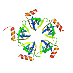
3HSA
DownloadVisualize
BU of 3hsa by Molmil
Crystal structure of pleckstrin homology domain (YP_926556.1) from SHEWANELLA AMAZONENSIS SB2B at 1.99 A resolution
分子名称: DI(HYDROXYETHYL)ETHER, GLYCEROL, pleckstrin homology domain
著者Joint Center for Structural Genomics (JCSG)
登録日2009-06-10
公開日2009-06-23
最終更新日2023-02-01
実験手法X-RAY DIFFRACTION (1.99 Å)
主引用文献Bacterial pleckstrin homology domains: a prokaryotic origin for the PH domain.
J.Mol.Biol., 396, 2010
3IRB
DownloadVisualize
BU of 3irb by Molmil
Crystal structure of protein with unknown function from DUF35 family (13815350) from SULFOLOBUS SOLFATARICUS at 1.80 A resolution
分子名称: ACETIC ACID, SULFATE ION, ZINC ION, ...
著者Joint Center for Structural Genomics (JCSG)
登録日2009-08-21
公開日2009-09-01
最終更新日2023-02-01
実験手法X-RAY DIFFRACTION (1.8 Å)
主引用文献The structure of SSO2064, the first representative of Pfam family PF01796, reveals a novel two-domain zinc-ribbon OB-fold architecture with a potential acyl-CoA-binding role.
Acta Crystallogr.,Sect.F, 66, 2010
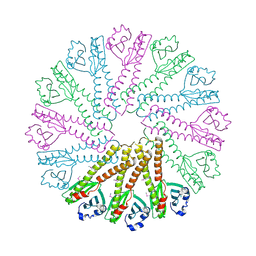
3GO5
DownloadVisualize
BU of 3go5 by Molmil
Crystal structure of a multidomain protein with nucleic acid binding domains (sp_0946) from streptococcus pneumoniae tigr4 at 1.40 A resolution
分子名称: 1,2-ETHANEDIOL, CHLORIDE ION, Multidomain protein with S1 RNA-binding domains
著者Joint Center for Structural Genomics (JCSG)
登録日2009-03-18
公開日2009-04-07
最終更新日2023-02-01
実験手法X-RAY DIFFRACTION (1.4 Å)
主引用文献Structure of a virulence regulatory factor CvfB reveals a novel winged helix RNA binding module.
Structure, 18, 2010
3HBZ
DownloadVisualize
BU of 3hbz by Molmil
Crystal structure of a putative glycoside hydrolase (bt_2081) from bacteroides thetaiotaomicron vpi-5482 at 2.05 A resolution
分子名称: ACETATE ION, CACODYLATE ION, CALCIUM ION, ...
著者Joint Center for Structural Genomics (JCSG)
登録日2009-05-05
公開日2009-05-26
最終更新日2023-02-01
実験手法X-RAY DIFFRACTION (2.05 Å)
主引用文献Structure of Bacteroides thetaiotaomicron BT2081 at 2.05 A resolution: the first structural representative of a new protein family that may play a role in carbohydrate metabolism.
Acta Crystallogr.,Sect.F, 66, 2010
3K5J
DownloadVisualize
BU of 3k5j by Molmil
Crystal structure of Putative SUFU (suppressor of fused protein) homolog (YP_208451.1) from Neisseria gonorrhoeae FA 1090 at 1.40 A resolution
分子名称: GLYCEROL, SULFATE ION, Suppressor of fused family protein
著者Joint Center for Structural Genomics (JCSG)
登録日2009-10-07
公開日2010-01-26
最終更新日2019-07-17
実験手法X-RAY DIFFRACTION (1.4 Å)
主引用文献The crystal structure of a bacterial Sufu-like protein defines a novel group of bacterial proteins that are similar to the N-terminal domain of human Sufu.
Protein Sci., 19, 2010
4QDG
DownloadVisualize
BU of 4qdg by Molmil
Crystal structure of a putative adhesin (BT2657) from Bacteroides thetaiotaomicron VPI-5482 at 2.20 A resolution (PSI Community Target, Nakayama)
分子名称: 1,2-ETHANEDIOL, MAGNESIUM ION, Putative adhesin
著者Joint Center for Structural Genomics (JCSG)
登録日2014-05-13
公開日2014-06-11
最終更新日2023-02-01
実験手法X-RAY DIFFRACTION (2.2 Å)
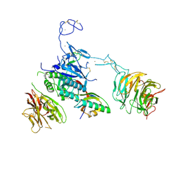
主引用文献A Distinct Type of Pilus from the Human Microbiome.
Cell, 165, 2016
4Z49
DownloadVisualize
BU of 4z49 by Molmil
Homo Sapiens Fatty Acid Synthetase, Thioesterase Domain at 1.7 Angstroms Resolution
分子名称: Fatty acid synthase, POTASSIUM ION
著者Spraggon, G.
登録日2015-04-01
公開日2016-03-16
最終更新日2024-03-06
実験手法X-RAY DIFFRACTION (1.7 Å)
主引用文献Estimation of Hydrogen-Exchange Protection Factors from MD Simulation Based on Amide Hydrogen Bonding Analysis.
J.Chem.Inf.Model., 55, 2015
3KK7
DownloadVisualize
BU of 3kk7 by Molmil
Crystal structure of Putative cell invasion protein with MAC/Perforin domain (NP_812351.1) from BACTERIODES THETAIOTAOMICRON VPI-5482 at 2.46 A resolution
分子名称: (4S)-2-METHYL-2,4-PENTANEDIOL, 1,2-ETHANEDIOL, CHLORIDE ION, ...
著者Joint Center for Structural Genomics (JCSG)
登録日2009-11-04
公開日2009-11-24
最終更新日2023-02-01
実験手法X-RAY DIFFRACTION (2.46 Å)
主引用文献Structure of a membrane-attack complex/perforin (MACPF) family protein from the human gut symbiont Bacteroides thetaiotaomicron.
Acta Crystallogr.,Sect.F, 66, 2010
3KW0
DownloadVisualize
BU of 3kw0 by Molmil
Crystal structure of Cysteine peptidase (NP_982244.1) from BACILLUS CEREUS ATCC 10987 at 2.50 A resolution
分子名称: CHLORIDE ION, Cysteine peptidase, LYSINE
著者Joint Center for Structural Genomics (JCSG)
登録日2009-11-30
公開日2009-12-15
最終更新日2023-02-01
実験手法X-RAY DIFFRACTION (2.5 Å)
主引用文献Structural Analysis of Papain-Like NlpC/P60 Superfamily Enzymes with a Circularly Permuted Topology Reveals Potential Lipid Binding Sites.
Plos One, 6, 2011
3LCT
DownloadVisualize
BU of 3lct by Molmil
Crystal Structure of the Anaplastic Lymphoma Kinase Catalytic Domain
分子名称: ADENOSINE-5'-DIPHOSPHATE, ALK tyrosine kinase receptor, GLYCEROL
著者Lee, C.C.
登録日2010-01-11
公開日2010-08-04
最終更新日2023-09-06
実験手法X-RAY DIFFRACTION (2.1 Å)
主引用文献Crystal structure of the ALK (anaplastic lymphoma kinase) catalytic domain.
Biochem.J., 430, 2010

225158

件を2024-09-18に公開中

PDB statisticsPDBj update infoContact PDBjnumon