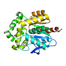
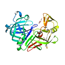
1YSO
 
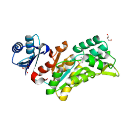
 | | YEAST CU, ZN SUPEROXIDE DISMUTASE WITH THE REDUCED BRIDGE BROKEN | | 分子名称: | COPPER (I) ION, YEAST CU, ZN SUPEROXIDE DISMUTASE, ... | | 著者 | Parge, H.E, Crane, B.R, Tsang, J, Tainer, J.A. | | 登録日 | 1995-12-21 | | 公開日 | 1996-06-10 | | 最終更新日 | 2024-10-09 | | 実験手法 | X-RAY DIFFRACTION (1.73 Å) | | 主引用文献 | Unusual trigonal-planar copper configuration revealed in the atomic structure of yeast copper-zinc superoxide dismutase.
Biochemistry, 35, 1996
|
|
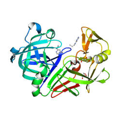
2ADW
 | |
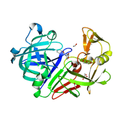
8OYH
 | | X-ray structure of furin (PCSK3) in complex with Guanidinomethyl-Phac-Can-Tle-Can-6-(aminomethyl)-3-amino-isoindol | | 分子名称: | CALCIUM ION, CHLORIDE ION, DIMETHYL SULFOXIDE, ... | | 著者 | Dahms, S.O, Brandstetter, H. | | 登録日 | 2023-05-04 | | 公開日 | 2024-03-13 | | 最終更新日 | 2024-05-15 | | 実験手法 | X-RAY DIFFRACTION (1.8 Å) | | 主引用文献 | Fragment-Based Design, Synthesis, and Characterization of Aminoisoindole-Derived Furin Inhibitors.
Chemmedchem, 19, 2024
|
|
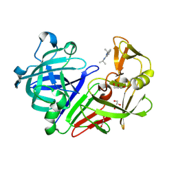
8QH6
 | | Crystal structure of IpgC in complex with a follow-up compound based on J20 | | 分子名称: | 3-azanyl-6-chloranyl-isoindol-1-one, CHLORIDE ION, Chaperone protein IpgC, ... | | 著者 | Wallbaum, J.E, Heine, A, Reuter, K. | | 登録日 | 2023-09-06 | | 公開日 | 2023-12-27 | | 実験手法 | X-RAY DIFFRACTION (1.8 Å) | | 主引用文献 | Crystallographic Fragment Screening on the Shigella Type III Secretion System Chaperone IpgC.
Acs Omega, 8, 2023
|
|
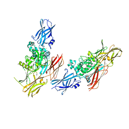
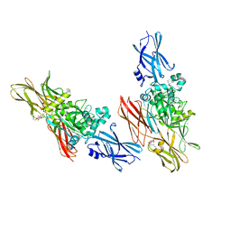
5DIS
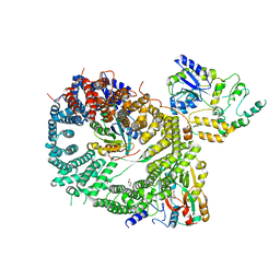 | | Crystal structure of a CRM1-RanGTP-SPN1 export complex bound to a 113 amino acid FG-repeat containing fragment of Nup214 | | 分子名称: | Exportin-1, GTP-binding nuclear protein Ran, GUANOSINE-5'-TRIPHOSPHATE, ... | | 著者 | Monecke, T, Port, S.A, Dickmanns, A, Kehlenbach, R.H, Ficner, R. | | 登録日 | 2015-09-01 | | 公開日 | 2015-11-04 | | 最終更新日 | 2024-01-10 | | 実験手法 | X-RAY DIFFRACTION (2.85 Å) | | 主引用文献 | Structural and Functional Characterization of CRM1-Nup214 Interactions Reveals Multiple FG-Binding Sites Involved in Nuclear Export.
Cell Rep, 13, 2015
|
|
4A6N
 | | STRUCTURE OF THE TETRACYCLINE DEGRADING MONOOXYGENASE TETX IN COMPLEX WITH TIGECYCLINE | | 分子名称: | FLAVIN-ADENINE DINUCLEOTIDE, SULFATE ION, TETX2 PROTEIN, ... | | 著者 | Volkers, G, Palm, G.J, Weiss, M.S, Hinrichs, W. | | 登録日 | 2011-11-07 | | 公開日 | 2012-11-21 | | 最終更新日 | 2023-12-20 | | 実験手法 | X-RAY DIFFRACTION (2.3 Å) | | 主引用文献 | Putative Dioxygen-Binding Sites and Recognition of Tigecycline and Minocycline in the Tetracycline-Degrading Monooxygenase Tetx
Acta Crystallogr.,Sect.D, 69, 2013
|
|
4A99
 | | STRUCTURE OF THE TETRACYCLINE DEGRADING MONOOXYGENASE TETX IN COMPLEX WITH MINOCYCLINE | | 分子名称: | (4S,4AS,5AR,12AS)-4,7-BIS(DIMETHYLAMINO)-3,10,12,12A-TETRAHYDROXY-1,11-DIOXO-1,4,4A,5,5A,6,11,12A-OCTAHYDROTETRACENE-2- CARBOXAMIDE, FLAVIN-ADENINE DINUCLEOTIDE, SULFATE ION, ... | | 著者 | Volkers, G, Palm, G.J, Weiss, M.S, Hinrichs, W. | | 登録日 | 2011-11-25 | | 公開日 | 2012-12-12 | | 最終更新日 | 2023-12-20 | | 実験手法 | X-RAY DIFFRACTION (2.18 Å) | | 主引用文献 | Putative Dioxygen-Binding Sites and Recognition of Tigecycline and Minocycline in the Tetracycline-Degrading Monooxygenase Tetx
Acta Crystallogr.,Sect.D, 69, 2013
|
|
1PHO
 | |
4HZG
 | | Structure of haloalkane dehalogenase DhaA from Rhodococcus rhodochrous | | 分子名称: | CHLORIDE ION, Haloalkane dehalogenase | | 著者 | Stsiapanava, A, Weiss, M.S, Mesters, J.R, Kuta Smatanova, I. | | 登録日 | 2012-11-15 | | 公開日 | 2013-10-02 | | 最終更新日 | 2023-09-20 | | 実験手法 | X-RAY DIFFRACTION (1.95 Å) | | 主引用文献 | Crystallographic analysis of 1,2,3-trichloropropane biodegradation by the haloalkane dehalogenase DhaA31.
Acta Crystallogr.,Sect.D, 70, 2014
|
|
1OPF
 | |
7ZYT
 | | Crystal structure of the I318T pathogenic variant of the human dihydrolipoamide dehydrogenase | | 分子名称: | 1,2-ETHANEDIOL, 2-[BIS-(2-HYDROXY-ETHYL)-AMINO]-2-HYDROXYMETHYL-PROPANE-1,3-DIOL, Dihydrolipoyl dehydrogenase, ... | | 著者 | Nemes-Nikodem, E, Szabo, E, Vass, K.R, Lennartz, F, Nagy, B, Torocsik, B, Weiss, M.S, Adam-Vizi, V, Ambrus, A. | | 登録日 | 2022-05-25 | | 公開日 | 2023-06-14 | | 最終更新日 | 2024-02-07 | | 実験手法 | X-RAY DIFFRACTION (2.892 Å) | | 主引用文献 | Structural and Biochemical Investigation of Selected Pathogenic Mutants of the Human Dihydrolipoamide Dehydrogenase.
Int J Mol Sci, 24, 2023
|
|
2W93
 | | Crystal structure of the Saccharomyces cerevisiae pyruvate decarboxylase variant E477Q in complex with the surrogate pyruvamide | | 分子名称: | (1S,2S)-1-amino-1,2-dihydroxypropan-1-olate, MAGNESIUM ION, PYRUVATE DECARBOXYLASE ISOZYME 1, ... | | 著者 | Kutter, S, Weiss, M.S, Konig, S. | | 登録日 | 2009-01-21 | | 公開日 | 2009-02-03 | | 最終更新日 | 2023-12-13 | | 実験手法 | X-RAY DIFFRACTION (1.6 Å) | | 主引用文献 | Allosteric Activation of Pyruvate Decarboxylases. A Never-Ending Story.
J.Mol.Catal., B Enzym., 61, 2014
|
|
8BHD
 | | N-terminal domain of Plasmodium berghei glutamyl-tRNA synthetase (Tbxo4 derivative crystal structure) | | 分子名称: | GLYCEROL, Glutamate--tRNA ligase, SULFATE ION, ... | | 著者 | Benas, P, Jaramillo Ponce, J.R, Legrand, P, Frugier, M, Sauter, C. | | 登録日 | 2022-10-31 | | 公開日 | 2023-01-25 | | 最終更新日 | 2024-06-19 | | 実験手法 | X-RAY DIFFRACTION (3.17 Å) | | 主引用文献 | Solution X-ray scattering highlights discrepancies in Plasmodium multi-aminoacyl-tRNA synthetase complexes.
Protein Sci., 32, 2023
|
|
8BM8
 | | Crystal Structure of Ephrin A2 (EphA2) Receptor Protein Kinase with Compound 11 | | 分子名称: | Ephrin type-A receptor 2, ~{N}-(3-cyanophenyl)-4-methyl-3-[(1-methyl-6-pyridin-3-yl-pyrazolo[3,4-d]pyrimidin-4-yl)amino]benzamide | | 著者 | Linhard, V, Witt, K, Gande, S, Wollenhaupt, J, Lennartz, F, Weiss, M.S, Schwalbe, H. | | 登録日 | 2022-11-10 | | 公開日 | 2024-06-05 | | 実験手法 | X-RAY DIFFRACTION (1.68 Å) | | 主引用文献 | (placeholder, will be filled after publication)
To Be Published
|
|
1L9M
 | |
1L9N
 | |
5VGI
 | | Crystal Structure of KDM4 with the Small Molecule Inhibitor QC6352 | | 分子名称: | 3-[({(1R)-6-[methyl(phenyl)amino]-1,2,3,4-tetrahydronaphthalen-1-yl}methyl)amino]pyridine-4-carboxylic acid, Lysine-specific demethylase 4A, NICKEL (II) ION, ... | | 著者 | Hosfield, D.J. | | 登録日 | 2017-04-11 | | 公開日 | 2017-09-27 | | 最終更新日 | 2024-03-13 | | 実験手法 | X-RAY DIFFRACTION (2.07 Å) | | 主引用文献 | Design of KDM4 Inhibitors with Antiproliferative Effects in Cancer Models.
ACS Med Chem Lett, 8, 2017
|
|
7PSC
 | | Crystal structure of the disease-causing I358T mutant of the human dihydrolipoamide dehydrogenase | | 分子名称: | 2-[BIS-(2-HYDROXY-ETHYL)-AMINO]-2-HYDROXYMETHYL-PROPANE-1,3-DIOL, Dihydrolipoyl dehydrogenase, mitochondrial, ... | | 著者 | Nemes-Nikodem, E, Szabo, E, Zambo, Z, Vass, K.R, Taberman, H, Torocsik, B, Weiss, M.S, Adam-Vizi, V, Ambrus, A. | | 登録日 | 2021-09-22 | | 公開日 | 2023-04-05 | | 最終更新日 | 2024-02-07 | | 実験手法 | X-RAY DIFFRACTION (2.436 Å) | | 主引用文献 | Structural and Biochemical Investigation of Selected Pathogenic Mutants of the Human Dihydrolipoamide Dehydrogenase.
Int J Mol Sci, 24, 2023
|
|
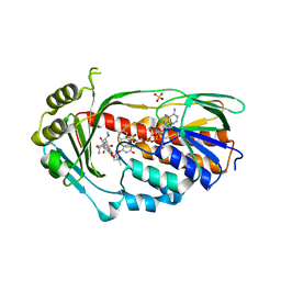
4Y36
 | | Endothiapepsin in complex with fragment 4 | | 分子名称: | 1,2-ETHANEDIOL, 3-methylbenzohydrazide, Endothiapepsin, ... | | 著者 | Radeva, N, Uehlein, M, Weiss, M.S, Heine, A, Klebe, G. | | 登録日 | 2015-02-10 | | 公開日 | 2016-02-17 | | 最終更新日 | 2024-01-10 | | 実験手法 | X-RAY DIFFRACTION (1.59 Å) | | 主引用文献 | Crystallographic Fragment Screening of an Entire Library
To Be Published
|
|
4Y3P
 | | Endothiapepsin in complex with fragment 17 | | 分子名称: | 1-(6-methylpyrimidin-4-yl)azepane, Endothiapepsin, GLYCEROL | | 著者 | Radeva, N, Uehlein, M, Weiss, M.S, Heine, A, Klebe, G. | | 登録日 | 2015-02-10 | | 公開日 | 2016-02-17 | | 最終更新日 | 2024-01-10 | | 実験手法 | X-RAY DIFFRACTION (1.55 Å) | | 主引用文献 | Crystallographic Fragment Screening of an Entire Library
To Be Published
|
|
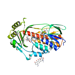
4Y3Z
 | | Endothiapepsin in complex with fragment 41 | | 分子名称: | 1,2-ETHANEDIOL, ACETATE ION, Endothiapepsin, ... | | 著者 | Radeva, N, Uehlein, M, Weiss, M.S, Heine, A, Klebe, G. | | 登録日 | 2015-02-10 | | 公開日 | 2016-02-17 | | 最終更新日 | 2024-10-16 | | 実験手法 | X-RAY DIFFRACTION (1.12 Å) | | 主引用文献 | Crystallographic Fragment Screening of an Entire Library
To Be Published
|
|
4Y43
 | | Endothiapepsin in complex with fragment 42 | | 分子名称: | (1E,2E)-N,N'-di(propan-2-yl)cyclohepta-3,5-diene-1,2-diimine, 1,2-ETHANEDIOL, Endothiapepsin, ... | | 著者 | Radeva, N, Uehlein, M, Weiss, M.S, Heine, A, Klebe, G. | | 登録日 | 2015-02-10 | | 公開日 | 2016-02-17 | | 最終更新日 | 2024-10-09 | | 実験手法 | X-RAY DIFFRACTION (1.48 Å) | | 主引用文献 | Crystallographic Fragment Screening of an Entire Library
To Be Published
|
|
8Q4S
 | |
4Y3E
 | | Endothiapepsin in complex with fragment 5 | | 分子名称: | 1H-isoindol-3-amine, ACETATE ION, DIMETHYL SULFOXIDE, ... | | 著者 | Radeva, N, Uehlein, M, Weiss, M.S, Heine, A, Klebe, G. | | 登録日 | 2015-02-10 | | 公開日 | 2016-02-17 | | 最終更新日 | 2024-01-10 | | 実験手法 | X-RAY DIFFRACTION (1.25 Å) | | 主引用文献 | Crystallographic Fragment Screening of an Entire Library
To Be Published
|
|
4Y4U
 | | Endothiapepsin in complex with fragment 14 | | 分子名称: | 1-(4-bromo-2-chlorophenyl)-3-methylthiourea, DI(HYDROXYETHYL)ETHER, Endothiapepsin, ... | | 著者 | Radeva, N, Uehlein, M, Weiss, M.S, Heine, A, Klebe, G. | | 登録日 | 2015-02-11 | | 公開日 | 2016-03-02 | | 最終更新日 | 2024-10-16 | | 実験手法 | X-RAY DIFFRACTION (1.754 Å) | | 主引用文献 | Crystallographic Fragment Screening of an Entire Library
To Be Published
|
|


