Loading
PDBj
メニューPDBj@FacebookPDBj@TwitterPDBj@YouTubewwPDB FoundationwwPDB
RCSB PDBPDBeBMRBAdv. SearchSearch help
Search by PDB author
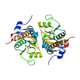
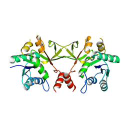
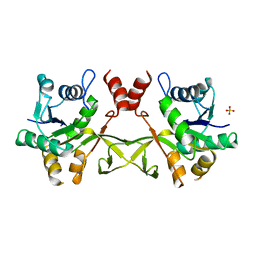
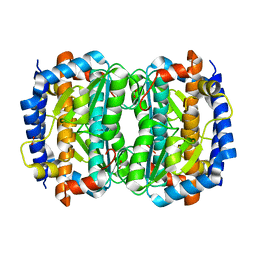
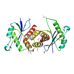
1VHM
DownloadVisualize
BU of 1vhm by Molmil
Crystal structure of an hypothetical protein
分子名称: 2-(N-MORPHOLINO)-ETHANESULFONIC ACID, Protein yebR
著者Structural GenomiX
登録日2003-12-01
公開日2003-12-30
最終更新日2023-12-27
実験手法X-RAY DIFFRACTION (2.1 Å)
主引用文献Structural analysis of a set of proteins resulting from a bacterial genomics project
Proteins, 60, 2005
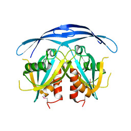
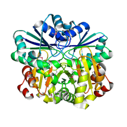
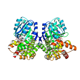
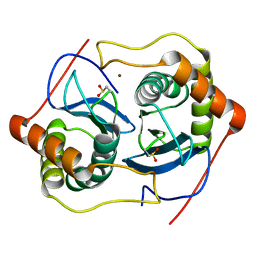
1VHX
DownloadVisualize
BU of 1vhx by Molmil
Crystal structure of Putative Holliday junction resolvase
分子名称: Putative Holliday junction resolvase
著者Structural GenomiX
登録日2003-12-01
公開日2003-12-30
最終更新日2023-12-27
実験手法X-RAY DIFFRACTION (1.96 Å)
主引用文献Structural analysis of a set of proteins resulting from a bacterial genomics project
Proteins, 60, 2005
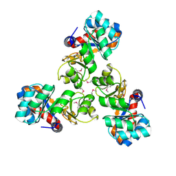
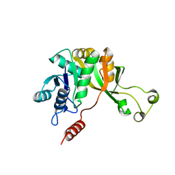
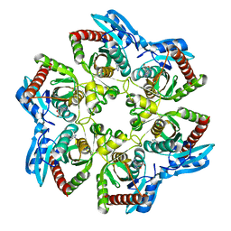
1VI6
DownloadVisualize
BU of 1vi6 by Molmil
Crystal structure of ribosomal protein S2P
分子名称: 30S ribosomal protein S2P, SODIUM ION
著者Structural GenomiX
登録日2003-12-01
公開日2003-12-30
最終更新日2023-12-27
実験手法X-RAY DIFFRACTION (1.95 Å)
主引用文献Structural analysis of a set of proteins resulting from a bacterial genomics project
Proteins, 60, 2005
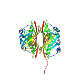
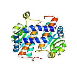
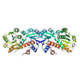
1VIU
DownloadVisualize
BU of 1viu by Molmil
Crystal structure of putative ADP ribose pyrophosphatase
分子名称: ADP-ribose pyrophosphatase
著者Structural GenomiX
登録日2003-12-01
公開日2003-12-30
最終更新日2023-12-27
実験手法X-RAY DIFFRACTION (2.4 Å)
主引用文献Structural analysis of a set of proteins resulting from a bacterial genomics project
Proteins, 60, 2005
1VHC
DownloadVisualize
BU of 1vhc by Molmil
Crystal structure of a putative KHG/KDPG aldolase
分子名称: Putative KHG/KDPG aldolase
著者Structural GenomiX
登録日2003-12-01
公開日2003-12-30
最終更新日2023-12-27
実験手法X-RAY DIFFRACTION (1.89 Å)
主引用文献Structural analysis of a set of proteins resulting from a bacterial genomics project
Proteins, 60, 2005
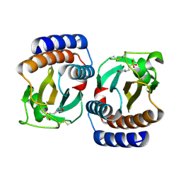
1VI8
DownloadVisualize
BU of 1vi8 by Molmil
Crystal structure of a putative thioesterase
分子名称: Hypothetical protein ydiI
著者Structural GenomiX, Burley, S.K, New York SGX Research Center for Structural Genomics (NYSGXRC)
登録日2003-12-01
公開日2003-12-30
最終更新日2023-12-27
実験手法X-RAY DIFFRACTION (2.2 Å)
主引用文献Structural analysis of a set of proteins resulting from a bacterial genomics project
Proteins, 60, 2005
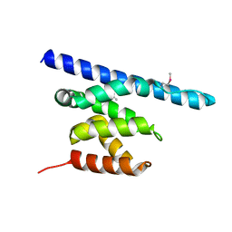
3NFQ
DownloadVisualize
BU of 3nfq by Molmil
Crystal structure of the conserved central domain of yeast Spn1/Iws1
分子名称: Transcription factor IWS1
著者Stargell, L.A, Luger, K.
登録日2010-06-10
公開日2010-10-13
最終更新日2011-07-13
実験手法X-RAY DIFFRACTION (1.85 Å)
主引用文献The Transcription Factor Spn1 Regulates Gene Expression via a Highly Conserved Novel Structural Motif.
J.Mol.Biol., 404, 2010
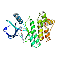
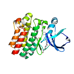
3OCT
DownloadVisualize
BU of 3oct by Molmil
Crystal structure of bruton's tyrosine kinase mutant V555R in complex with dasatinib
分子名称: N-(2-CHLORO-6-METHYLPHENYL)-2-({6-[4-(2-HYDROXYETHYL)PIPERAZIN-1-YL]-2-METHYLPYRIMIDIN-4-YL}AMINO)-1,3-THIAZOLE-5-CARBOXAMIDE, Tyrosine-protein kinase BTK
著者Davies, D.R, Staker, B.L.
登録日2010-08-10
公開日2010-11-03
最終更新日2023-09-06
実験手法X-RAY DIFFRACTION (1.95 Å)
主引用文献A novel, specific Btk inhibitor antagonizes BCR and Fc[gamma]R signaling and suppresses inflammatory arthritis
To be Published
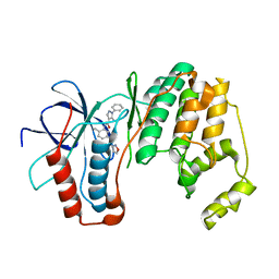
3OCG
DownloadVisualize
BU of 3ocg by Molmil
P38 Alpha kinase complexed with a 5-amino-pyrazole based inhibitor
分子名称: 5-amino-N-[5-(isoxazol-3-ylcarbamoyl)-2-methylphenyl]-1-phenyl-1H-pyrazole-4-carboxamide, Mitogen-activated protein kinase 14
著者Sack, J.S.
登録日2010-08-10
公開日2010-11-17
最終更新日2024-02-21
実験手法X-RAY DIFFRACTION (2.21 Å)
主引用文献5-Amino-pyrazoles as potent and selective p38α inhibitors
Bioorg.Med.Chem.Lett., 20, 2010
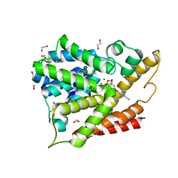
3O0J
DownloadVisualize
BU of 3o0j by Molmil
PDE4B In complex with ligand an2898
分子名称: 1,2-ETHANEDIOL, 4-[(1-hydroxy-1,3-dihydro-2,1-benzoxaborol-5-yl)oxy]benzene-1,2-dicarbonitrile, MAGNESIUM ION, ...
著者Alley, M.R.K, Zhou, Y.
登録日2010-07-19
公開日2011-08-10
最終更新日2024-02-21
実験手法X-RAY DIFFRACTION (1.95 Å)
主引用文献Boron-based phosphodiesterase inhibitors show novel binding of boron to PDE4 bimetal center.
Febs Lett., 586, 2012
1VGW
DownloadVisualize
BU of 1vgw by Molmil
Crystal structure of 4-diphosphocytidyl-2C-methyl-D-erythritol synthase
分子名称: 4-diphosphocytidyl-2C-methyl-D-erythritol synthase
著者Structural GenomiX
登録日2003-11-03
公開日2003-11-25
最終更新日2023-12-27
実験手法X-RAY DIFFRACTION (2.35 Å)
主引用文献Structural analysis of a set of proteins resulting from a bacterial genomics project
Proteins, 60, 2005
1VHV
DownloadVisualize
BU of 1vhv by Molmil
Crystal structure of diphthine synthase
分子名称: diphthine synthase
著者Structural GenomiX
登録日2003-12-01
公開日2003-12-30
最終更新日2023-12-27
実験手法X-RAY DIFFRACTION (1.75 Å)
主引用文献Structural analysis of a set of proteins resulting from a bacterial genomics project
Proteins, 60, 2005
1VIC
DownloadVisualize
BU of 1vic by Molmil
Crystal structure of CMP-KDO synthetase
分子名称: 3-deoxy-manno-octulosonate cytidylyltransferase
著者Structural GenomiX
登録日2003-12-01
公開日2003-12-30
最終更新日2023-12-27
実験手法X-RAY DIFFRACTION (1.8 Å)
主引用文献Structural analysis of a set of proteins resulting from a bacterial genomics project
Proteins, 60, 2005
1VIV
DownloadVisualize
BU of 1viv by Molmil
Crystal structure of a hypothetical protein
分子名称: Hypothetical protein yckF
著者Structural GenomiX
登録日2003-12-01
公開日2003-12-30
最終更新日2023-12-27
実験手法X-RAY DIFFRACTION (2.6 Å)
主引用文献Structural analysis of a set of proteins resulting from a bacterial genomics project
Proteins, 60, 2005
1VGZ
DownloadVisualize
BU of 1vgz by Molmil
Crystal structure of 4-diphosphocytidyl-2C-methyl-D-erythritol synthase
分子名称: 4-diphosphocytidyl-2C-methyl-D-erythritol synthase, SULFATE ION
著者Structural GenomiX
登録日2003-11-03
公開日2003-11-25
最終更新日2023-12-27
実験手法X-RAY DIFFRACTION (3 Å)
主引用文献Structural analysis of a set of proteins resulting from a bacterial genomics project
Proteins, 60, 2005
1VHD
DownloadVisualize
BU of 1vhd by Molmil
Crystal structure of an iron containing alcohol dehydrogenase
分子名称: CACODYLATE ION, NADP NICOTINAMIDE-ADENINE-DINUCLEOTIDE PHOSPHATE, ZINC ION, ...
著者Structural GenomiX
登録日2003-12-01
公開日2003-12-30
最終更新日2023-12-27
実験手法X-RAY DIFFRACTION (1.6 Å)
主引用文献Structural analysis of a set of proteins resulting from a bacterial genomics project
Proteins, 60, 2005
1VHW
DownloadVisualize
BU of 1vhw by Molmil
Crystal structure of purine nucleoside phosphorylase with adenosine
分子名称: ADENOSINE, purine nucleoside phosphorylase
著者Structural GenomiX
登録日2003-12-01
公開日2003-12-30
最終更新日2023-12-27
実験手法X-RAY DIFFRACTION (1.54 Å)
主引用文献Structural analysis of a set of proteins resulting from a bacterial genomics project
Proteins, 60, 2005
1VI2
DownloadVisualize
BU of 1vi2 by Molmil
Crystal structure of shikimate-5-dehydrogenase with NAD
分子名称: NICOTINAMIDE-ADENINE-DINUCLEOTIDE, SULFATE ION, Shikimate 5-dehydrogenase 2
著者Structural GenomiX
登録日2003-12-01
公開日2003-12-30
最終更新日2023-12-27
実験手法X-RAY DIFFRACTION (2.1 Å)
主引用文献Structural analysis of a set of proteins resulting from a bacterial genomics project
Proteins, 60, 2005
1VIM
DownloadVisualize
BU of 1vim by Molmil
Crystal structure of an hypothetical protein
分子名称: FORMIC ACID, Hypothetical protein AF1796
著者Structural GenomiX
登録日2003-12-01
公開日2003-12-30
最終更新日2023-12-27
実験手法X-RAY DIFFRACTION (1.36 Å)
主引用文献Structural analysis of a set of proteins resulting from a bacterial genomics project
Proteins, 60, 2005
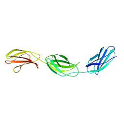
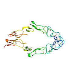
1FNH
DownloadVisualize
BU of 1fnh by Molmil
CRYSTAL STRUCTURE OF HEPARIN AND INTEGRIN BINDING SEGMENT OF HUMAN FIBRONECTIN
分子名称: PROTEIN (FIBRONECTIN)
著者Sharma, A, Askari, J, Humphries, M, Jones, E.Y, Stuart, D.I.
登録日1999-01-28
公開日1999-03-16
最終更新日2023-12-27
実験手法X-RAY DIFFRACTION (2.8 Å)
主引用文献Crystal structure of a heparin- and integrin-binding segment of human fibronectin.
EMBO J., 18, 1999
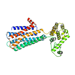
3PBL
DownloadVisualize
BU of 3pbl by Molmil
Structure of the human dopamine D3 receptor in complex with eticlopride
分子名称: 3-chloro-5-ethyl-N-{[(2S)-1-ethylpyrrolidin-2-yl]methyl}-6-hydroxy-2-methoxybenzamide, D(3) dopamine receptor, Lysozyme chimera, ...
著者Chien, E.Y.T, Liu, W, Han, G.W, Katritch, V, Zhao, Q, Cherezov, V, Stevens, R.C, Accelerated Technologies Center for Gene to 3D Structure (ATCG3D), GPCR Network (GPCR)
登録日2010-10-20
公開日2010-11-03
最終更新日2023-09-06
実験手法X-RAY DIFFRACTION (2.89 Å)
主引用文献Structure of the human dopamine d3 receptor in complex with a d2/d3 selective antagonist.
Science, 330, 2010
3OCS
DownloadVisualize
BU of 3ocs by Molmil
Crystal structure of bruton's tyrosine kinase in complex with inhibitor CGI1746
分子名称: 4-tert-butyl-N-[2-methyl-3-(4-methyl-6-{[4-(morpholin-4-ylcarbonyl)phenyl]amino}-5-oxo-4,5-dihydropyrazin-2-yl)phenyl]benzamide, BETA-MERCAPTOETHANOL, SULFATE ION, ...
著者Davies, D.R, Gallion, S.L, Staker, B.L.
登録日2010-08-10
公開日2010-11-03
最終更新日2023-09-06
実験手法X-RAY DIFFRACTION (1.8 Å)
主引用文献A novel, specific Btk inhibitor antagonizes BCR and Fc[gamma]R signaling and suppresses inflammatory arthritis
To be Published
1FT4
DownloadVisualize
BU of 1ft4 by Molmil
PHOTOCHEMICALLY-ENHANCED BINDING OF SMALL MOLECULES TO THE TUMOR NECROSIS FACTOR RECEPTOR-1
分子名称: 5-(3-MORPHOLIN-4-YL-PROPYL)-2-(3-NITRO-PHENYL)-4-THIOXO-4,5-DIHYDRO-1-THIA-3B,5-DIAZA-CYCLOPENTA[A]PENTALEN-6-ONE, SOLUBLE TUMOR NECROSIS FACTOR RECEPTOR 1
著者Muckelbauer, J.K, Chang, C.-H.
登録日2000-09-11
公開日2001-10-12
最終更新日2017-10-04
実験手法X-RAY DIFFRACTION (2.9 Å)
主引用文献Photochemically enhanced binding of small molecules to the tumor necrosis factor receptor-1 inhibits the binding of TNF-alpha.
Proc.Natl.Acad.Sci.USA, 98, 2001
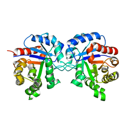
1TRE
DownloadVisualize
BU of 1tre by Molmil
THE STRUCTURE OF TRIOSEPHOSPHATE ISOMERASE FROM ESCHERICHIA COLI DETERMINED AT 2.6 ANGSTROM RESOLUTION
分子名称: TRIOSEPHOSPHATE ISOMERASE
著者Noble, M.E.M, Wierenga, R.K.
登録日1992-10-12
公開日1993-10-31
最終更新日2024-02-14
実験手法X-RAY DIFFRACTION (2.6 Å)
主引用文献Structure of triosephosphate isomerase from Escherichia coli determined at 2.6 A resolution.
Acta Crystallogr.,Sect.D, 49, 1993
1VGX
DownloadVisualize
BU of 1vgx by Molmil
Crystal structure of a autoinducer-2 synthesis protein
分子名称: S-ribosylhomocysteinase, ZINC ION
著者Structural GenomiX
登録日2003-11-03
公開日2003-11-25
最終更新日2023-12-27
実験手法X-RAY DIFFRACTION (1.9 Å)
主引用文献Structural analysis of a set of proteins resulting from a bacterial genomics project
Proteins, 60, 2005

225399

件を2024-09-25に公開中

PDB statisticsPDBj update infoContact PDBjnumon