4HB2
 
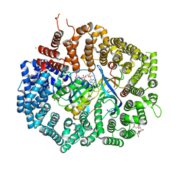
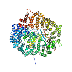 | | Crystal structure of CRM1-Ran-RanBP1 | | 分子名称: | 1,2-ETHANEDIOL, CHLORIDE ION, Exportin-1, ... | | 著者 | Sun, Q, Chook, Y.M. | | 登録日 | 2012-09-27 | | 公開日 | 2013-01-09 | | 最終更新日 | 2024-02-28 | | 実験手法 | X-RAY DIFFRACTION (1.8 Å) | | 主引用文献 | Nuclear export inhibition through covalent conjugation and hydrolysis of Leptomycin B by CRM1.
Proc.Natl.Acad.Sci.USA, 110, 2013
|
|
4HAU
 
 | |
7KBQ
 
 | |
7K15
 
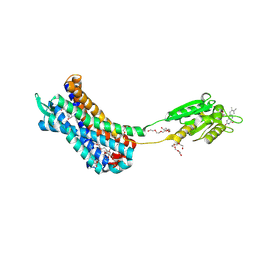 | | Crystal structure of the Human Leukotriene B4 Receptor 1 in Complex with Selective Antagonist MK-D-046 | | 分子名称: | (2R)-2,3-dihydroxypropyl (9Z)-octadec-9-enoate, FLAVIN MONONUCLEOTIDE, HEXAETHYLENE GLYCOL, ... | | 著者 | Michaelian, N, Han, G.W, Cherezov, V. | | 登録日 | 2020-09-07 | | 公開日 | 2021-02-17 | | 最終更新日 | 2024-10-16 | | 実験手法 | X-RAY DIFFRACTION (2.88 Å) | | 主引用文献 | Structural insights on ligand recognition at the human leukotriene B4 receptor 1.
Nat Commun, 12, 2021
|
|
5F0V
 
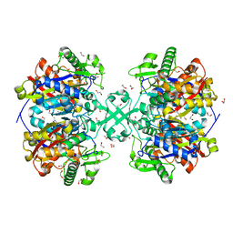 | | X-ray crystal structure of a thiolase from Escherichia coli at 1.8 A resolution | | 分子名称: | 1,2-ETHANEDIOL, Acetyl-CoA acetyltransferase | | 著者 | Ithayaraja, M, Neelanjana, J, Wierenga, R, Savithri, H.S, Murthy, M.R.N. | | 登録日 | 2015-11-28 | | 公開日 | 2016-07-13 | | 最終更新日 | 2023-11-08 | | 実験手法 | X-RAY DIFFRACTION (1.8 Å) | | 主引用文献 | Crystal structure of a thiolase from Escherichia coli at 1.8 angstrom resolution.
Acta Crystallogr.,Sect.F, 72, 2016
|
|
5F38
 
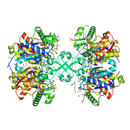 | | X-ray crystal structure of a thiolase from Escherichia coli at 1.8 A resolution | | 分子名称: | 1,2-ETHANEDIOL, Acetyl-CoA acetyltransferase, COENZYME A, ... | | 著者 | Ithayaraja, M, Neelanjana, J, Wierenga, R, Savithri, H.S, Murthy, M.R.N. | | 登録日 | 2015-12-02 | | 公開日 | 2016-07-13 | | 最終更新日 | 2023-11-08 | | 実験手法 | X-RAY DIFFRACTION (1.9 Å) | | 主引用文献 | Crystal structure of a thiolase from Escherichia coli at 1.8 angstrom resolution.
Acta Crystallogr.,Sect.F, 72, 2016
|
|
1AOD
 
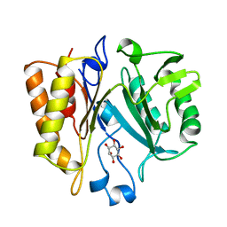 | | PHOSPHATIDYLINOSITOL-SPECIFIC PHOSPHOLIPASE C FROM LISTERIA MONOCYTOGENES | | 分子名称: | 1,2,3,4,5,6-HEXAHYDROXY-CYCLOHEXANE, PHOSPHATIDYLINOSITOL-SPECIFIC PHOSPHOLIPASE C | | 著者 | Heinz, D.W, Moser, J. | | 登録日 | 1997-07-02 | | 公開日 | 1998-01-07 | | 最終更新日 | 2024-05-22 | | 実験手法 | X-RAY DIFFRACTION (2.6 Å) | | 主引用文献 | Crystal structure of the phosphatidylinositol-specific phospholipase C from the human pathogen Listeria monocytogenes.
J.Mol.Biol., 273, 1997
|
|
1T1F
 
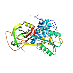 | |
1TB4
 
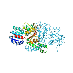 | |
1BRX
 
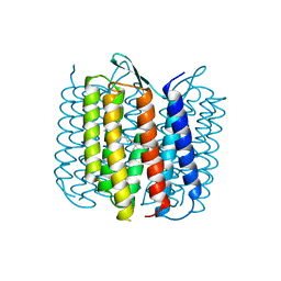 | |
1TA4
 
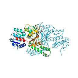 | |
1QC6
 
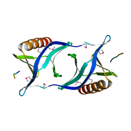 | | EVH1 domain from ENA/VASP-like protein in complex with ACTA peptide | | 分子名称: | EVH1 DOMAIN FROM ENA/VASP-LIKE PROTEIN, PHE-GLU-PHE-PRO-PRO-PRO-PRO-THR-ASP-GLU-GLU | | 著者 | Fedorov, A.A, Fedorov, E.V, Gertler, F.B, Almo, S.C. | | 登録日 | 1999-05-17 | | 公開日 | 1999-05-25 | | 最終更新日 | 2018-02-07 | | 実験手法 | X-RAY DIFFRACTION (2.6 Å) | | 主引用文献 | Structure of EVH1, a novel proline-rich ligand-binding module involved in cytoskeletal dynamics and neural function
Nat.Struct.Biol., 6, 1999
|
|
8S85
 
 | | Crystal structure of JAK1 JH1 domain in complex with an inhibitor | | 分子名称: | SODIUM ION, Tyrosine-protein kinase JAK1, ~{N}-(2-ethylphenyl)-~{N},1-dimethyl-imidazo[4,5-c]pyridin-6-amine | | 著者 | Flower, T.F, Lopez-Ramos, M, Geney, R, Jestel, A. | | 登録日 | 2024-03-05 | | 公開日 | 2024-11-06 | | 実験手法 | X-RAY DIFFRACTION (1.75 Å) | | 主引用文献 | Design of a potent and selective dual JAK1/TYK2 inhibitor.
Bioorg.Med.Chem., 114, 2024
|
|
1BVA
 
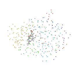 | |
