3E3S
 
 | | Structure of thaumatin with the magic triangle I3C | | 分子名称: | 5-amino-2,4,6-triiodobenzene-1,3-dicarboxylic acid, L(+)-TARTARIC ACID, POTASSIUM ION, ... | | 著者 | Beck, T, Gruene, T, Sheldrick, G.M. | | 登録日 | 2008-08-08 | | 公開日 | 2008-10-28 | | 最終更新日 | 2017-10-25 | | 実験手法 | X-RAY DIFFRACTION (1.73 Å) | | 主引用文献 | A magic triangle for experimental phasing of macromolecules
Acta Crystallogr.,Sect.D, 64, 2008
|
|
3E3D
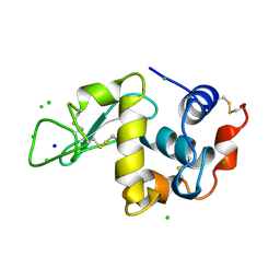
 | | Structure of hen egg white lysozyme with the magic triangle I3C | | 分子名称: | 4-(2-HYDROXYETHYL)-1-PIPERAZINE ETHANESULFONIC ACID, 5-amino-2,4,6-triiodobenzene-1,3-dicarboxylic acid, Lysozyme C | | 著者 | Beck, T, Gruene, T, Sheldrick, G.M. | | 登録日 | 2008-08-07 | | 公開日 | 2008-10-28 | | 最終更新日 | 2017-10-25 | | 実験手法 | X-RAY DIFFRACTION (1.55 Å) | | 主引用文献 | A magic triangle for experimental phasing of macromolecules
Acta Crystallogr.,Sect.D, 64, 2008
|
|
3E3T
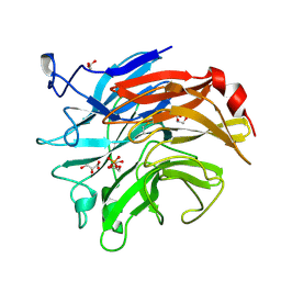
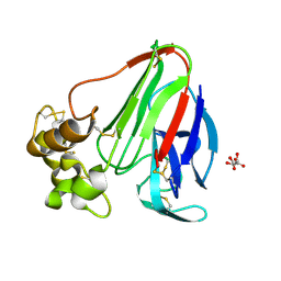
 | | Structure of porcine pancreatic elastase with the magic triangle I3C | | 分子名称: | 5-amino-2,4,6-triiodobenzene-1,3-dicarboxylic acid, Elastase-1, IODIDE ION, ... | | 著者 | Beck, T, Gruene, T, Sheldrick, G.M. | | 登録日 | 2008-08-08 | | 公開日 | 2008-10-28 | | 最終更新日 | 2012-04-11 | | 実験手法 | X-RAY DIFFRACTION (1.6 Å) | | 主引用文献 | A magic triangle for experimental phasing of macromolecules
Acta Crystallogr.,Sect.D, 64, 2008
|
|
1VTR
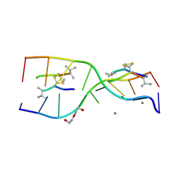
 | | STRUCTURE OF THE DEOXYTETRANUCLEOTIDE D-PAPTPAPT AND A SEQUENCE-DEPENDENT MODEL FOR POLY(DA-DT) | | 分子名称: | DNA (5'-D(*AP*TP*AP*T)-3') | | 著者 | Viswamitra, M.A, Shakked, Z, Jones, P.G, Sheldrick, G.M, Salisbury, S.A, Kennard, O. | | 登録日 | 1988-08-18 | | 公開日 | 2011-07-13 | | 最終更新日 | 2023-12-27 | | 実験手法 | X-RAY DIFFRACTION (1.04 Å) | | 主引用文献 | Structure of the Deoxytetranucleotide d-pApTpApT and a Sequence-Dependent Model for Poly(dA-dT)
Biopolymers, 21, 1982
|
|
3JU4
 | | Crystal Structure Analysis of EndosialidaseNF at 0.98 A Resolution | | 分子名称: | CHLORIDE ION, Endo-N-acetylneuraminidase, N-acetyl-beta-neuraminic acid, ... | | 著者 | Schulz, E.C, Neuman, P, Gerardy-Schahn, R, Sheldrick, G.M, Ficner, R. | | 登録日 | 2009-09-14 | | 公開日 | 2010-02-02 | | 最終更新日 | 2023-11-01 | | 実験手法 | X-RAY DIFFRACTION (0.98 Å) | | 主引用文献 | Structure analysis of endosialidase NF at 0.98 A resolution.
Acta Crystallogr.,Sect.D, 66, 2010
|
|
2J80
 | | Structure of Citrate-bound Periplasmic Domain of Sensor Histidine Kinase CitA | | 分子名称: | CITRATE ANION, GLYCEROL, SENSOR KINASE CITA, ... | | 著者 | Sevvana, M, Vijayan, V, Zweckstetter, M, Reinelt, S, Madden, D.R, Sheldrick, G.M, Bott, M, Griesinger, C, Becker, S. | | 登録日 | 2006-10-18 | | 公開日 | 2007-10-23 | | 最終更新日 | 2019-05-08 | | 実験手法 | X-RAY DIFFRACTION (1.6 Å) | | 主引用文献 | A Ligand-Induced Switch in the Periplasmic Domain of Sensor Histidine Kinase Cita.
J.Mol.Biol., 377, 2008
|
|
2J8T
 | | Human aldose reductase in complex with NADP and citrate at 0.82 angstrom | | 分子名称: | ALDO-KETO REDUCTASE FAMILY 1, MEMBER B1, CITRATE ANION, ... | | 著者 | Biadene, M, Hazemann, I, Cousido, A, Ginell, S, Sheldrick, G.M, Podjarny, A, Schneider, T.R. | | 登録日 | 2006-10-27 | | 公開日 | 2007-05-29 | | 最終更新日 | 2023-12-13 | | 実験手法 | X-RAY DIFFRACTION (0.82 Å) | | 主引用文献 | The Atomic Resolution Structure of Human Aldose Reductase Reveals that Rearrangement of a Bound Ligand Allows the Opening of the Safety-Belt Loop.
Acta Crystallogr.,Sect.D, 63, 2007
|
|
3SIL
 | |
3SZS
 | |
3UFE
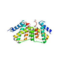 | | Structure of transcriptional antiterminator (BGLG-family) at 1.5 A resolution | | 分子名称: | CHLORIDE ION, GLYCEROL, PHOSPHATE ION, ... | | 著者 | Grosse, C, Himmel, S, Becker, S, Sheldrick, G.M, Uson, I. | | 登録日 | 2011-11-01 | | 公開日 | 2012-02-01 | | 最終更新日 | 2023-09-13 | | 実験手法 | X-RAY DIFFRACTION (1.5 Å) | | 主引用文献 | Structure of transcriptional antiterminator (BGLG-family) at 1.5 A resolution
To be Published
|
|
2C0F
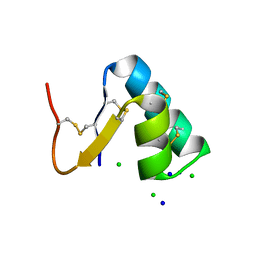
 | | Structure of Wind Y53F mutant | | 分子名称: | WINDBEUTEL PROTEIN | | 著者 | Sevvana, M, Ma, Q, Barnewitz, K, Guo, C, Soling, H.-D, Ferrari, D.M, Sheldrick, G.M. | | 登録日 | 2005-09-02 | | 公開日 | 2006-08-29 | | 最終更新日 | 2023-12-13 | | 実験手法 | X-RAY DIFFRACTION (2.28 Å) | | 主引用文献 | Structural Elucidation of the Pdi-Related Chaperone Wind with the Help of Mutants.
Acta Crystallogr.,Sect.D, 62, 2006
|
|
2C0G
 | | Structure of PDI-related Chaperone, Wind mutant-Y53S | | 分子名称: | CHLORIDE ION, SODIUM ION, WINDBEUTEL PROTEIN | | 著者 | Sevvana, M, Ma, Q, Barnewitz, K, Guo, C, Soling, H.-D, Ferrari, D.M, Sheldrick, G.M. | | 登録日 | 2005-09-02 | | 公開日 | 2006-08-29 | | 最終更新日 | 2023-12-13 | | 実験手法 | X-RAY DIFFRACTION (1.75 Å) | | 主引用文献 | Structural Elucidation of the Pdi-Related Chaperone Wind with the Help of Mutants.
Acta Crystallogr.,Sect.D, 62, 2006
|
|
2C0E
 | | Structure of PDI-related Chaperone, Wind with his-tag on C-terminus. | | 分子名称: | WINDBEUTEL PROTEIN | | 著者 | Sevvana, M, Ma, Q, Barnewitz, K, Guo, C, Soling, H.-D, Ferrari, D.M, Sheldrick, G.M. | | 登録日 | 2005-09-01 | | 公開日 | 2006-08-29 | | 最終更新日 | 2023-12-13 | | 実験手法 | X-RAY DIFFRACTION (2.35 Å) | | 主引用文献 | Structural Elucidation of the Pdi-Related Chaperone Wind with the Help of Mutants.
Acta Crystallogr.,Sect.D, 62, 2006
|
|
2C1Y
 | | Structure of PDI-related Chaperone, Wind mutant-Y55K | | 分子名称: | WINDBEUTEL PROTEIN | | 著者 | Sevvana, M, Ma, Q, Barnewitz, K, Guo, C, Soling, H.-D, Ferrari, D.M, Sheldrick, G.M. | | 登録日 | 2005-09-22 | | 公開日 | 2006-08-29 | | 最終更新日 | 2023-12-13 | | 実験手法 | X-RAY DIFFRACTION (2.25 Å) | | 主引用文献 | Structural Elucidation of the Pdi-Related Chaperone Wind with the Help of Mutants.
Acta Crystallogr.,Sect.D, 62, 2006
|
|
3MBS
 | | Crystal structure of 8mer PNA | | 分子名称: | 1,2-ETHANEDIOL, Peptide Nucleic Acid | | 著者 | Yeh, J.I, Pohl, E, Truan, D, He, W, Sheldrick, G.M, Achim, C. | | 登録日 | 2010-03-26 | | 公開日 | 2011-03-30 | | 最終更新日 | 2023-11-15 | | 実験手法 | X-RAY DIFFRACTION (1.27 Å) | | 主引用文献 | The crystal structure of non-modified and bipyridine-modified PNA duplexes.
Chemistry, 16, 2010
|
|
3EE6
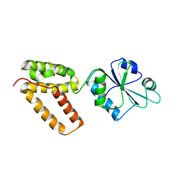
 | | Crystal Structure Analysis of Tripeptidyl peptidase -I | | 分子名称: | 2-acetamido-2-deoxy-beta-D-glucopyranose, CALCIUM ION, CHLORIDE ION, ... | | 著者 | Pal, A, Kraetzner, R, Grapp, M, Gruene, T, Schreiber, K, Granborg, M, Urlaub, H, Asif, A.R, Becker, S, Gartner, J, Sheldrick, G.M, Steinfeld, R. | | 登録日 | 2008-09-04 | | 公開日 | 2008-11-25 | | 最終更新日 | 2020-07-29 | | 実験手法 | X-RAY DIFFRACTION (2.35 Å) | | 主引用文献 | Structure of tripeptidyl-peptidase I provides insight into the molecular basis of late infantile neuronal ceroid lipofuscinosis
J.Biol.Chem., 284, 2009
|
|
3DU1
 | |
3GO3
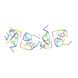
 | | Interactions of an echinomycin-DNA complex with manganese(II) ions | | 分子名称: | 2-CARBOXYQUINOXALINE, 5'-D(*AP*CP*GP*TP*AP*CP*GP*T)-3', DI(HYDROXYETHYL)ETHER, ... | | 著者 | Pfoh, R, Cuesta-Seijo, J.A, Sheldrick, G.M. | | 登録日 | 2009-03-18 | | 公開日 | 2009-03-31 | | 最終更新日 | 2012-12-12 | | 実験手法 | X-RAY DIFFRACTION (1.1 Å) | | 主引用文献 | Interaction of an Echinomycin-DNA Complex with Manganese Ion
Acta Crystallogr.,Sect.F, 65, 2009
|
|
3GWH
 | | Crystallographic Ab Initio protein solution far below atomic resolution | | 分子名称: | PHOSPHATE ION, Transcriptional antiterminator (BglG family) | | 著者 | Rodriguez, D.D, Grosse, C, Himmel, S, Gonzalez, C, Becker, S, Sheldrick, G.M, Uson, I. | | 登録日 | 2009-04-01 | | 公開日 | 2010-04-07 | | 最終更新日 | 2024-03-20 | | 実験手法 | X-RAY DIFFRACTION (1.95 Å) | | 主引用文献 | Crystallographic ab initio protein structure solution below atomic resolution
Nat.Methods, 6, 2009
|
|
1LU0
 | | Atomic Resolution Structure of Squash Trypsin Inhibitor: Unexpected Metal Coordination | | 分子名称: | (4R)-2-METHYLPENTANE-2,4-DIOL, GLYCEROL, SULFATE ION, ... | | 著者 | Thaimattam, R, Tykarska, E, Bierzynski, A, Sheldrick, G.M, Jaskolski, M. | | 登録日 | 2002-05-21 | | 公開日 | 2002-08-28 | | 最終更新日 | 2021-10-27 | | 実験手法 | X-RAY DIFFRACTION (1.03 Å) | | 主引用文献 | Atomic resolution structure of squash trypsin inhibitor: unexpected metal coordination.
Acta Crystallogr.,Sect.D, 58, 2002
|
|
1LZ8
 | | LYSOZYME PHASED ON ANOMALOUS SIGNAL OF SULFURS AND CHLORINES | | 分子名称: | CHLORIDE ION, PROTEIN (LYSOZYME), SODIUM ION | | 著者 | Dauter, Z, Dauter, M, De La Fortelle, E, Bricogne, G, Sheldrick, G.M. | | 登録日 | 1999-03-14 | | 公開日 | 1999-05-26 | | 最終更新日 | 2023-12-27 | | 実験手法 | X-RAY DIFFRACTION (1.53 Å) | | 主引用文献 | Can anomalous signal of sulfur become a tool for solving protein crystal structures?
J.Mol.Biol., 289, 1999
|
|
1M32
 | | Crystal Structure of 2-aminoethylphosphonate Transaminase | | 分子名称: | 2-aminoethylphosphonate-pyruvate aminotransferase, PHOSPHATE ION, PHOSPHONOACETALDEHYDE, ... | | 著者 | Chen, C.C.H, Zhang, H, Kim, A.D, Howard, A, Sheldrick, G.M, Mariano-Dunnaway, D, Herzberg, O. | | 登録日 | 2002-06-26 | | 公開日 | 2002-11-20 | | 最終更新日 | 2017-10-11 | | 実験手法 | X-RAY DIFFRACTION (2.2 Å) | | 主引用文献 | Degradation Pathway of the Phosphonate Ciliatine: Crystal Structure of 2-Aminoethylphosphonate Transaminase
Biochemistry, 41, 2002
|
|
1RQW
 | |
1UNJ
 | | Crystal structure of a 7-Aminoactinomycin D complex with non-complementary DNA | | 分子名称: | 5'-D(*TP*TP*AP*GP*BRU*TP)-3', 7-AMINO-ACTINOMYCIN D | | 著者 | Alexopoulos, E.C, Klement, R, Jares-Erijman, E.A, Uson, I, Jovin, T.M, Sheldrick, G.M. | | 登録日 | 2003-09-10 | | 公開日 | 2004-12-16 | | 最終更新日 | 2024-07-10 | | 実験手法 | X-RAY DIFFRACTION (2.5 Å) | | 主引用文献 | Crystal and Solution Structures of 7-Amino-Actinomycin D Complexes with D(Ttagbrut), D(Ttagtt) and D(Tttagttt)
Acta Crystallogr.,Sect.D, 61, 2005
|
|
1UNM
 | | Crystal structure of 7-Aminoactinomycin D with non-complementary DNA | | 分子名称: | 5'-D(*TP*TP*AP*GP*BRU*TP)-3', 7-AMINOACTINOMYCIN D | | 著者 | Alexopoulos, E.C, Klement, R, Jares-Erijman, E.A, Uson, I, Jovin, T.M, Sheldrick, G.M. | | 登録日 | 2003-09-11 | | 公開日 | 2004-09-24 | | 最終更新日 | 2024-07-10 | | 実験手法 | X-RAY DIFFRACTION (2 Å) | | 主引用文献 | Crystal and Solution Structures of 7-Amino-Actinomycin D Complexes with D(Ttagbrut), D(Ttagtt) and D(Tttagttt)
Acta Crystallogr.,Sect.D, 61, 2005
|
|


