3O4B
 
 | |
3O4A
 | |
3O3Q
 | |
3O49
 | |
3OL0
 | |
3O4D
 | |
3OGF
 | |
3PD7
 | |
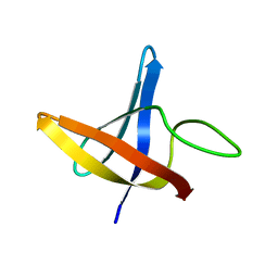
2LXJ
 | | Backbone 1H, 13C, and 15N Chemical Shift Assignments for cold shock protein, LmCsp with dT7 | | 分子名称: | Cold shock-like protein CspLA | | 著者 | Lee, J, Jeong, K, Kim, Y. | | 登録日 | 2012-08-27 | | 公開日 | 2013-08-07 | | 最終更新日 | 2024-05-15 | | 実験手法 | SOLUTION NMR | | 主引用文献 | Structural and Dynamic Features of Cold-Shock Proteins of Listeriamonocytogenes, a Psychrophilic Bacterium
Biochemistry, 52, 2013
|
|
2LXK
 | | Backbone 1H, 13C, and 15N Chemical Shift Assignments for cold shock protein, LmCsp | | 分子名称: | Cold shock-like protein CspLA | | 著者 | Lee, J, Jeong, K, Kim, Y. | | 登録日 | 2012-08-27 | | 公開日 | 2013-08-07 | | 最終更新日 | 2024-05-15 | | 実験手法 | SOLUTION NMR | | 主引用文献 | Structural and Dynamic Features of Cold-Shock Proteins of Listeriamonocytogenes, a Psychrophilic Bacterium
Biochemistry, 52, 2013
|
|
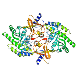
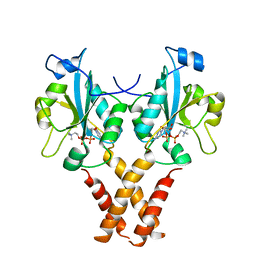
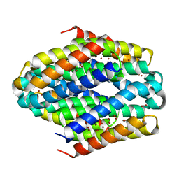
4MVD
 | | Crystal Structure of a Mammalian Cytidylyltransferase | | 分子名称: | Choline-phosphate cytidylyltransferase A, [2-CYTIDYLATE-O'-PHOSPHONYLOXYL]-ETHYL-TRIMETHYL-AMMONIUM | | 著者 | Lee, J, Cornell, R.B. | | 登録日 | 2013-09-23 | | 公開日 | 2013-12-11 | | 最終更新日 | 2024-02-28 | | 実験手法 | X-RAY DIFFRACTION (8 Å) | | 主引用文献 | Structural Basis for Autoinhibition of CTP:Phosphocholine Cytidylyltransferase (CCT), the Regulatory Enzyme in Phosphatidylcholine Synthesis, by Its Membrane-binding Amphipathic Helix.
J.Biol.Chem., 289, 2014
|
|
2PLK
 | |
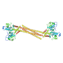
5T42
 | | Structure of the Ebola virus envelope protein MPER/TM domain and its interaction with the fusion loop explains their fusion activity | | 分子名称: | Envelope glycoprotein | | 著者 | Lee, J, Nyenhuis, D.A, Nelson, E.A, Cafiso, D.S, White, J.M, Tamm, L.K. | | 登録日 | 2016-08-28 | | 公開日 | 2017-08-30 | | 最終更新日 | 2024-05-15 | | 実験手法 | SOLUTION NMR | | 主引用文献 | Structure of the Ebola virus envelope protein MPER/TM domain and its interaction with the fusion loop explains their fusion activity.
Proc. Natl. Acad. Sci. U.S.A., 114, 2017
|
|
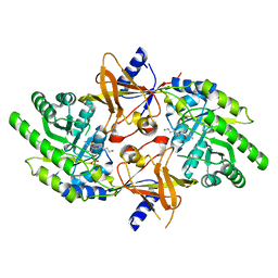
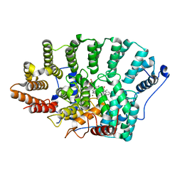
4MVC
 | | Crystal Structure of a Mammalian Cytidylyltransferase | | 分子名称: | Choline-phosphate cytidylyltransferase A, [2-CYTIDYLATE-O'-PHOSPHONYLOXYL]-ETHYL-TRIMETHYL-AMMONIUM | | 著者 | Lee, J, Cornell, R.B. | | 登録日 | 2013-09-23 | | 公開日 | 2013-12-11 | | 最終更新日 | 2023-09-20 | | 実験手法 | X-RAY DIFFRACTION (3 Å) | | 主引用文献 | Structural Basis for Autoinhibition of CTP:Phosphocholine Cytidylyltransferase (CCT), the Regulatory Enzyme in Phosphatidylcholine Synthesis, by Its Membrane-binding Amphipathic Helix.
J.Biol.Chem., 289, 2014
|
|
2PLJ
 | |
7Y7O
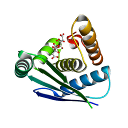 | |
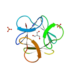
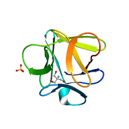
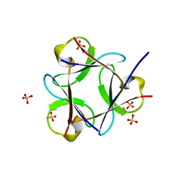
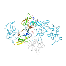
2QQR
 | | JMJD2A hybrid tudor domains | | 分子名称: | JmjC domain-containing histone demethylation protein 3A, SULFATE ION | | 著者 | Lee, J, Botuyan, M.V, Mer, G. | | 登録日 | 2007-07-26 | | 公開日 | 2007-12-11 | | 最終更新日 | 2023-11-15 | | 実験手法 | X-RAY DIFFRACTION (1.8 Å) | | 主引用文献 | Distinct binding modes specify the recognition of methylated histones H3K4 and H4K20 by JMJD2A-tudor.
Nat.Struct.Mol.Biol., 15, 2008
|
|
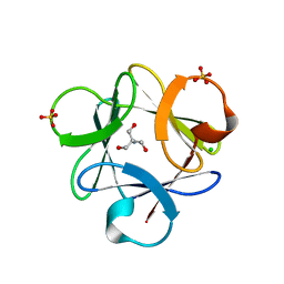
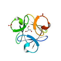
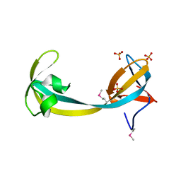
2G3R
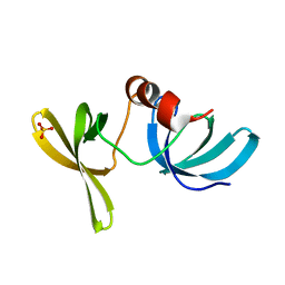 | | Crystal Structure of 53BP1 tandem tudor domains at 1.2 A resolution | | 分子名称: | SULFATE ION, Tumor suppressor p53-binding protein 1 | | 著者 | Lee, J, Botuyan, M.V, Thompson, J.R, Mer, G. | | 登録日 | 2006-02-20 | | 公開日 | 2007-01-02 | | 最終更新日 | 2023-08-30 | | 実験手法 | X-RAY DIFFRACTION (1.25 Å) | | 主引用文献 | Structural Basis for the Methylation State-Specific Recognition of Histone H4-K20 by 53BP1 and Crb2 in DNA Repair.
Cell(Cambridge,Mass.), 127, 2006
|
|
6ZIF
 | |
2F0Y
 | |
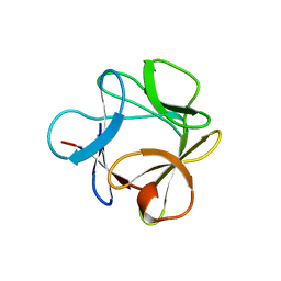
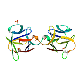
2FHD
 | | Crystal structure of Crb2 tandem tudor domains | | 分子名称: | DNA repair protein rhp9/CRB2, PHOSPHATE ION | | 著者 | Lee, J, Botuyan, M.V, Thompson, J.R, Mer, G. | | 登録日 | 2005-12-23 | | 公開日 | 2007-01-02 | | 最終更新日 | 2011-07-13 | | 実験手法 | X-RAY DIFFRACTION (2.4 Å) | | 主引用文献 | Structural basis for the methylation state-specific recognition of histone H4-K20 by 53BP1 and Crb2 in DNA repair.
Cell(Cambridge,Mass.), 127, 2006
|
|
3HAL
 | |
3BA5
 | |
3BAD
 | |
3BA4
 | |


