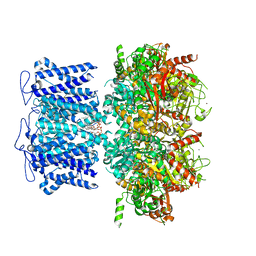
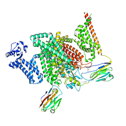
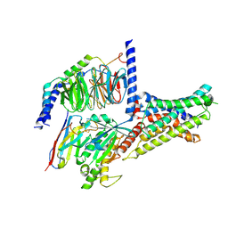
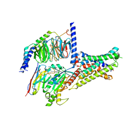
8JT9
 
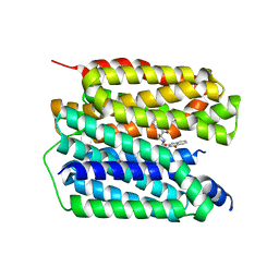 | | Human VMAT2 complex with ketanserin | | 分子名称: | 3-[2-[4-(4-fluorophenyl)carbonylpiperidin-1-yl]ethyl]-1~{H}-quinazoline-2,4-dione, Synaptic vesicular amine transporter | | 著者 | Jiang, D.H, Wu, D. | | 登録日 | 2023-06-21 | | 公開日 | 2023-11-29 | | 最終更新日 | 2024-02-21 | | 実験手法 | ELECTRON MICROSCOPY (2.97 Å) | | 主引用文献 | Transport and inhibition mechanisms of human VMAT2.
Nature, 626, 2024
|
|
6O35
 | |
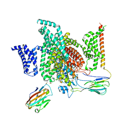
8J6O
 | | transport T2 | | 分子名称: | 2-acetamido-2-deoxy-beta-D-glucopyranose, 2-acetamido-2-deoxy-beta-D-glucopyranose-(1-4)-2-acetamido-2-deoxy-beta-D-glucopyranose, Green fluorescent protein (Fragment),SID1 transmembrane family member 2, ... | | 著者 | Jiang, D.H, Zhang, J.T. | | 登録日 | 2023-04-26 | | 公開日 | 2024-05-01 | | 最終更新日 | 2024-07-31 | | 実験手法 | ELECTRON MICROSCOPY (3.25 Å) | | 主引用文献 | Structural insights into double-stranded RNA recognition and transport by SID-1.
Nat.Struct.Mol.Biol., 31, 2024
|
|
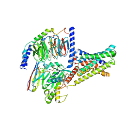
8J6M
 | | SIDT1 protein | | 分子名称: | CHOLESTEROL, Green fluorescent protein,SID1 transmembrane family member 1, OLEIC ACID, ... | | 著者 | Zhang, J.T, Jiang, D.H. | | 登録日 | 2023-04-26 | | 公開日 | 2024-05-01 | | 最終更新日 | 2024-07-31 | | 実験手法 | ELECTRON MICROSCOPY (2.77 Å) | | 主引用文献 | Structural insights into double-stranded RNA recognition and transport by SID-1.
Nat.Struct.Mol.Biol., 31, 2024
|
|
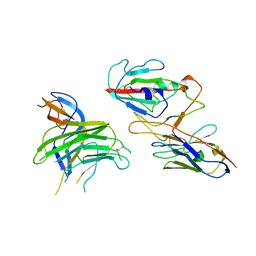
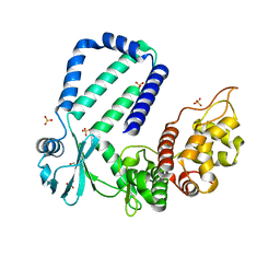
4PZA
 | | The complex structure of mycobacterial glucosyl-3-phosphoglycerate phosphatase Rv2419c with inorganic phosphate | | 分子名称: | Glucosyl-3-phosphoglycerate phosphatase, PHOSPHATE ION | | 著者 | Zhou, W.H, Zheng, Q.Q, Jiang, D.Q, Zhang, W, Zhang, Q.Q, Jin, J, Li, X, Yang, H.T, Shaw, N, Rao, Z. | | 登録日 | 2014-03-29 | | 公開日 | 2014-06-11 | | 最終更新日 | 2023-11-08 | | 実験手法 | X-RAY DIFFRACTION (1.776 Å) | | 主引用文献 | Mechanism of dephosphorylation of glucosyl-3-phosphoglycerate by a histidine phosphatase
J.Biol.Chem., 289, 2014
|
|
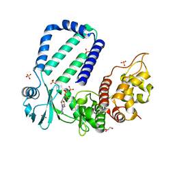
4PZ9
 | | The native structure of mycobacterial glucosyl-3-phosphoglycerate phosphatase Rv2419c | | 分子名称: | Glucosyl-3-phosphoglycerate phosphatase | | 著者 | Zhou, W.H, Zheng, Q.Q, Jiang, D.Q, Zhang, W, Zhang, Q.Q, Jin, J, Li, X, Yang, H.T, Shaw, N, Rao, Z. | | 登録日 | 2014-03-28 | | 公開日 | 2014-06-11 | | 最終更新日 | 2023-11-08 | | 実験手法 | X-RAY DIFFRACTION (1.94 Å) | | 主引用文献 | Mechanism of dephosphorylation of glucosyl-3-phosphoglycerate by a histidine phosphatase
J.Biol.Chem., 289, 2014
|
|
4QIH
 | | The structure of mycobacterial glucosyl-3-phosphoglycerate phosphatase Rv2419c complexes with VO3 | | 分子名称: | Glucosyl-3-phosphoglycerate phosphatase, VANADATE ION | | 著者 | Zhou, W.H, Zheng, Q.Q, Jiang, D.Q, Zhang, W, Zhang, Q.Q, Jin, J, Li, X, Yang, H.T, Shaw, N, Rao, Z. | | 登録日 | 2014-05-30 | | 公開日 | 2014-06-11 | | 最終更新日 | 2023-11-08 | | 実験手法 | X-RAY DIFFRACTION (2.299 Å) | | 主引用文献 | Mechanism of dephosphorylation of glucosyl-3-phosphoglycerate by a histidine phosphatase
J.Biol.Chem., 289, 2014
|
|
7XRP
 | | Cryo-EM structure of SARS-CoV-2 spike protein in complex with nanobody C5G2 (localized refinement) | | 分子名称: | 2-acetamido-2-deoxy-beta-D-glucopyranose, C5G2 nanobody, Spike protein S1 | | 著者 | Liu, L, Sun, H, Jiang, Y, Liu, X, Zhao, D, Zheng, Q, Li, S, Xia, N. | | 登録日 | 2022-05-11 | | 公開日 | 2022-10-05 | | 実験手法 | ELECTRON MICROSCOPY (3.88 Å) | | 主引用文献 | A potent synthetic nanobody with broad-spectrum activity neutralizes SARS-CoV-2 virus and the Omicron variant BA.1 through a unique binding mode.
J Nanobiotechnology, 20, 2022
|
|
8HKQ
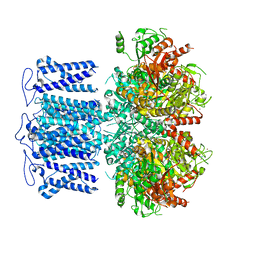 | | ion channel | | 分子名称: | POTASSIUM ION, Potassium channel subfamily T member 1, SODIUM ION, ... | | 著者 | Jiang, D.H, Zhang, J.T. | | 登録日 | 2022-11-27 | | 公開日 | 2023-08-16 | | 実験手法 | ELECTRON MICROSCOPY (2.9 Å) | | 主引用文献 | Structural basis of human Slo2.2 channel gating and modulation.
Cell Rep, 42, 2023
|
|
8HKK
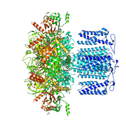 | | ion channel | | 分子名称: | POTASSIUM ION, Potassium channel subfamily T member 1, SODIUM ION, ... | | 著者 | Jiang, D.H, Zhang, J.T. | | 登録日 | 2022-11-27 | | 公開日 | 2023-08-16 | | 実験手法 | ELECTRON MICROSCOPY (2.84 Å) | | 主引用文献 | Structural basis of human Slo2.2 channel gating and modulation.
Cell Rep, 42, 2023
|
|
8HIR
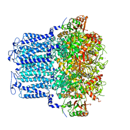 | | potassium channels | | 分子名称: | POTASSIUM ION, Potassium channel subfamily T member 1, SODIUM ION, ... | | 著者 | Jiang, D.H, Zhang, J.T. | | 登録日 | 2022-11-21 | | 公開日 | 2023-08-16 | | 実験手法 | ELECTRON MICROSCOPY (3.18 Å) | | 主引用文献 | Structural basis of human Slo2.2 channel gating and modulation.
Cell Rep, 42, 2023
|
|
8HKM
 | | ion channel | | 分子名称: | POTASSIUM ION, Potassium channel subfamily T member 1, ZINC ION, ... | | 著者 | Jiang, D.H, Zhang, J.T. | | 登録日 | 2022-11-27 | | 公開日 | 2023-08-16 | | 実験手法 | ELECTRON MICROSCOPY (2.95 Å) | | 主引用文献 | Structural basis of human Slo2.2 channel gating and modulation.
Cell Rep, 42, 2023
|
|
8HK6
 | | potassium channel | | 分子名称: | POTASSIUM ION, Potassium channel subfamily T member 1, ZINC ION | | 著者 | Jiang, D.H, Zhang, J.T. | | 登録日 | 2022-11-25 | | 公開日 | 2023-08-16 | | 実験手法 | ELECTRON MICROSCOPY (2.64 Å) | | 主引用文献 | Structural basis of human Slo2.2 channel gating and modulation.
Cell Rep, 42, 2023
|
|
8HKF
 | | ion channel | | 分子名称: | POTASSIUM ION, Potassium channel subfamily T member 1, ZINC ION | | 著者 | Jiang, D.H, Zhang, Z.T. | | 登録日 | 2022-11-25 | | 公開日 | 2023-08-16 | | 実験手法 | ELECTRON MICROSCOPY (2.66 Å) | | 主引用文献 | Structural basis of human Slo2.2 channel gating and modulation.
Cell Rep, 42, 2023
|
|
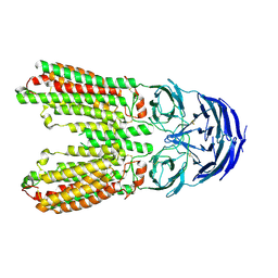
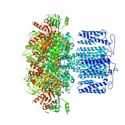
7XMF
 | | Cryo-EM structure of human NaV1.7/beta1/beta2-Nav1.7-IN2 | | 分子名称: | 2-acetamido-2-deoxy-beta-D-glucopyranose, 2-acetamido-2-deoxy-beta-D-glucopyranose-(1-4)-2-acetamido-2-deoxy-beta-D-glucopyranose, 3-[[4-[3-(4-fluoranyl-2-methyl-phenoxy)azetidin-1-yl]pyrimidin-2-yl]amino]-~{N}-methyl-benzamide, ... | | 著者 | Zhang, J.T, Jiang, D.H. | | 登録日 | 2022-04-25 | | 公開日 | 2022-11-30 | | 最終更新日 | 2022-12-28 | | 実験手法 | ELECTRON MICROSCOPY (3.07 Å) | | 主引用文献 | Structural basis for Na V 1.7 inhibition by pore blockers.
Nat.Struct.Mol.Biol., 29, 2022
|
|
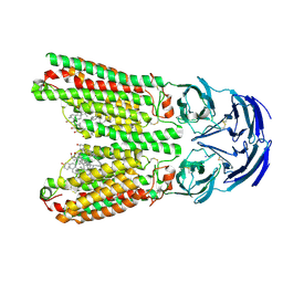
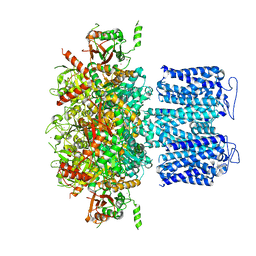
7XM9
 | | Cryo-EM structure of human NaV1.7/beta1/beta2-XEN907 | | 分子名称: | (7~{R})-1'-pentylspiro[6~{H}-furo[3,2-f][1,3]benzodioxole-7,3'-indole]-2'-one, 2-acetamido-2-deoxy-beta-D-glucopyranose, 2-acetamido-2-deoxy-beta-D-glucopyranose-(1-4)-2-acetamido-2-deoxy-beta-D-glucopyranose, ... | | 著者 | zhang, J.T, Jiang, D.H. | | 登録日 | 2022-04-25 | | 公開日 | 2022-11-30 | | 最終更新日 | 2022-12-28 | | 実験手法 | ELECTRON MICROSCOPY (3.22 Å) | | 主引用文献 | Structural basis for Na V 1.7 inhibition by pore blockers.
Nat.Struct.Mol.Biol., 29, 2022
|
|
7XMG
 | | Cryo-EM structure of human NaV1.7/beta1/beta2-TCN-1752 | | 分子名称: | (1~{Z})-~{N}-[2-methyl-3-[(~{E})-[6-[4-[[4-(trifluoromethyloxy)phenyl]methoxy]piperidin-1-yl]-1~{H}-1,3,5-triazin-2-ylidene]amino]phenyl]ethanimidic acid, 2-acetamido-2-deoxy-beta-D-glucopyranose, 2-acetamido-2-deoxy-beta-D-glucopyranose-(1-4)-2-acetamido-2-deoxy-beta-D-glucopyranose, ... | | 著者 | Jiang, D.H, Zhang, J.T. | | 登録日 | 2022-04-25 | | 公開日 | 2022-11-30 | | 最終更新日 | 2022-12-28 | | 実験手法 | ELECTRON MICROSCOPY (3.09 Å) | | 主引用文献 | Structural basis for Na V 1.7 inhibition by pore blockers.
Nat.Struct.Mol.Biol., 29, 2022
|
|
4MH1
 | |
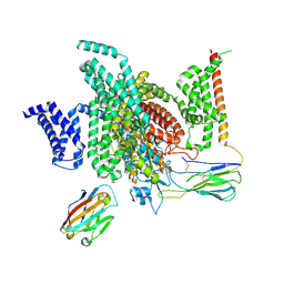
7XKE
 | | Cryo-EM structure of DHEA-ADGRG2-FL-Gs complex | | 分子名称: | 3-BETA-HYDROXY-5-ANDROSTEN-17-ONE, Adhesion G-protein coupled receptor G2, Guanine nucleotide-binding protein G(I)/G(S)/G(O) subunit gamma-2, ... | | 著者 | Guo, S.C, Xiao, P, Lin, H, Sun, J.P, Yu, X. | | 登録日 | 2022-04-19 | | 公開日 | 2022-08-10 | | 最終更新日 | 2023-03-15 | | 実験手法 | ELECTRON MICROSCOPY (2.9 Å) | | 主引用文献 | Structures of the ADGRG2-G s complex in apo and ligand-bound forms.
Nat.Chem.Biol., 18, 2022
|
|
7XKF
 | | Cryo-EM structure of DHEA-ADGRG2-BT-Gs complex at lower state | | 分子名称: | 3-BETA-HYDROXY-5-ANDROSTEN-17-ONE, Adhesion G-protein coupled receptor G2, Guanine nucleotide-binding protein G(I)/G(S)/G(O) subunit gamma-2, ... | | 著者 | Guo, S.C, Xiao, P, Lin, H, Sun, J.P, Yu, X. | | 登録日 | 2022-04-19 | | 公開日 | 2022-08-10 | | 最終更新日 | 2023-03-15 | | 実験手法 | ELECTRON MICROSCOPY (2.4 Å) | | 主引用文献 | Structures of the ADGRG2-G s complex in apo and ligand-bound forms.
Nat.Chem.Biol., 18, 2022
|
|
7XKD
 | | Cryo-EM structure of DHEA-ADGRG2-BT-Gs complex | | 分子名称: | 3-BETA-HYDROXY-5-ANDROSTEN-17-ONE, Adhesion G-protein coupled receptor G2, Guanine nucleotide-binding protein G(I)/G(S)/G(O) subunit gamma-2, ... | | 著者 | Guo, S.C, Xiao, P, Lin, H, Sun, J.P, Yu, X. | | 登録日 | 2022-04-19 | | 公開日 | 2022-08-10 | | 最終更新日 | 2023-03-15 | | 実験手法 | ELECTRON MICROSCOPY (2.4 Å) | | 主引用文献 | Structures of the ADGRG2-G s complex in apo and ligand-bound forms.
Nat.Chem.Biol., 18, 2022
|
|
5XLL
 | | Dimer form of M. tuberculosis PknI sensor domain | | 分子名称: | Serine/threonine-protein kinase PknI | | 著者 | Rao, Z, Yan, Q. | | 登録日 | 2017-05-10 | | 公開日 | 2018-05-16 | | 最終更新日 | 2021-10-27 | | 実験手法 | X-RAY DIFFRACTION (2.201 Å) | | 主引用文献 | Structural Insight into the Activation of PknI Kinase from M. tuberculosis via Dimerization of the Extracellular Sensor Domain.
Structure, 25, 2017
|
|
5XLM
 | | Monomer form of M.tuberculosis PknI sensor domain | | 分子名称: | Serine/threonine-protein kinase PknI | | 著者 | Rao, Z, Yan, Q. | | 登録日 | 2017-05-10 | | 公開日 | 2018-05-16 | | 最終更新日 | 2021-10-27 | | 実験手法 | X-RAY DIFFRACTION (2.2 Å) | | 主引用文献 | Structural Insight into the Activation of PknI Kinase from M. tuberculosis via Dimerization of the Extracellular Sensor Domain.
Structure, 25, 2017
|
|
5YJZ
 | |
5YK1
 | |


