5E95
 
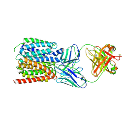
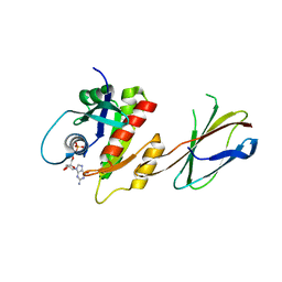 | | Crystal Structure of Mb(NS1)/H-Ras Complex | | 分子名称: | GTPase HRas, GUANOSINE-5'-DIPHOSPHATE, MAGNESIUM ION, ... | | 著者 | Eguchi, R.R, Sha, F, Gupta, A, Koide, A, Koide, S. | | 登録日 | 2015-10-14 | | 公開日 | 2016-11-02 | | 最終更新日 | 2023-09-27 | | 実験手法 | X-RAY DIFFRACTION (1.402 Å) | | 主引用文献 | Inhibition of RAS function through targeting an allosteric regulatory site.
Nat. Chem. Biol., 13, 2017
|
|
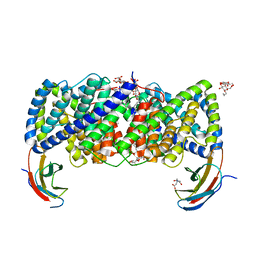
5CWU
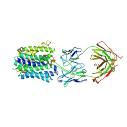
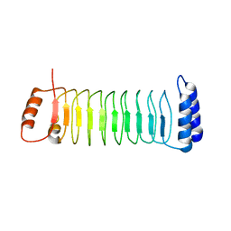
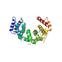 | | Crystal structure of Chaetomium thermophilum Nup188 TAIL domain | | 分子名称: | GLYCEROL, Nucleoporin NUP188 | | 著者 | Stuwe, T, Bley, C.J, Thierbach, K, Petrovic, S, Schilbach, S, Mayo, D.J, Perriches, T, Rundlet, E.J, Jeon, Y.E, Collins, L.N, Lin, D.H, Paduch, M, Koide, A, Lu, V, Fischer, J, Hurt, E, Koide, S, Kossiakoff, A.A, Hoelz, A. | | 登録日 | 2015-07-28 | | 公開日 | 2015-09-16 | | 最終更新日 | 2019-12-25 | | 実験手法 | X-RAY DIFFRACTION (3.35 Å) | | 主引用文献 | Architecture of the fungal nuclear pore inner ring complex.
Science, 350, 2015
|
|
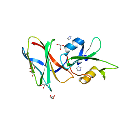
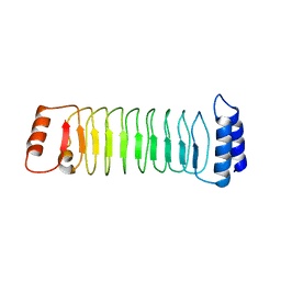
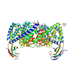
5CWV
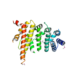 | | Crystal structure of Chaetomium thermophilum Nup192 TAIL domain | | 分子名称: | Nucleoporin NUP192 | | 著者 | Stuwe, T, Bley, C.J, Thierbach, K, Petrovic, S, Schilbach, S, Mayo, D.J, Perriches, T, Rundlet, E.J, Jeon, Y.E, Collins, L.N, Lin, D.H, Paduch, M, Koide, A, Lu, V, Fischer, J, Hurt, E, Koide, S, Kossiakoff, A.A, Hoelz, A. | | 登録日 | 2015-07-28 | | 公開日 | 2015-09-16 | | 最終更新日 | 2019-12-25 | | 実験手法 | X-RAY DIFFRACTION (3.155 Å) | | 主引用文献 | Architecture of the fungal nuclear pore inner ring complex.
Science, 350, 2015
|
|
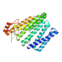
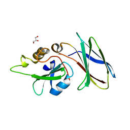
5G15
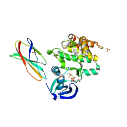 | | Structure Aurora A (122-403) bound to activating monobody Mb1 and AMPPCP | | 分子名称: | AURORA A KINASE, MAGNESIUM ION, MB1 MONOBODY, ... | | 著者 | Zorba, A, Kutter, S, Kern, D, Koide, S, Koide, A. | | 登録日 | 2016-03-23 | | 公開日 | 2018-03-14 | | 最終更新日 | 2024-01-10 | | 実験手法 | X-RAY DIFFRACTION (2.06 Å) | | 主引用文献 | Allosteric modulation of a human protein kinase with monobodies.
Proc.Natl.Acad.Sci.USA, 116, 2019
|
|
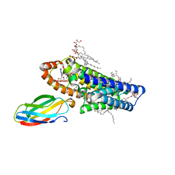
5L4K
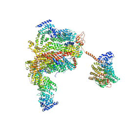 | | The human 26S proteasome lid | | 分子名称: | 26S proteasome complex subunit DSS1, 26S proteasome non-ATPase regulatory subunit 1, 26S proteasome non-ATPase regulatory subunit 11, ... | | 著者 | Schweitzer, A, Aufderheide, A, Rudack, T, Beck, F. | | 登録日 | 2016-05-25 | | 公開日 | 2016-09-07 | | 最終更新日 | 2024-05-08 | | 実験手法 | ELECTRON MICROSCOPY (3.9 Å) | | 主引用文献 | Structure of the human 26S proteasome at a resolution of 3.9 angstrom.
Proc.Natl.Acad.Sci.USA, 113, 2016
|
|
1XSO
 | | THREE-DIMENSIONAL STRUCTURE OF XENOPUS LAEVIS CU,ZN SUPEROXIDE DISMUTASE B DETERMINED BY X-RAY CRYSTALLOGRAPHY AT 1.5 ANGSTROMS RESOLUTION | | 分子名称: | COPPER (II) ION, COPPER,ZINC SUPEROXIDE DISMUTASE, ZINC ION | | 著者 | Djinovic Carugo, K, Coda, A, Battistoni, A, Carri, M.T, Polticelli, F, Desideri, A, Rotilio, G, Wilson, K.S, Bolognesi, M. | | 登録日 | 1995-03-14 | | 公開日 | 1995-07-10 | | 最終更新日 | 2019-08-14 | | 実験手法 | X-RAY DIFFRACTION (1.49 Å) | | 主引用文献 | Three-dimensional structure of Xenopus laevis Cu,Zn superoxide dismutase b determined by X-ray crystallography at 1.5 A resolution.
Acta Crystallogr.,Sect.D, 52, 1996
|
|
5L4G
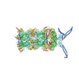 | | The human 26S proteasome at 3.9 A | | 分子名称: | 26S protease regulatory subunit 10B, 26S protease regulatory subunit 4, 26S protease regulatory subunit 6A, ... | | 著者 | Schweitzer, A, Aufderheide, A, Rudack, T, Beck, F. | | 登録日 | 2016-05-25 | | 公開日 | 2016-09-07 | | 最終更新日 | 2024-05-08 | | 実験手法 | ELECTRON MICROSCOPY (3.9 Å) | | 主引用文献 | Structure of the human 26S proteasome at a resolution of 3.9 angstrom.
Proc.Natl.Acad.Sci.USA, 113, 2016
|
|
7KZX
 | | Cryo-EM structure of YiiP-Fab complex in Apo state | | 分子名称: | Cadmium and zinc efflux pump FieF, Fab2R heavy chain, Fab2R light chain | | 著者 | Lopez-Redondo, M.L, Fan, S, Koide, A, Koide, S, Beckstein, O, Stokes, D.L. | | 登録日 | 2020-12-10 | | 公開日 | 2021-01-06 | | 最終更新日 | 2021-07-28 | | 実験手法 | ELECTRON MICROSCOPY (4 Å) | | 主引用文献 | Zinc binding alters the conformational dynamics and drives the transport cycle of the cation diffusion facilitator YiiP.
J.Gen.Physiol., 153, 2021
|
|
7KZZ
 | | Cryo-EM structure of YiiP-Fab complex in Holo state | | 分子名称: | Cadmium and zinc efflux pump FieF, Fab2R heavy chain, Fab2R light chain, ... | | 著者 | Lopez-Redondo, M.L, Fan, S, Koide, A, Koide, S, Beckstein, O, Stokes, D.L. | | 登録日 | 2020-12-10 | | 公開日 | 2021-01-06 | | 最終更新日 | 2021-07-28 | | 実験手法 | ELECTRON MICROSCOPY (3.42 Å) | | 主引用文献 | Zinc binding alters the conformational dynamics and drives the transport cycle of the cation diffusion facilitator YiiP.
J.Gen.Physiol., 153, 2021
|
|
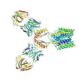
7LO8
 | | NorA in complex with Fab36 | | 分子名称: | Fab36 Heavy Chain, Fab36 Light Chain, Quinolone resistance protein NorA | | 著者 | Brawley, D.N, Sauer, D.B, Song, J.M, Koide, A, Koide, S, Traaseth, N.J, Wang, D.N. | | 登録日 | 2021-02-09 | | 公開日 | 2022-04-20 | | 最終更新日 | 2022-07-13 | | 実験手法 | ELECTRON MICROSCOPY (3.16 Å) | | 主引用文献 | Structural basis for inhibition of the drug efflux pump NorA from Staphylococcus aureus.
Nat.Chem.Biol., 18, 2022
|
|
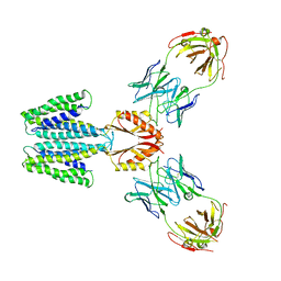
7LO7
 | | NorA in complex with Fab25 | | 分子名称: | Fab25 Heavy Chain, Fab25 Light Chain, Quinolone resistance protein NorA | | 著者 | Brawley, D.N, Sauer, D.B, Song, J.M, Koide, A, Koide, S, Traaseth, N.J, Wang, D.N. | | 登録日 | 2021-02-09 | | 公開日 | 2022-04-20 | | 最終更新日 | 2022-07-13 | | 実験手法 | ELECTRON MICROSCOPY (3.74 Å) | | 主引用文献 | Structural basis for inhibition of the drug efflux pump NorA from Staphylococcus aureus.
Nat.Chem.Biol., 18, 2022
|
|
5DC9
 | | CRYSTAL STRUCTURE OF MONOBODY AS25/ABL1 SH2 DOMAIN COMPLEX, CRYSTAL B | | 分子名称: | AS25 monobody, GLYCEROL, IMIDAZOLE, ... | | 著者 | Wojcik, J.B, Koide, A, Koide, S. | | 登録日 | 2015-08-23 | | 公開日 | 2016-03-02 | | 最終更新日 | 2024-03-06 | | 実験手法 | X-RAY DIFFRACTION (1.56 Å) | | 主引用文献 | Allosteric Inhibition of Bcr-Abl Kinase by High Affinity Monobody Inhibitors Directed to the Src Homology 2 (SH2)-Kinase Interface.
J.Biol.Chem., 291, 2016
|
|
5VG9
 | | Structure of the eukaryotic intramembrane Ras methyltransferase ICMT (isoprenylcysteine carboxyl methyltransferase) without a monobody | | 分子名称: | Protein-S-isoprenylcysteine O-methyltransferase, S-ADENOSYL-L-HOMOCYSTEINE | | 著者 | Long, S.B, Diver, M.M, Pedi, L, Koide, A, Koide, S. | | 登録日 | 2017-04-10 | | 公開日 | 2018-01-17 | | 最終更新日 | 2024-03-13 | | 実験手法 | X-RAY DIFFRACTION (4 Å) | | 主引用文献 | Atomic structure of the eukaryotic intramembrane RAS methyltransferase ICMT.
Nature, 553, 2018
|
|
7YA8
 | | The crystal structure of IpaH2.5 LRR domain | | 分子名称: | RING-type E3 ubiquitin transferase | | 著者 | Hiragi, K, Nishide, A, Takagi, K, Iwai, K, Kim, M, Mizushima, T. | | 登録日 | 2022-06-27 | | 公開日 | 2023-02-08 | | 最終更新日 | 2023-11-29 | | 実験手法 | X-RAY DIFFRACTION (3.4 Å) | | 主引用文献 | Structural insight into the recognition of the linear ubiquitin assembly complex by Shigella E3 ligase IpaH1.4/2.5.
J.Biochem., 173, 2023
|
|
7YA7
 | | The crystal structure of IpaH1.4 LRR domain | | 分子名称: | RING-type E3 ubiquitin transferase | | 著者 | Hiragi, K, Nishide, A, Takagi, K, Iwai, K, Kim, M, Mizushima, T. | | 登録日 | 2022-06-27 | | 公開日 | 2023-02-08 | | 最終更新日 | 2023-11-29 | | 実験手法 | X-RAY DIFFRACTION (1.4 Å) | | 主引用文献 | Structural insight into the recognition of the linear ubiquitin assembly complex by Shigella E3 ligase IpaH1.4/2.5.
J.Biochem., 173, 2023
|
|
5DC4
 | | CRYSTAL STRUCTURE OF MONOBODY AS25/ABL1 SH2 DOMAIN COMPLEX, CRYSTAL A | | 分子名称: | AS25 monobody, GLYCEROL, Tyrosine-protein kinase ABL1 | | 著者 | Wojcik, J.B, Koide, A, Koide, S. | | 登録日 | 2015-08-23 | | 公開日 | 2016-03-02 | | 最終更新日 | 2024-03-06 | | 実験手法 | X-RAY DIFFRACTION (1.48 Å) | | 主引用文献 | Allosteric Inhibition of Bcr-Abl Kinase by High Affinity Monobody Inhibitors Directed to the Src Homology 2 (SH2)-Kinase Interface.
J.Biol.Chem., 291, 2016
|
|
5V7P
 | | Atomic structure of the eukaryotic intramembrane Ras methyltransferase ICMT (isoprenylcysteine carboxyl methyltransferase), in complex with a monobody | | 分子名称: | DECANE, Protein-S-isoprenylcysteine O-methyltransferase, S-ADENOSYL-L-HOMOCYSTEINE, ... | | 著者 | Long, S.B, Diver, M.M, Pedi, L, Koide, A, Koide, S. | | 登録日 | 2017-03-20 | | 公開日 | 2018-01-17 | | 最終更新日 | 2024-03-06 | | 実験手法 | X-RAY DIFFRACTION (2.3 Å) | | 主引用文献 | Atomic structure of the eukaryotic intramembrane RAS methyltransferase ICMT.
Nature, 553, 2018
|
|
6D0N
 | | Crystal structure of a CLC-type fluoride/proton antiporter, V319G mutant | | 分子名称: | (CARBAMOYLMETHYL-CARBOXYMETHYL-AMINO)-ACETIC ACID, CLC-type fluoride/proton antiporter, DECYL-BETA-D-MALTOPYRANOSIDE, ... | | 著者 | Last, N.B, Stockbridge, R.B, Wilson, A.E, Shane, T, Kolmakova-Partensky, L, Koide, A, Koide, S, Miller, C. | | 登録日 | 2018-04-10 | | 公開日 | 2018-07-04 | | 最終更新日 | 2023-10-04 | | 実験手法 | X-RAY DIFFRACTION (3.12 Å) | | 主引用文献 | A CLC-type F-/H+antiporter in ion-swapped conformations.
Nat. Struct. Mol. Biol., 25, 2018
|
|
6D0K
 | | Crystal structure of a CLC-type fluoride/proton antiporter, E118Q mutant | | 分子名称: | (CARBAMOYLMETHYL-CARBOXYMETHYL-AMINO)-ACETIC ACID, CLC-type fluoride/proton antiporter, DECYL-BETA-D-MALTOPYRANOSIDE, ... | | 著者 | Last, N.B, Stockbridge, R.B, Wilson, A.E, Shane, T, Kolmakova-Partensky, L, Koide, A, Koide, S, Miller, C. | | 登録日 | 2018-04-10 | | 公開日 | 2018-07-04 | | 最終更新日 | 2023-10-04 | | 実験手法 | X-RAY DIFFRACTION (3.35 Å) | | 主引用文献 | A CLC-type F-/H+antiporter in ion-swapped conformations.
Nat. Struct. Mol. Biol., 25, 2018
|
|
6D0J
 | | Crystal structure of a CLC-type fluoride/proton antiporter | | 分子名称: | (CARBAMOYLMETHYL-CARBOXYMETHYL-AMINO)-ACETIC ACID, CLC-type fluoride/proton antiporter, DECYL-BETA-D-MALTOPYRANOSIDE, ... | | 著者 | Last, N.B, Stockbridge, R.B, Wilson, A.E, Shane, T, Kolmakova-Partensky, L, Koide, A, Koide, S, Miller, C. | | 登録日 | 2018-04-10 | | 公開日 | 2018-07-04 | | 最終更新日 | 2023-10-04 | | 実験手法 | X-RAY DIFFRACTION (3 Å) | | 主引用文献 | A CLC-type F-/H+antiporter in ion-swapped conformations.
Nat. Struct. Mol. Biol., 25, 2018
|
|
1VDQ
 | | The crystal structure of the orthorhombic form of hen egg white lysozyme at 1.5 angstroms resolution | | 分子名称: | Lysozyme C | | 著者 | Aibara, S, Suzuki, A, Kidera, A, Shibata, K, Yamane, T, DeLucas, L.J, Hirose, M. | | 登録日 | 2004-03-24 | | 公開日 | 2004-04-13 | | 最終更新日 | 2023-12-27 | | 実験手法 | X-RAY DIFFRACTION (1.5 Å) | | 主引用文献 | The crystal structure of the orthorhombic form of hen egg white lysozyme at 1.5 angstroms resolution
to be published
|
|
1VDT
 | | The crystal structure of the tetragonal form of hen egg white lysozyme at 1.7 angstroms resolution under basic conditions in space | | 分子名称: | Lysozyme C | | 著者 | Aibara, S, Suzuki, A, Kidera, A, Shibata, K, Yamane, T, DeLucas, L.J, Hirose, M. | | 登録日 | 2004-03-24 | | 公開日 | 2004-04-13 | | 最終更新日 | 2023-12-27 | | 実験手法 | X-RAY DIFFRACTION (1.7 Å) | | 主引用文献 | The crystal structure of the tetragonal form of hen egg white lysozyme at 1.7 angstroms resolution under basic conditions in space
to be published
|
|
1VDP
 | | The crystal structure of the monoclinic form of hen egg white lysozyme at 1.7 angstroms resolution in space | | 分子名称: | Lysozyme C | | 著者 | Aibara, S, Suzuki, A, Kidera, A, Shibata, K, Yamane, T, DeLucas, L.J, Hirose, M. | | 登録日 | 2004-03-24 | | 公開日 | 2004-04-13 | | 最終更新日 | 2023-12-27 | | 実験手法 | X-RAY DIFFRACTION (1.7 Å) | | 主引用文献 | The crystal structure of the monoclinic form of hen egg white lysozyme at 1.7 angstroms resolution in space
to be published
|
|
1VED
 | | The crystal structure of the orthorhombic form of hen egg white lysozyme at 1.9 angstroms resolution in space | | 分子名称: | Lysozyme C | | 著者 | Aibara, S, Suzuki, A, Kidera, A, Shibata, K, Yamane, T, DeLucas, L.J, Hirose, M. | | 登録日 | 2004-03-30 | | 公開日 | 2004-04-13 | | 最終更新日 | 2023-12-27 | | 実験手法 | X-RAY DIFFRACTION (1.9 Å) | | 主引用文献 | The crystal structure of the orthorhombic form of hen egg white lysozyme at 1.9 angstroms resolution in space
To be Published
|
|
1VDS
 | | The crystal structure of the tetragonal form of hen egg white lysozyme at 1.6 angstroms resolution in space | | 分子名称: | Lysozyme C | | 著者 | Aibara, S, Suzuki, A, Kidera, A, Shibata, K, Yamane, T, DeLucas, L.J, Hirose, M. | | 登録日 | 2004-03-24 | | 公開日 | 2004-04-13 | | 最終更新日 | 2023-12-27 | | 実験手法 | X-RAY DIFFRACTION (1.6 Å) | | 主引用文献 | The crystal structure of the tetragonal form of hen egg white lysozyme at 1.6 angstroms resolution in space
to be published
|
|


