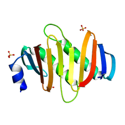
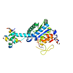
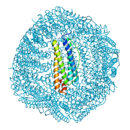
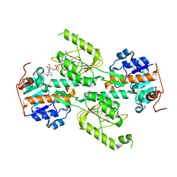
4FTX
 
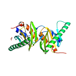 | | Crystal structure of Ego3 homodimer | | 分子名称: | Protein SLM4, SUCCINIC ACID | | 著者 | Zhang, T, Peli-Gulli, M.P, Yang, H, De Virgilio, C, Ding, J. | | 登録日 | 2012-06-28 | | 公開日 | 2012-11-28 | | 最終更新日 | 2024-03-20 | | 実験手法 | X-RAY DIFFRACTION (2.1 Å) | | 主引用文献 | Ego3 functions as a homodimer to mediate the interaction between Gtr1-Gtr2 and Ego1 in the ego complex to activate TORC1.
Structure, 20, 2012
|
|
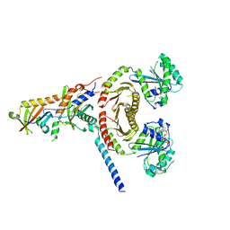
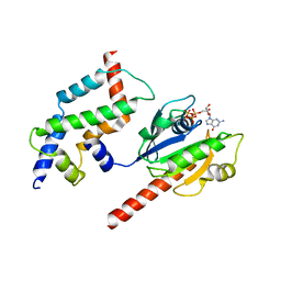
6D3U
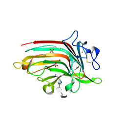 | | Complex structure of Ulvan lyase from Nonlaben Ulvanivorans- NLR48 | | 分子名称: | 1,2-ETHANEDIOL, 4-deoxy-alpha-L-threo-hex-4-enopyranuronic acid-(1-4)-3-O-sulfo-alpha-L-rhamnopyranose-(1-4)-beta-D-glucopyranuronic acid-(1-4)-3-O-sulfo-alpha-L-rhamnopyranose, CALCIUM ION, ... | | 著者 | Ulaganathan, T, Cygler, M. | | 登録日 | 2018-04-16 | | 公開日 | 2018-06-06 | | 最終更新日 | 2020-07-29 | | 実験手法 | X-RAY DIFFRACTION (2.21 Å) | | 主引用文献 | Structural and functional characterization of PL28 family ulvan lyase NLR48 fromNonlabens ulvanivorans.
J. Biol. Chem., 293, 2018
|
|
5Y42
 | |
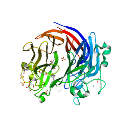
5Y97
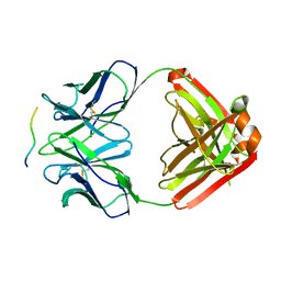
 | | Crystal structure of snake gourd seed lectin in complex with lactose | | 分子名称: | 2-acetamido-2-deoxy-beta-D-glucopyranose, Seed lectin, beta-D-galactopyranose-(1-4)-beta-D-glucopyranose | | 著者 | Chandran, T, Vijayan, M, Sivaji, N, Surolia, A. | | 登録日 | 2017-08-23 | | 公開日 | 2018-08-29 | | 最終更新日 | 2023-11-22 | | 実験手法 | X-RAY DIFFRACTION (3.05 Å) | | 主引用文献 | Ligand binding and retention in snake gourd seed lectin (SGSL). A crystallographic, thermodynamic and molecular dynamics study
Glycobiology, 28, 2018
|
|
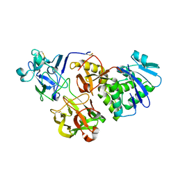
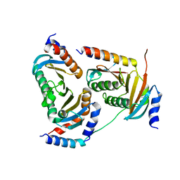
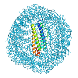
6JWP
 | | crystal structure of EGOC | | 分子名称: | Ego2, GTP-binding protein GTR1, GTP-binding protein GTR2, ... | | 著者 | Zhang, T, Ding, J. | | 登録日 | 2019-04-21 | | 公開日 | 2019-12-11 | | 最終更新日 | 2023-11-22 | | 実験手法 | X-RAY DIFFRACTION (3.2 Å) | | 主引用文献 | Structural insights into the EGO-TC-mediated membrane tethering of the TORC1-regulatory Rag GTPases.
Sci Adv, 5, 2019
|
|
8J0G
 | |
8J27
 | |
8J28
 | |
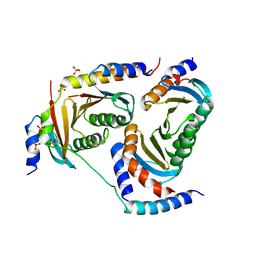
5Y38
 | | Crystal structure of C7orf59-HBXIP complex | | 分子名称: | Ragulator complex protein LAMTOR4, Ragulator complex protein LAMTOR5, SULFATE ION | | 著者 | Zhang, T, Ding, J. | | 登録日 | 2017-07-28 | | 公開日 | 2017-12-06 | | 最終更新日 | 2023-11-22 | | 実験手法 | X-RAY DIFFRACTION (2.8 Å) | | 主引用文献 | Structural basis for Ragulator functioning as a scaffold in membrane-anchoring of Rag GTPases and mTORC1
Nat Commun, 8, 2017
|
|
5Y39
 | | Crystal structure of Ragulator complex (p18 76-145) | | 分子名称: | Ragulator complex protein LAMTOR1, Ragulator complex protein LAMTOR2, Ragulator complex protein LAMTOR3, ... | | 著者 | Zhang, T, Ding, J. | | 登録日 | 2017-07-28 | | 公開日 | 2017-12-06 | | 最終更新日 | 2024-03-27 | | 実験手法 | X-RAY DIFFRACTION (2.65 Å) | | 主引用文献 | Structural basis for Ragulator functioning as a scaffold in membrane-anchoring of Rag GTPases and mTORC1
Nat Commun, 8, 2017
|
|
5Y3A
 | | Crystal structure of Ragulator complex (p18 49-161) | | 分子名称: | Ragulator complex protein LAMTOR1, Ragulator complex protein LAMTOR2, Ragulator complex protein LAMTOR3, ... | | 著者 | Zhang, T, Ding, J. | | 登録日 | 2017-07-28 | | 公開日 | 2017-12-27 | | 最終更新日 | 2023-11-22 | | 実験手法 | X-RAY DIFFRACTION (2.9 Å) | | 主引用文献 | Structural basis for Ragulator functioning as a scaffold in membrane-anchoring of Rag GTPases and mTORC1.
Nat Commun, 8, 2017
|
|
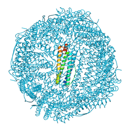
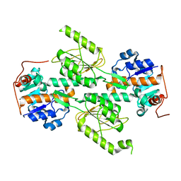
3DOE
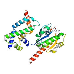 | | Complex of ARL2 and BART, Crystal Form 1 | | 分子名称: | ADP-ribosylation factor-like protein 2, ADP-ribosylation factor-like protein 2-binding protein, GUANOSINE-5'-TRIPHOSPHATE, ... | | 著者 | Zhang, T, Li, S, Ding, J. | | 登録日 | 2008-07-04 | | 公開日 | 2009-03-03 | | 最終更新日 | 2023-11-01 | | 実験手法 | X-RAY DIFFRACTION (2.25 Å) | | 主引用文献 | Crystal structure of the ARL2-GTP-BART complex reveals a novel recognition and binding mode of small GTPase with effector
Structure, 17, 2009
|
|
8J0E
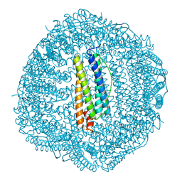
 | | GK monomer complexes with catalytic intermediate | | 分子名称: | (2~{R})-5-[[[[(2~{R},3~{S},4~{S},5~{R})-5-(6-aminopurin-9-yl)-3,4-bis(oxidanyl)oxolan-2-yl]methoxy-oxidanyl-phosphoryl]oxy-oxidanyl-phosphoryl]oxy-oxidanyl-phosphoryl]oxy-2-azanyl-5-oxidanylidene-pentanoic acid, Delta-1-pyrroline-5-carboxylate synthase B, MAGNESIUM ION | | 著者 | Zhang, T, Guo, C.J, Liu, J.L. | | 登録日 | 2023-04-10 | | 公開日 | 2024-04-17 | | 実験手法 | ELECTRON MICROSCOPY (3.4 Å) | | 主引用文献 | GK monomer complexes with catalytic intermediate
To Be Published
|
|
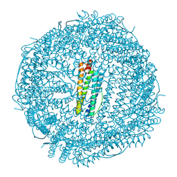
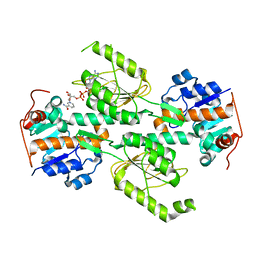
3DOF
 | | Complex of ARL2 and BART, Crystal Form 2 | | 分子名称: | ADP-ribosylation factor-like protein 2, ADP-ribosylation factor-like protein 2-binding protein, GUANOSINE-5'-TRIPHOSPHATE, ... | | 著者 | Zhang, T, Li, S, Ding, J. | | 登録日 | 2008-07-04 | | 公開日 | 2009-03-03 | | 最終更新日 | 2023-11-01 | | 実験手法 | X-RAY DIFFRACTION (3.3 Å) | | 主引用文献 | Crystal structure of the ARL2-GTP-BART complex reveals a novel recognition and binding mode of small GTPase with effector
Structure, 17, 2009
|
|
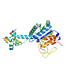
6BYX
 | | Complex structure of LOR107 mutant (R259N) with tetrasaccharide substrate | | 分子名称: | 4-deoxy-alpha-L-threo-hex-4-enopyranuronic acid-(1-4)-3-O-sulfo-alpha-L-rhamnopyranose-(1-4)-beta-D-glucopyranuronic acid-(1-4)-3-O-sulfo-alpha-L-rhamnopyranose, CALCIUM ION, GLYCEROL, ... | | 著者 | Ulaganathan, T, Cygler, M. | | 登録日 | 2017-12-21 | | 公開日 | 2018-02-07 | | 最終更新日 | 2024-03-13 | | 実験手法 | X-RAY DIFFRACTION (2.208 Å) | | 主引用文献 | Structure-function analyses of a PL24 family ulvan lyase reveal key features and suggest its catalytic mechanism.
J. Biol. Chem., 293, 2018
|
|
8W91
 | |
8W93
 | |
8W95
 | |
8W92
 | |
8WB3
 | |
8W94
 | |
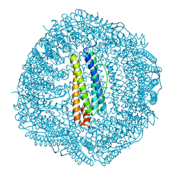
1X13
 | | Crystal structure of E. coli transhydrogenase domain I | | 分子名称: | NAD(P) transhydrogenase subunit alpha | | 著者 | Johansson, T, Oswald, C, Pedersen, A, Tornroth, S, Okvist, M, Karlsson, B.G, Rydstrom, J, Krengel, U. | | 登録日 | 2005-03-31 | | 公開日 | 2005-09-13 | | 最終更新日 | 2024-03-13 | | 実験手法 | X-RAY DIFFRACTION (1.9 Å) | | 主引用文献 | X-ray Structure of Domain I of the Proton-pumping Membrane Protein Transhydrogenase from Escherichia coli
J.Mol.Biol., 352, 2005
|
|
1X14
 | | Crystal structure of E. coli transhydrogenase domain I with bound NAD | | 分子名称: | NAD(P) transhydrogenase subunit alpha, NICOTINAMIDE-ADENINE-DINUCLEOTIDE | | 著者 | Johansson, T, Oswald, C, Pedersen, A, Tornroth, S, Okvist, M, Karlsson, B.G, Rydstrom, J, Krengel, U. | | 登録日 | 2005-03-31 | | 公開日 | 2005-09-13 | | 最終更新日 | 2024-03-13 | | 実験手法 | X-RAY DIFFRACTION (1.94 Å) | | 主引用文献 | X-ray Structure of Domain I of the Proton-pumping Membrane Protein Transhydrogenase from Escherichia coli
J.Mol.Biol., 352, 2005
|
|
2Y06
 | |
1X15
 | | Crystal structure of E. coli transhydrogenase domain I with bound NADH | | 分子名称: | NAD(P) transhydrogenase subunit alpha, NICOTINAMIDE-ADENINE-DINUCLEOTIDE | | 著者 | Johansson, T, Oswald, C, Pedersen, A, Tornroth, S, Okvist, M, Karlsson, B.G, Rydstrom, J, Krengel, U. | | 登録日 | 2005-03-31 | | 公開日 | 2005-09-13 | | 最終更新日 | 2024-03-13 | | 実験手法 | X-RAY DIFFRACTION (2.04 Å) | | 主引用文献 | X-ray Structure of Domain I of the Proton-pumping Membrane Protein Transhydrogenase from Escherichia coli
J.Mol.Biol., 352, 2005
|
|


