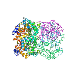
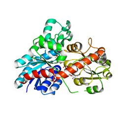
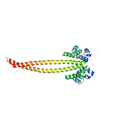
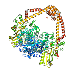
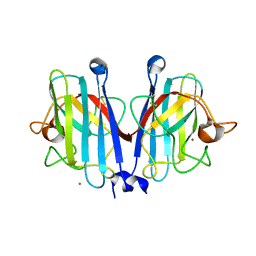
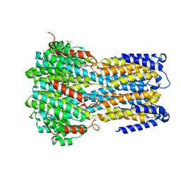
5YXW
 
 | | Crystal structure of the prefusion form of measles virus fusion protein | | 分子名称: | 2-acetamido-2-deoxy-beta-D-glucopyranose, 2-acetamido-2-deoxy-beta-D-glucopyranose-(1-4)-2-acetamido-2-deoxy-beta-D-glucopyranose, glycoprotein F1,measles virus fusion protein, ... | | 著者 | Hashiguchi, T, Fukuda, Y, Matsuoka, R, Kuroda, D, Kubota, M, Shirogane, Y, Watanabe, S, Tsumoto, K, Kohda, D, Plemper, R.K, Yanagi, Y. | | 登録日 | 2017-12-07 | | 公開日 | 2018-02-21 | | 最終更新日 | 2022-03-23 | | 実験手法 | X-RAY DIFFRACTION (2.776 Å) | | 主引用文献 | Structures of the prefusion form of measles virus fusion protein in complex with inhibitors.
Proc. Natl. Acad. Sci. U.S.A., 115, 2018
|
|
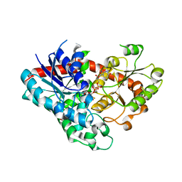
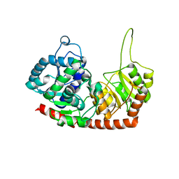
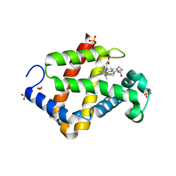
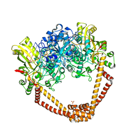
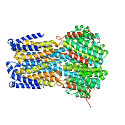
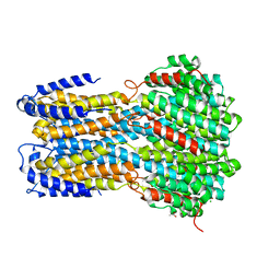
5YZC
 | | Crystal structure of the prefusion form of measles virus fusion protein in complex with a fusion inhibitor compound (AS-48) | | 分子名称: | 2-acetamido-2-deoxy-beta-D-glucopyranose, 2-acetamido-2-deoxy-beta-D-glucopyranose-(1-4)-2-acetamido-2-deoxy-beta-D-glucopyranose, 4-nitro-2-[(phenylacetyl)amino]benzamide, ... | | 著者 | Hashiguchi, T, Fukuda, Y, Matsuoka, R, Kuroda, D, Kubota, M, Shirogane, Y, Watanabe, S, Tsumoto, K, Kohda, D, Plemper, R.K, Yanagi, Y. | | 登録日 | 2017-12-14 | | 公開日 | 2018-02-21 | | 最終更新日 | 2023-11-22 | | 実験手法 | X-RAY DIFFRACTION (2.334 Å) | | 主引用文献 | Structures of the prefusion form of measles virus fusion protein in complex with inhibitors.
Proc. Natl. Acad. Sci. U.S.A., 115, 2018
|
|
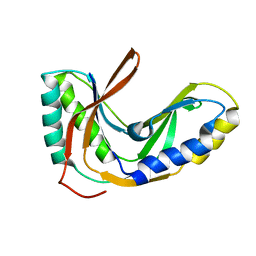
3WE7
 | | Crystal Structure of Diacetylchitobiose Deacetylase from Pyrococcus horikoshii | | 分子名称: | ACETIC ACID, GLYCEROL, HEXANE-1,6-DIOL, ... | | 著者 | Mine, S, Nakamura, T, Fukuda, Y, Inoue, T, Uegaki, K, Sato, T. | | 登録日 | 2013-07-01 | | 公開日 | 2014-05-07 | | 最終更新日 | 2014-08-20 | | 実験手法 | X-RAY DIFFRACTION (1.55 Å) | | 主引用文献 | Expression from engineered Escherichia coli chromosome and crystallographic study of archaeal N,N'-diacetylchitobiose deacetylase
Febs J., 281, 2014
|
|
7VEL
 | | Crystal structure of Phytolacca americana UGT3 with UDP-2fluoroglucose | | 分子名称: | 1,2-ETHANEDIOL, 1,4,7,10,13,16-HEXAOXACYCLOOCTADECANE, Glycosyltransferase, ... | | 著者 | Maharjan, R, Fukuda, Y, Nakayama, T, Nakayama, T, Hamada, H, Ozaki, S, Inoue, T. | | 登録日 | 2021-09-09 | | 公開日 | 2022-03-02 | | 最終更新日 | 2023-11-29 | | 実験手法 | X-RAY DIFFRACTION (2.15 Å) | | 主引用文献 | Structural basis for substrate recognition in the Phytolacca americana glycosyltransferase PaGT3.
Acta Crystallogr D Struct Biol, 78, 2022
|
|
7VEK
 | | Crystal structure of Phytolacca americana UGT3 with capsaicin and UDP-2fluoroglucose | | 分子名称: | (6E)-N-(4-hydroxy-3-methoxybenzyl)-8-methylnon-6-enamide, 1,4,7,10,13,16-HEXAOXACYCLOOCTADECANE, Glycosyltransferase, ... | | 著者 | Maharjan, R, Fukuda, Y, Nakayama, T, Nakayama, T, Hamada, H, Ozaki, S, Inoue, T. | | 登録日 | 2021-09-09 | | 公開日 | 2022-03-02 | | 最終更新日 | 2023-11-29 | | 実験手法 | X-RAY DIFFRACTION (2.6 Å) | | 主引用文献 | Structural basis for substrate recognition in the Phytolacca americana glycosyltransferase PaGT3.
Acta Crystallogr D Struct Biol, 78, 2022
|
|
7VEJ
 | | Crystal structure of Phytolacca americana UGT3 with kaempferol and UDP-2fluoroglucose | | 分子名称: | 1,2-ETHANEDIOL, 1,4,7,10,13,16-HEXAOXACYCLOOCTADECANE, 3,5,7-TRIHYDROXY-2-(4-HYDROXYPHENYL)-4H-CHROMEN-4-ONE, ... | | 著者 | Maharjan, R, Fukuda, Y, Nakayama, T, Nakayama, T, Hamada, H, Ozaki, S, Inoue, T. | | 登録日 | 2021-09-09 | | 公開日 | 2022-03-02 | | 最終更新日 | 2023-11-29 | | 実験手法 | X-RAY DIFFRACTION (1.85 Å) | | 主引用文献 | Structural basis for substrate recognition in the Phytolacca americana glycosyltransferase PaGT3.
Acta Crystallogr D Struct Biol, 78, 2022
|
|
6JEN
 | | Structure of Phytolacca americana UGT2 complexed with UDP-2fluoro-glucose and pterostilbene | | 分子名称: | Glycosyltransferase, Pterostilbene, URIDINE-5'-DIPHOSPHATE-2-DEOXY-2-FLUORO-ALPHA-D-GLUCOSE | | 著者 | Maharjan, R, Fukuda, Y, Nakayama, T, Hamada, H, Ozaki, S, Inoue, T. | | 登録日 | 2019-02-06 | | 公開日 | 2020-03-11 | | 最終更新日 | 2023-11-22 | | 実験手法 | X-RAY DIFFRACTION (2.65 Å) | | 主引用文献 | An Ambidextrous Polyphenol GlycosyltransferasePaGT2 fromPhytolacca americana.
Biochemistry, 59, 2020
|
|
6JEL
 | | Structure of Phytolacca americana apo UGT2 | | 分子名称: | Glycosyltransferase | | 著者 | Maharjan, R, Fukuda, Y, Nakayama, T, Hamada, H, Ozaki, S, Inoue, T. | | 登録日 | 2019-02-06 | | 公開日 | 2020-03-11 | | 最終更新日 | 2023-11-22 | | 実験手法 | X-RAY DIFFRACTION (2.3 Å) | | 主引用文献 | An Ambidextrous Polyphenol GlycosyltransferasePaGT2 fromPhytolacca americana.
Biochemistry, 59, 2020
|
|
7X6G
 | |
7X6H
 | |
7X6F
 | | Outer membrane lipoprotein QseG of Escherichia coli O157:H7 | | 分子名称: | 1,2-ETHANEDIOL, ACETATE ION, CITRATE ANION, ... | | 著者 | Matsumoto, K, Fukuda, Y, Inoue, T. | | 登録日 | 2022-03-07 | | 公開日 | 2023-03-15 | | 最終更新日 | 2023-11-15 | | 実験手法 | X-RAY DIFFRACTION (2.3 Å) | | 主引用文献 | Crystal structures of QseE and QseG: elements of a three-component system from Escherichia coli.
Acta Crystallogr.,Sect.F, 79, 2023
|
|
5ZIQ
 | | Crystal structure of hexacoordinated heme protein from anhydrobiotic tardigrade at pH 4 | | 分子名称: | 1,2-ETHANEDIOL, Globin protein, PROTOPORPHYRIN IX CONTAINING FE, ... | | 著者 | Kim, J, Fukuda, Y, Inoue, T. | | 登録日 | 2018-03-16 | | 公開日 | 2019-01-02 | | 最終更新日 | 2024-03-27 | | 実験手法 | X-RAY DIFFRACTION (1.5 Å) | | 主引用文献 | Crystal structure of Kumaglobin: a hexacoordinated heme protein from an anhydrobiotic tardigrade, Ramazzottius varieornatus.
FEBS J., 286, 2019
|
|
5ZM9
 | | Crystal structure of hexacoordinated heme protein from anhydrobiotic tardigrade at pH 7 | | 分子名称: | CHLORIDE ION, Globin Protein, PROTOPORPHYRIN IX CONTAINING FE | | 著者 | Kim, J, Fukuda, Y, Inoue, T. | | 登録日 | 2018-04-02 | | 公開日 | 2019-01-02 | | 最終更新日 | 2024-03-27 | | 実験手法 | X-RAY DIFFRACTION (2.7 Å) | | 主引用文献 | Crystal structure of Kumaglobin: a hexacoordinated heme protein from an anhydrobiotic tardigrade, Ramazzottius varieornatus.
FEBS J., 286, 2019
|
|
6JEM
 | | Structure of Phytolacca americana UGT2 complexed with UDP-2fluoro-glucose and resveratrol | | 分子名称: | Glycosyltransferase, RESVERATROL, URIDINE-5'-DIPHOSPHATE-2-DEOXY-2-FLUORO-ALPHA-D-GLUCOSE | | 著者 | Maharjan, R, Fukuda, Y, Nakayama, T, Hamada, H, Ozaki, S, Inoue, T. | | 登録日 | 2019-02-06 | | 公開日 | 2020-03-11 | | 最終更新日 | 2023-11-22 | | 実験手法 | X-RAY DIFFRACTION (2.6 Å) | | 主引用文献 | An Ambidextrous Polyphenol GlycosyltransferasePaGT2 fromPhytolacca americana.
Biochemistry, 59, 2020
|
|
4PLB
 | | Crystal Structure of S.A. gyrase-AM8191 complex | | 分子名称: | 6-[({(1r,4S)-1-[(1S)-2-(3-fluoro-6-methoxy-1,5-naphthyridin-4-yl)-1-hydroxyethyl]-2-oxabicyclo[2.2.2]oct-4-yl}amino)methyl]-2H-pyrido[3,2-b][1,4]oxazin-3(4H)-one, Chimera protein of DNA gyrase subunits B and A, DNA (5'-D(P*AP*GP*CP*CP*GP*TP*AP*GP*GP*GP*CP*CP*CP*TP*AP*CP*GP*GP*CP*T)-3'), ... | | 著者 | Lu, J, Patel, S, Soisson, S. | | 登録日 | 2014-05-16 | | 公開日 | 2014-06-18 | | 最終更新日 | 2023-12-27 | | 実験手法 | X-RAY DIFFRACTION (2.69 Å) | | 主引用文献 | Oxabicyclooctane-linked novel bacterial topoisomerase inhibitors as broad spectrum antibacterial agents.
Acs Med.Chem.Lett., 5, 2014
|
|
2FYH
 | | Solution structure of the 2'-5' RNA ligase-like protein from Pyrococcus furiosus | | 分子名称: | putative integral membrane transport protein | | 著者 | Okada, K, Matsuda, T, Sakamoto, T, Muto, Y, Yokoyama, S, Kanai, A, Kawai, G, RIKEN Structural Genomics/Proteomics Initiative (RSGI) | | 登録日 | 2006-02-08 | | 公開日 | 2007-02-20 | | 最終更新日 | 2024-05-01 | | 実験手法 | SOLUTION NMR | | 主引用文献 | Characterization of a heat-stable enzyme possessing GTP-dependent RNA ligase activity from a hyperthermophilic archaeon, Pyrococcus furiosus
Rna, 15, 2009
|
|
5BS3
 | | Crystal Structure of S.A. gyrase in complex with Compound 7 | | 分子名称: | (4R)-3-fluoro-4-hydroxy-4-{[(1r,4R)-4-{[(3-oxo-3,4-dihydro-2H-pyrido[3,2-b][1,4]oxazin-6-yl)methyl]amino}-2-oxabicyclo[2.2.2]oct-1-yl]methyl}-4,5-dihydro-7H-pyrrolo[3,2,1-de][1,5]naphthyridin-7-one, DNA gyrase subunit A and B, DNA/RNA (5'-R(P*AP*GP*CP*CP*G)-D(P*T)-R(P*AP*GP*GP*GP*CP*CP*C)-D(P*T)-R(P*AP*CP*GP*GP*C)-D(P*T)-3'), ... | | 著者 | Lu, J, Patel, S, Soisson, S. | | 登録日 | 2015-06-01 | | 公開日 | 2015-06-17 | | 最終更新日 | 2024-03-06 | | 実験手法 | X-RAY DIFFRACTION (2.65 Å) | | 主引用文献 | Tricyclic 1,5-naphthyridinone oxabicyclooctane-linked novel bacterial topoisomerase inhibitors as broad-spectrum antibacterial agents-SAR of left-hand-side moiety (Part-2).
Bioorg.Med.Chem.Lett., 25, 2015
|
|
7YPP
 | | Structural basis of a superoxide dismutase from a tardigrade, Ramazzottius varieornatus strain YOKOZUNA-1. | | 分子名称: | CALCIUM ION, COPPER (II) ION, Superoxide dismutase [Cu-Zn], ... | | 著者 | Sim, K.-S, Fukuda, Y, Inoue, T. | | 登録日 | 2022-08-04 | | 公開日 | 2023-07-26 | | 実験手法 | X-RAY DIFFRACTION (2.2 Å) | | 主引用文献 | Structure of a superoxide dismutase from a tardigrade: Ramazzottius varieornatus strain YOKOZUNA-1.
Acta Crystallogr.,Sect.F, 79, 2023
|
|
7YPR
 | | Structural basis of a superoxide dismutase from a tardigrade, Ramazzottius varieornatus strain YOKOZUNA-1. | | 分子名称: | COPPER (II) ION, POTASSIUM ION, Superoxide dismutase [Cu-Zn], ... | | 著者 | Sim, K.-S, Fukuda, Y, Inoue, T. | | 登録日 | 2022-08-04 | | 公開日 | 2023-12-06 | | 実験手法 | X-RAY DIFFRACTION (2.101 Å) | | 主引用文献 | Structure of a superoxide dismutase from a tardigrade: Ramazzottius varieornatus strain YOKOZUNA-1.
Acta Crystallogr.,Sect.F, 79, 2023
|
|
6IVQ
 | | Crystal structure of a membrane protein S19A | | 分子名称: | 1,2-ETHANEDIOL, ACETIC ACID, CHLORIDE ION, ... | | 著者 | Kittredge, A, Fukuda, F, Zhang, Y, Yang, T. | | 登録日 | 2018-12-04 | | 公開日 | 2019-11-06 | | 最終更新日 | 2024-05-29 | | 実験手法 | X-RAY DIFFRACTION (2.65 Å) | | 主引用文献 | Dual Ca2+-dependent gates in human Bestrophin1 underlie disease-causing mechanisms of gain-of-function mutations.
Commun Biol, 2, 2019
|
|
6IVK
 | | Crystal structure of a membrane protein G175A | | 分子名称: | 1,2-ETHANEDIOL, ACETIC ACID, CHLORIDE ION, ... | | 著者 | Kittredge, A, Fukuda, F, Zhang, Y, Yang, T. | | 登録日 | 2018-12-04 | | 公開日 | 2019-11-06 | | 最終更新日 | 2024-05-29 | | 実験手法 | X-RAY DIFFRACTION (2.65 Å) | | 主引用文献 | Dual Ca2+-dependent gates in human Bestrophin1 underlie disease-causing mechanisms of gain-of-function mutations.
Commun Biol, 2, 2019
|
|
6IVN
 | | Crystal structure of a membrane protein G264A | | 分子名称: | CHLORIDE ION, GLUTAMIC ACID, Ibestrophin, ... | | 著者 | Kittredge, A, Fukuda, F, Zhang, Y, Yang, T. | | 登録日 | 2018-12-04 | | 公開日 | 2019-11-06 | | 最終更新日 | 2024-05-29 | | 実験手法 | X-RAY DIFFRACTION (3.1 Å) | | 主引用文献 | Dual Ca2+-dependent gates in human Bestrophin1 underlie disease-causing mechanisms of gain-of-function mutations.
Commun Biol, 2, 2019
|
|
6IVL
 | | Crystal structure of a membrane protein L259A | | 分子名称: | ACETIC ACID, CHLORIDE ION, Ibestrophin, ... | | 著者 | Kittredge, A, Fukuda, F, Zhang, Y, Yang, T. | | 登録日 | 2018-12-04 | | 公開日 | 2019-11-06 | | 最終更新日 | 2024-05-29 | | 実験手法 | X-RAY DIFFRACTION (3.4 Å) | | 主引用文献 | Dual Ca2+-dependent gates in human Bestrophin1 underlie disease-causing mechanisms of gain-of-function mutations.
Commun Biol, 2, 2019
|
|
6IVJ
 | | Crystal structure of a membrane protein G18A | | 分子名称: | ACETIC ACID, CHLORIDE ION, Ibestrophin, ... | | 著者 | Kittredge, A, Fukuda, F, Zhang, Y, Yang, T. | | 登録日 | 2018-12-04 | | 公開日 | 2019-11-06 | | 最終更新日 | 2024-05-29 | | 実験手法 | X-RAY DIFFRACTION (2.77 Å) | | 主引用文献 | Dual Ca2+-dependent gates in human Bestrophin1 underlie disease-causing mechanisms of gain-of-function mutations.
Commun Biol, 2, 2019
|
|
6IVP
 | | Crystal structure of a membrane protein P262A | | 分子名称: | CHLORIDE ION, ZINC ION, bestrophin | | 著者 | Kittredge, A, Fukuda, F, Zhang, Y, Yang, T. | | 登録日 | 2018-12-04 | | 公開日 | 2019-11-06 | | 最終更新日 | 2024-05-29 | | 実験手法 | X-RAY DIFFRACTION (3.8 Å) | | 主引用文献 | Dual Ca2+-dependent gates in human Bestrophin1 underlie disease-causing mechanisms of gain-of-function mutations.
Commun Biol, 2, 2019
|
|


