8OTY
 
 | |
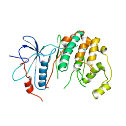
5BMN
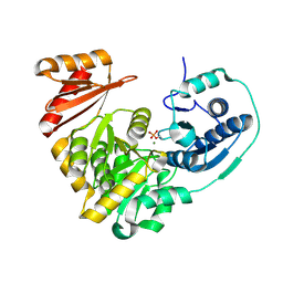 | | Crystal Structure of APO form of Phosphoglucomutase from Xanthomonas citri | | 分子名称: | MAGNESIUM ION, Phosphoglucomutase | | 著者 | Goto, L.S, Pereira, H.M, Novo Mansur, M.T.M, Brandao-Neto, J. | | 登録日 | 2015-05-22 | | 公開日 | 2016-06-01 | | 最終更新日 | 2023-09-27 | | 実験手法 | X-RAY DIFFRACTION (1.27 Å) | | 主引用文献 | Structural and functional characterization of the phosphoglucomutase from Xanthomonas citri subsp. citri.
Biochim.Biophys.Acta, 1864, 2016
|
|
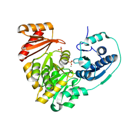
5BMP
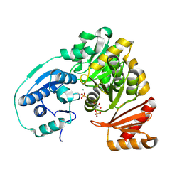 | |
5KL0
 | | Crystal Structure of Phosphoglucomutase from Xanthomonas citri citri complexed with Glucose-1,6-biphosphate | | 分子名称: | 1,6-di-O-phosphono-alpha-D-glucopyranose, MAGNESIUM ION, Phosphoglucomutase | | 著者 | Goto, L.S, Pereira, H.M, Novo Mansur, M.T.M. | | 登録日 | 2016-06-23 | | 公開日 | 2016-07-13 | | 最終更新日 | 2023-09-27 | | 実験手法 | X-RAY DIFFRACTION (1.853 Å) | | 主引用文献 | Structural and functional characterization of the phosphoglucomutase from Xanthomonas citri subsp. citri.
Biochim.Biophys.Acta, 1864, 2016
|
|
4IC7
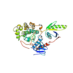 | |
4IC8
 | | Crystal structure of the apo ERK5 kinase domain | | 分子名称: | Mitogen-activated protein kinase 7 | | 著者 | Gogl, G, Remenyi, A. | | 登録日 | 2012-12-10 | | 公開日 | 2013-02-13 | | 最終更新日 | 2024-02-28 | | 実験手法 | X-RAY DIFFRACTION (2.8 Å) | | 主引用文献 | Structural mechanism for the specific assembly and activation of the extracellular signal regulated kinase 5 (ERK5) module.
J.Biol.Chem., 288, 2013
|
|


