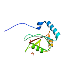
4LJQ
 
 | | Crystal structure of the catalytic core of E3 ligase HOIP | | 分子名称: | E3 ubiquitin-protein ligase RNF31, ZINC ION | | 著者 | Stieglitz, B, Rana, R.R, Koliopoulos, M.G, Morris-Davies, A.C, Christodoulou, E, Howell, S, Brown, N.R, Rittinger, K. | | 登録日 | 2013-07-05 | | 公開日 | 2013-10-16 | | 最終更新日 | 2013-12-18 | | 実験手法 | X-RAY DIFFRACTION (2.45 Å) | | 主引用文献 | Structural basis for ligase-specific conjugation of linear ubiquitin chains by HOIP.
Nature, 503, 2013
|
|
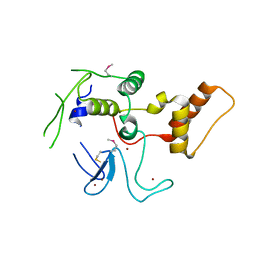
2LUE
 | | LC3B OPTN-LIR Ptot complex structure | | 分子名称: | Microtubule-associated proteins 1A/1B light chain 3B, Optineurin | | 著者 | Rogov, V.V, Rozenknop, A, Loehr, F, Guentert, P, Doetsch, V. | | 登録日 | 2012-06-13 | | 公開日 | 2013-07-17 | | 最終更新日 | 2022-08-24 | | 実験手法 | SOLUTION NMR | | 主引用文献 | Structural basis for phosphorylation-triggered autophagic clearance of Salmonella.
Biochem.J., 454, 2013
|
|
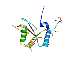
3VTW
 | | Crystal structure of T7-tagged Optineurin LIR-fused human LC3B_2-119 | | 分子名称: | Optineurin, microtubule-associated proteins 1A/1B light chain 3B, SULFATE ION | | 著者 | Suzuki, H, Kawasaki, M, Kato, R, Wakatsuki, S. | | 登録日 | 2012-06-08 | | 公開日 | 2013-06-26 | | 最終更新日 | 2023-11-08 | | 実験手法 | X-RAY DIFFRACTION (2.52 Å) | | 主引用文献 | Structural basis for phosphorylation-triggered autophagic clearance of Salmonella
Biochem.J., 454, 2013
|
|
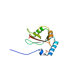
3VTU
 | | Crystal structure of human LC3B_2-119 | | 分子名称: | Microtubule-associated proteins 1A/1B light chain 3B, SULFATE ION | | 著者 | Suzuki, H, Kawasaki, M, Kato, R, Wakatsuki, S. | | 登録日 | 2012-06-08 | | 公開日 | 2013-06-26 | | 最終更新日 | 2023-11-08 | | 実験手法 | X-RAY DIFFRACTION (1.6 Å) | | 主引用文献 | Structural basis for phosphorylation-triggered autophagic clearance of Salmonella
Biochem.J., 454, 2013
|
|
3VHS
 | |
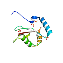
3VTV
 | | Crystal structure of Optineurin LIR-fused human LC3B_2-119 | | 分子名称: | Optineurin, microtubule-associated proteins 1A/1B light chain 3B, SULFATE ION | | 著者 | Suzuki, H, Kawasaki, M, Kato, R, Wakatsuki, S. | | 登録日 | 2012-06-08 | | 公開日 | 2013-06-26 | | 最終更新日 | 2023-11-08 | | 実験手法 | X-RAY DIFFRACTION (1.7 Å) | | 主引用文献 | Structural basis for phosphorylation-triggered autophagic clearance of Salmonella
Biochem.J., 454, 2013
|
|
3WUP
 | | Crystal Structure of the Ubiquitin-Binding Zinc Finger (UBZ) Domain of the Human DNA Polymerase Eta | | 分子名称: | CHLORIDE ION, DNA polymerase eta, GLYCEROL, ... | | 著者 | Suzuki, N, Wakatsuki, S, Kawasaki, S. | | 登録日 | 2014-05-01 | | 公開日 | 2015-06-17 | | 最終更新日 | 2024-05-29 | | 実験手法 | X-RAY DIFFRACTION (1.6 Å) | | 主引用文献 | A novel mode of ubiquitin recognition by the ubiquitin-binding zinc finger domain of WRNIP1.
Febs J., 283, 2016
|
|
3AI4
 | | Crystal structure of yeast enhanced green fluorescent protein - mouse polymerase iota ubiquitin binding motif fusion protein | | 分子名称: | SULFATE ION, yeast enhanced green fluorescent protein,DNA polymerase iota | | 著者 | Suzuki, N, Wakatsuki, S, Kawasaki, M. | | 登録日 | 2010-05-10 | | 公開日 | 2010-09-29 | | 最終更新日 | 2023-11-15 | | 実験手法 | X-RAY DIFFRACTION (1.6 Å) | | 主引用文献 | Crystallization of small proteins assisted by green fluorescent protein
Acta Crystallogr.,Sect.D, 66, 2010
|
|
3AI5
 | | Crystal structure of yeast enhanced green fluorescent protein-ubiquitin fusion protein | | 分子名称: | 1,2-ETHANEDIOL, yeast enhanced green fluorescent protein,Ubiquitin | | 著者 | Suzuki, N, Wakatsuki, S, Kawasaki, M. | | 登録日 | 2010-05-10 | | 公開日 | 2010-09-29 | | 最終更新日 | 2023-11-15 | | 実験手法 | X-RAY DIFFRACTION (1.4 Å) | | 主引用文献 | Crystallization of small proteins assisted by green fluorescent protein
Acta Crystallogr.,Sect.D, 66, 2010
|
|
2Z59
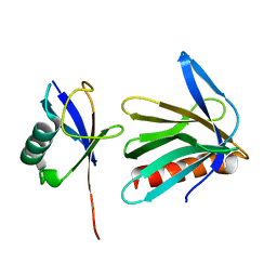 | | Complex Structures of Mouse Rpn13 (22-130aa) and ubiquitin | | 分子名称: | Protein ADRM1, Ubiquitin | | 著者 | Chen, X, Schreiner, P, Groll, M, Walters, K.J. | | 登録日 | 2007-07-01 | | 公開日 | 2008-05-20 | | 最終更新日 | 2024-05-29 | | 実験手法 | SOLUTION NMR | | 主引用文献 | Ubiquitin docking at the proteasome through a novel pleckstrin-homology domain interaction.
Nature, 453, 2008
|
|
2Z4D
 | |
3VHT
 | | Crystal structure of GFP-Wrnip1 UBZ domain fusion protein in complex with ubiquitin | | 分子名称: | Green fluorescent protein, Green fluorescent protein,ATPase WRNIP1, Ubiquitin, ... | | 著者 | Suzuki, N, Wakatsuki, S, Kawasaki, M. | | 登録日 | 2011-09-06 | | 公開日 | 2012-10-10 | | 最終更新日 | 2023-12-06 | | 実験手法 | X-RAY DIFFRACTION (2.4 Å) | | 主引用文献 | A novel mode of ubiquitin recognition by the ubiquitin-binding zinc finger domain of WRNIP1.
Febs J., 2016
|
|


