8PW7
 
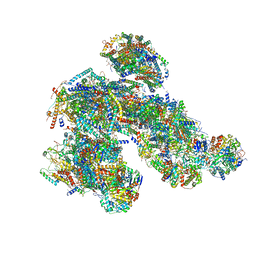 | | A respirasome from murine liver | | 分子名称: | 1,2-DIACYL-SN-GLYCERO-3-PHOSPHOCHOLINE, 1,2-Distearoyl-sn-glycerophosphoethanolamine, 2'-DEOXYGUANOSINE-5'-TRIPHOSPHATE, ... | | 著者 | Vercellino, I, Sazanov, L.A. | | 登録日 | 2023-07-19 | | 公開日 | 2024-04-24 | | 実験手法 | ELECTRON MICROSCOPY (3.5 Å) | | 主引用文献 | SCAF1 drives the compositional diversity of mammalian respirasomes.
Nat.Struct.Mol.Biol., 2024
|
|
8PW5
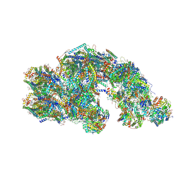 | | CS respirasome from murine liver | | 分子名称: | 1,2-DIACYL-SN-GLYCERO-3-PHOSPHOCHOLINE, 1,2-Distearoyl-sn-glycerophosphoethanolamine, 2'-DEOXYGUANOSINE-5'-TRIPHOSPHATE, ... | | 著者 | Vercellino, I, Sazanov, L.A. | | 登録日 | 2023-07-19 | | 公開日 | 2024-04-24 | | 実験手法 | ELECTRON MICROSCOPY (3.6 Å) | | 主引用文献 | SCAF1 drives the compositional diversity of mammalian respirasomes.
Nat.Struct.Mol.Biol., 2024
|
|
8PW6
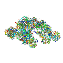 | | C respirasome from murine liver | | 分子名称: | 1,2-DIACYL-SN-GLYCERO-3-PHOSPHOCHOLINE, 1,2-Distearoyl-sn-glycerophosphoethanolamine, 2'-DEOXYGUANOSINE-5'-TRIPHOSPHATE, ... | | 著者 | Vercellino, I, Sazanov, L.A. | | 登録日 | 2023-07-19 | | 公開日 | 2024-04-24 | | 実験手法 | ELECTRON MICROSCOPY (3.3 Å) | | 主引用文献 | SCAF1 drives the compositional diversity of mammalian respirasomes.
Nat.Struct.Mol.Biol., 2024
|
|
8QB8
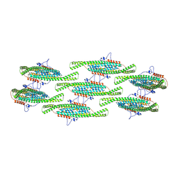 | |
8QBF
 | | Compact state - Pil1 dimer with lipid headgroups fitted in native eisosome lattice bound to plasma membrane microdomain | | 分子名称: | D-MYO-INOSITOL-1,4,5-TRIPHOSPHATE, PHOSPHOSERINE, Sphingolipid long chain base-responsive protein PIL1 | | 著者 | Kefauver, J.M, Zou, L, Desfosses, A, Loewith, R.J. | | 登録日 | 2023-08-24 | | 公開日 | 2024-07-24 | | 最終更新日 | 2024-08-07 | | 実験手法 | ELECTRON MICROSCOPY (3.67 Å) | | 主引用文献 | Cryo-EM architecture of a near-native stretch-sensitive membrane microdomain.
Nature, 2024
|
|
8QB7
 | |
8QBD
 | | Helical reconstruction of yeast eisosome protein Pil1 bound to membrane composed of lipid mixture +PIP2/+sterol (DOPC, DOPE, DOPS, cholesterol, PI(4,5)P2 35:20:20:15:10) | | 分子名称: | D-MYO-INOSITOL-1,4,5-TRIPHOSPHATE, O-[(R)-{[(2R)-2,3-bis(octadecanoyloxy)propyl]oxy}(hydroxy)phosphoryl]-L-serine, Sphingolipid long chain base-responsive protein PIL1 | | 著者 | Kefauver, J.M, Zou, L, Desfosses, A, Loewith, R.J. | | 登録日 | 2023-08-24 | | 公開日 | 2024-07-24 | | 最終更新日 | 2024-08-07 | | 実験手法 | ELECTRON MICROSCOPY (3.61 Å) | | 主引用文献 | Cryo-EM architecture of a near-native stretch-sensitive membrane microdomain.
Nature, 2024
|
|
8QBG
 | |
8QBE
 | |
6TX4
 | |
8RGR
 | | Closed Complex I from murine liver | | 分子名称: | 1,2-DIACYL-SN-GLYCERO-3-PHOSPHOCHOLINE, 1,2-Distearoyl-sn-glycerophosphoethanolamine, 2'-DEOXYGUANOSINE-5'-TRIPHOSPHATE, ... | | 著者 | Vercellino, I, Sazanov, L.A. | | 登録日 | 2023-12-14 | | 公開日 | 2024-04-24 | | 最終更新日 | 2024-07-31 | | 実験手法 | ELECTRON MICROSCOPY (2.9 Å) | | 主引用文献 | SCAF1 drives the compositional diversity of mammalian respirasomes.
Nat.Struct.Mol.Biol., 31, 2024
|
|
8RGT
 | | Open Complex I from murine brain | | 分子名称: | 1,2-DIACYL-SN-GLYCERO-3-PHOSPHOCHOLINE, 1,2-Distearoyl-sn-glycerophosphoethanolamine, 2'-DEOXYGUANOSINE-5'-TRIPHOSPHATE, ... | | 著者 | Vercellino, I, Sazanov, L.A. | | 登録日 | 2023-12-14 | | 公開日 | 2024-04-24 | | 最終更新日 | 2024-07-31 | | 実験手法 | ELECTRON MICROSCOPY (3.1 Å) | | 主引用文献 | SCAF1 drives the compositional diversity of mammalian respirasomes.
Nat.Struct.Mol.Biol., 31, 2024
|
|
6D54
 | |
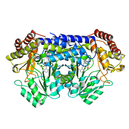
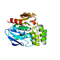
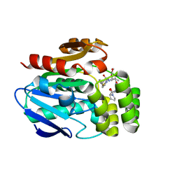
6E21
 | | Joint X-ray/neutron structure of PKAc with products Sr2-ADP and phosphorylated peptide SP20 | | 分子名称: | ADENOSINE-5'-DIPHOSPHATE, STRONTIUM ION, cAMP-dependent protein kinase catalytic subunit alpha, ... | | 著者 | Kovalevsky, A, Gerlits, O.O, Taylor, S. | | 登録日 | 2018-07-10 | | 公開日 | 2019-04-03 | | 最終更新日 | 2023-10-25 | | 実験手法 | NEUTRON DIFFRACTION (2 Å), X-RAY DIFFRACTION | | 主引用文献 | Zooming in on protons: Neutron structure of protein kinase A trapped in a product complex.
Sci Adv, 5, 2019
|
|
8SUJ
 | |
8SO0
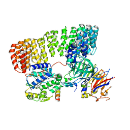 | | Cryo-EM structure of the PP2A:B55-FAM122A complex | | 分子名称: | FE (III) ION, PPP2R1A-PPP2R2A-interacting phosphatase regulator 1, Serine/threonine-protein phosphatase 2A 55 kDa regulatory subunit B alpha isoform, ... | | 著者 | Fuller, J.R, Padi, S.K.R, Peti, W, Page, R. | | 登録日 | 2023-04-28 | | 公開日 | 2023-10-25 | | 最終更新日 | 2024-01-17 | | 実験手法 | ELECTRON MICROSCOPY (2.79961 Å) | | 主引用文献 | Cryo-EM structures of PP2A:B55-FAM122A and PP2A:B55-ARPP19.
Nature, 625, 2024
|
|
8RGQ
 | | Open Complex I from murine liver | | 分子名称: | 1,2-DIACYL-SN-GLYCERO-3-PHOSPHOCHOLINE, 1,2-Distearoyl-sn-glycerophosphoethanolamine, 2'-DEOXYGUANOSINE-5'-TRIPHOSPHATE, ... | | 著者 | Vercellino, I, Sazanov, L.A. | | 登録日 | 2023-12-14 | | 公開日 | 2024-04-24 | | 最終更新日 | 2024-07-31 | | 実験手法 | ELECTRON MICROSCOPY (3 Å) | | 主引用文献 | SCAF1 drives the compositional diversity of mammalian respirasomes.
Nat.Struct.Mol.Biol., 31, 2024
|
|
8RGP
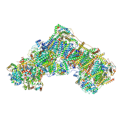 | | Closed Complex I from murine brain | | 分子名称: | 1,2-DIACYL-SN-GLYCERO-3-PHOSPHOCHOLINE, 1,2-Distearoyl-sn-glycerophosphoethanolamine, 2'-DEOXYGUANOSINE-5'-TRIPHOSPHATE, ... | | 著者 | Vercellino, I, Sazanov, L.A. | | 登録日 | 2023-12-14 | | 公開日 | 2024-04-24 | | 最終更新日 | 2024-07-31 | | 実験手法 | ELECTRON MICROSCOPY (3 Å) | | 主引用文献 | SCAF1 drives the compositional diversity of mammalian respirasomes.
Nat.Struct.Mol.Biol., 31, 2024
|
|
8QY4
 | |
8QY6
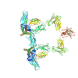 | |
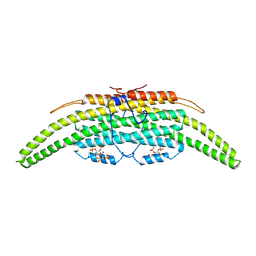
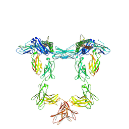
8QY5
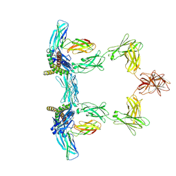 | | Structure of interleukin 6. | | 分子名称: | 2-acetamido-2-deoxy-beta-D-glucopyranose, 2-acetamido-2-deoxy-beta-D-glucopyranose-(1-4)-2-acetamido-2-deoxy-beta-D-glucopyranose, Interleukin-6, ... | | 著者 | Gardner, S, Bubeck, D, Jin, Y. | | 登録日 | 2023-10-25 | | 公開日 | 2024-02-21 | | 実験手法 | ELECTRON MICROSCOPY (3.1 Å) | | 主引用文献 | Structure of interleukin 6.
To Be Published
|
|
6BQ8
 | | Joint X-ray/neutron structure of PKG II CNB-B domain in complex with 8-pCPT-cGMP | | 分子名称: | (4S)-2-METHYL-2,4-PENTANEDIOL, 2-(~2~H_2_)amino-8-[(4-chlorophenyl)sulfanyl]-9-[(2S,4aR,6R,7R,7aS)-2-hydroxy-7-(~2~H)hydroxy-2-oxotetrahydro-2H,4H-2lambda~5~-furo[3,2-d][1,3,2]dioxaphosphinin-6-yl](~2~H)-1,9-dihydro-6H-purin-6-one, STRONTIUM ION, ... | | 著者 | Kim, C, Kovalevsky, A, Gerlits, O. | | 登録日 | 2017-11-27 | | 公開日 | 2018-03-21 | | 最終更新日 | 2024-04-03 | | 実験手法 | NEUTRON DIFFRACTION (2 Å), X-RAY DIFFRACTION | | 主引用文献 | Neutron Crystallography Detects Differences in Protein Dynamics: Structure of the PKG II Cyclic Nucleotide Binding Domain in Complex with an Activator.
Biochemistry, 57, 2018
|
|
6D4L
 | |
6T6Z
 | |
6T6Y
 | |


