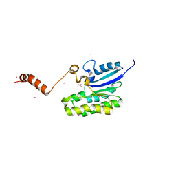
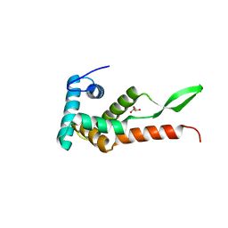
3FVS
 
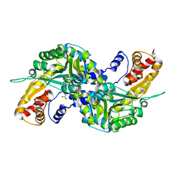 | | Human Kynurenine Aminotransferase I in complex with Glycerol | | 分子名称: | GLYCEROL, Kynurenine--oxoglutarate transaminase 1, SODIUM ION | | 著者 | Han, Q, Robinson, H, Cai, T, Tagle, D.A, Li, J. | | 登録日 | 2009-01-16 | | 公開日 | 2009-05-19 | | 最終更新日 | 2023-11-22 | | 実験手法 | X-RAY DIFFRACTION (1.5 Å) | | 主引用文献 | Structural insight into the inhibition of human kynurenine aminotransferase I/glutamine transaminase K
J.Med.Chem., 52, 2009
|
|
6KOC
 | | X-ray Structure of the proton-pumping cytochrome aa3-600 menaquinol oxidase from Bacillus subtilis complexed with 3-iodo-N-oxo-2-heptyl-4-hydroxyquinoline | | 分子名称: | 2-heptyl-3-iodanyl-1-oxidanyl-quinolin-4-one, AA3-600 quinol oxidase subunit I, AA3-600 quinol oxidase subunit IIII, ... | | 著者 | Xu, J, Ding, Z, Liu, B, Li, J, Gennis, R.B, Zhu, J. | | 登録日 | 2019-08-09 | | 公開日 | 2020-01-15 | | 最終更新日 | 2023-11-22 | | 実験手法 | X-RAY DIFFRACTION (3.8 Å) | | 主引用文献 | Structure of the cytochromeaa3-600 heme-copper menaquinol oxidase bound to inhibitor HQNO shows TM0 is part of the quinol binding site.
Proc.Natl.Acad.Sci.USA, 117, 2020
|
|
6KOE
 | | X-ray Structure of the proton-pumping cytochrome aa3-600 menaquinol oxidase from Bacillus subtilis | | 分子名称: | 2-HEPTYL-4-HYDROXY QUINOLINE N-OXIDE, AA3-600 quinol oxidase subunit I, AA3-600 quinol oxidase subunit IIII, ... | | 著者 | Xu, J, Ding, Z, Liu, B, Li, J, Gennis, R.B, Zhu, J. | | 登録日 | 2019-08-09 | | 公開日 | 2020-01-15 | | 最終更新日 | 2023-11-22 | | 実験手法 | X-RAY DIFFRACTION (3.75 Å) | | 主引用文献 | Structure of the cytochromeaa3-600 heme-copper menaquinol oxidase bound to inhibitor HQNO shows TM0 is part of the quinol binding site.
Proc.Natl.Acad.Sci.USA, 117, 2020
|
|
7C82
 | |
7C84
 | | Esterase AlinE4 mutant, D162A | | 分子名称: | ACETATE ION, CADMIUM ION, GLYCEROL, ... | | 著者 | Li, Z, Li, J. | | 登録日 | 2020-05-28 | | 公開日 | 2020-07-08 | | 最終更新日 | 2023-11-29 | | 実験手法 | X-RAY DIFFRACTION (1.552 Å) | | 主引用文献 | Structure-guided protein engineering increases enzymatic activities of the SGNH family esterases.
Biotechnol Biofuels, 13, 2020
|
|
7MU8
 | | Structure of the minimally glycosylated human CEACAM1 N-terminal domain | | 分子名称: | 2-acetamido-2-deoxy-beta-D-glucopyranose, Carcinoembryonic antigen-related cell adhesion molecule 1, GLYCEROL, ... | | 著者 | Belcher Dufrisne, M, Swope, N, Kieber, M, Yang, J.Y, Han, J, Li, J, Moremen, K.W, Prestegard, J.H, Columbus, L. | | 登録日 | 2021-05-14 | | 公開日 | 2022-02-09 | | 最終更新日 | 2024-10-30 | | 実験手法 | X-RAY DIFFRACTION (1.7 Å) | | 主引用文献 | Human CEACAM1 N-domain dimerization is independent from glycan modifications.
Structure, 30, 2022
|
|
7C85
 | | Esterase AlinE4 mutant-S13A | | 分子名称: | ACETATE ION, CADMIUM ION, SGNH-hydrolase family esterase | | 著者 | Li, Z, Li, J. | | 登録日 | 2020-05-28 | | 公開日 | 2020-07-08 | | 最終更新日 | 2023-11-29 | | 実験手法 | X-RAY DIFFRACTION (1.75 Å) | | 主引用文献 | Structure-guided protein engineering increases enzymatic activities of the SGNH family esterases.
Biotechnol Biofuels, 13, 2020
|
|
6KOB
 | | X-ray Structure of the proton-pumping cytochrome aa3-600 menaquinol oxidase from Bacillus subtilis | | 分子名称: | AA3-600 quinol oxidase subunit I, AA3-600 quinol oxidase subunit IIII, AA3-600 quinol oxidase subunit IV,Quinol oxidase subunit 4, ... | | 著者 | Xu, J, Ding, Z, Liu, B, Li, J, Gennis, R.B, Zhu, J. | | 登録日 | 2019-08-09 | | 公開日 | 2020-01-15 | | 最終更新日 | 2024-03-27 | | 実験手法 | X-RAY DIFFRACTION (3.6 Å) | | 主引用文献 | Structure of the cytochromeaa3-600 heme-copper menaquinol oxidase bound to inhibitor HQNO shows TM0 is part of the quinol binding site.
Proc.Natl.Acad.Sci.USA, 117, 2020
|
|
7KQ2
 | |
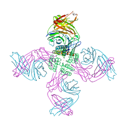
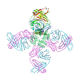
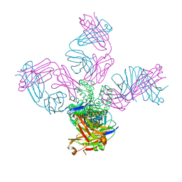
6LTJ
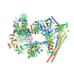 | | Structure of nucleosome-bound human BAF complex | | 分子名称: | AT-rich interactive domain-containing protein 1A, Actin, cytoplasmic 1, ... | | 著者 | He, S, Wu, Z, Tian, Y, Yu, Z, Yu, J, Wang, X, Li, J, Liu, B, Xu, Y. | | 登録日 | 2020-01-22 | | 公開日 | 2020-02-12 | | 最終更新日 | 2024-05-29 | | 実験手法 | ELECTRON MICROSCOPY (3.7 Å) | | 主引用文献 | Structure of nucleosome-bound human BAF complex.
Science, 367, 2020
|
|
5V7Z
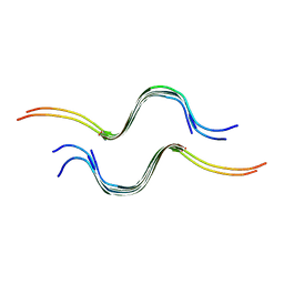 | | SSNMR Structure of the Human RIP1/RIP3 Necrosome | | 分子名称: | PRO-LEU-VAL-ASN-ILE-TYR-ASN-CYS-SER-GLY-VAL-GLN-VAL-GLY-ASP, THR-ILE-TYR-ASN-SER-THR-GLY-ILE-GLN-ILE-GLY-ALA-TYR-ASN-TYR-MET-GLU-ILE | | 著者 | Mompean, M, Li, W, Li, J, Laage, S, Siemer, A.B, Wu, H, McDermott, A.E. | | 登録日 | 2017-03-21 | | 公開日 | 2018-03-28 | | 最終更新日 | 2024-05-01 | | 実験手法 | SOLID-STATE NMR | | 主引用文献 | The Structure of the Necrosome RIPK1-RIPK3 Core, a Human Hetero-Amyloid Signaling Complex.
Cell, 173, 2018
|
|
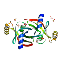
6JMK
 | | Ribosomal protein S7 from Mycobacterium tuberculosis | | 分子名称: | 1,2-ETHANEDIOL, 30S ribosomal protein S7, GLYCEROL | | 著者 | Li, Z, Li, J. | | 登録日 | 2019-03-11 | | 公開日 | 2019-03-20 | | 最終更新日 | 2023-11-22 | | 実験手法 | X-RAY DIFFRACTION (1.8 Å) | | 主引用文献 | Structural insights into the complex of trigger factor chaperone and ribosomal protein S7 from Mycobacterium tuberculosis.
Biochem. Biophys. Res. Commun., 512, 2019
|
|
7MJT
 | | KcsA open gate E71V mutant with Barium | | 分子名称: | BARIUM ION, Fab heavy chain, Fab light chain, ... | | 著者 | Rohaim, A, Li, J, Weingarth, M, Roux, B. | | 登録日 | 2021-04-20 | | 公開日 | 2022-03-23 | | 最終更新日 | 2024-11-06 | | 実験手法 | X-RAY DIFFRACTION (3.3 Å) | | 主引用文献 | A distinct mechanism of C-type inactivation in the Kv-like KcsA mutant E71V.
Nat Commun, 13, 2022
|
|
7MK6
 | | KcsA open gate E71V mutant with sodium | | 分子名称: | Fab heavy chain, Fab light chain, pH-gated potassium channel KcsA | | 著者 | Rohaim, A, Li, J, Weingarth, M, Roux, B. | | 登録日 | 2021-04-21 | | 公開日 | 2022-03-23 | | 最終更新日 | 2024-10-30 | | 実験手法 | X-RAY DIFFRACTION (3.1 Å) | | 主引用文献 | A distinct mechanism of C-type inactivation in the Kv-like KcsA mutant E71V.
Nat Commun, 13, 2022
|
|
7MHR
 | | KcsA E71V closed gate with K+ | | 分子名称: | Fab heavy chain, Fab light chain, POTASSIUM ION, ... | | 著者 | Rohaim, A, Li, J, Weingarth, M, Roux, B. | | 登録日 | 2021-04-15 | | 公開日 | 2022-03-23 | | 最終更新日 | 2024-10-09 | | 実験手法 | X-RAY DIFFRACTION (2.77 Å) | | 主引用文献 | A distinct mechanism of C-type inactivation in the Kv-like KcsA mutant E71V.
Nat Commun, 13, 2022
|
|
7MHX
 | | KcsA E71V closed gate with Ba2+ | | 分子名称: | BARIUM ION, DIACYL GLYCEROL, Fab heavy chain, ... | | 著者 | Rohaim, A, Li, J, Weingarth, M, Roux, B. | | 登録日 | 2021-04-15 | | 公開日 | 2022-03-23 | | 最終更新日 | 2024-11-20 | | 実験手法 | X-RAY DIFFRACTION (2.85 Å) | | 主引用文献 | A distinct mechanism of C-type inactivation in the Kv-like KcsA mutant E71V.
Nat Commun, 13, 2022
|
|
5ET0
 | | Crystal structure of Myo3b-ARB2 in complex with Espin1-AR | | 分子名称: | Espin, Myosin-IIIb | | 著者 | Liu, H, Li, J, Liu, W, Zhang, M. | | 登録日 | 2015-11-17 | | 公開日 | 2016-02-03 | | 最終更新日 | 2023-11-08 | | 実験手法 | X-RAY DIFFRACTION (2.3 Å) | | 主引用文献 | Myosin III-mediated cross-linking and stimulation of actin bundling activity of Espin
Elife, 5, 2016
|
|
5ET1
 | | Crystal structure of Myo3b-ARB1 in complex with Espin1-AR | | 分子名称: | Espin, GLYCEROL, Myosin-IIIb | | 著者 | Liu, H, Li, J, liu, W, Zhang, M. | | 登録日 | 2015-11-17 | | 公開日 | 2016-02-03 | | 最終更新日 | 2023-11-08 | | 実験手法 | X-RAY DIFFRACTION (1.65 Å) | | 主引用文献 | Myosin III-mediated cross-linking and stimulation of actin bundling activity of Espin
Elife, 5, 2016
|
|
2LXP
 | | NMR structure of two domains in ubiquitin ligase gp78, RING and G2BR, bound to its conjugating enzyme Ube2g | | 分子名称: | E3 ubiquitin-protein ligase AMFR, Ubiquitin-conjugating enzyme E2 G2, ZINC ION | | 著者 | Das, R, Linag, Y, Mariano, J, Li, J, Huang, T, King, A, Weissman, A, Ji, X, Byrd, R. | | 登録日 | 2012-08-30 | | 公開日 | 2013-08-28 | | 最終更新日 | 2024-05-15 | | 実験手法 | SOLUTION NMR | | 主引用文献 | Allosteric regulation of E2:E3 interactions promote a processive ubiquitination machine.
Embo J., 32, 2013
|
|
8H91
 | |
8YKN
 | |
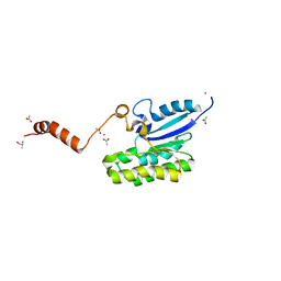
3UOT
 | | Crystal Structure of MDC1 FHA Domain in Complex with a Phosphorylated Peptide from the MDC1 N-terminus | | 分子名称: | Mediator of DNA damage checkpoint protein 1 | | 著者 | Clapperton, J.A, Lloyd, J, Haire, L.F, Li, J, Smerdon, S.J. | | 登録日 | 2011-11-17 | | 公開日 | 2011-12-28 | | 最終更新日 | 2024-10-16 | | 実験手法 | X-RAY DIFFRACTION (1.8 Å) | | 主引用文献 | The molecular basis of ATM-dependent dimerization of the Mdc1 DNA damage checkpoint mediator.
Nucleic Acids Res., 40, 2012
|
|
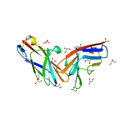
1DC2
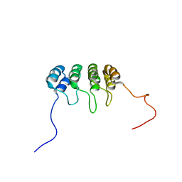 | | SOLUTION NMR STRUCTURE OF TUMOR SUPPRESSOR P16INK4A, 20 STRUCTURES | | 分子名称: | CYCLIN-DEPENDENT KINASE 4 INHIBITOR A (P16INK4A) | | 著者 | Byeon, I.-J.L, Li, J, Yuan, C, Tsai, M.-D. | | 登録日 | 1999-11-04 | | 公開日 | 1999-12-23 | | 最終更新日 | 2024-05-22 | | 実験手法 | SOLUTION NMR | | 主引用文献 | Tumor suppressor INK4: refinement of p16INK4A structure and determination of p15INK4B structure by comparative modeling and NMR data.
Protein Sci., 9, 2000
|
|
4GF6
 | | crystal structure of GFP with cuprum bound at the Incorporated metal Chelating Amino Acid PYZ151 | | 分子名称: | CALCIUM ION, COPPER (II) ION, green fluorescent protein | | 著者 | Dong, J, Liu, X, Li, J, Wang, J, Gong, W. | | 登録日 | 2012-08-03 | | 公開日 | 2012-08-29 | | 最終更新日 | 2023-11-15 | | 実験手法 | X-RAY DIFFRACTION (1.1 Å) | | 主引用文献 | Genetic incorporation of a metal-chelating amino Acid as a probe for protein electron transfer.
Angew.Chem.Int.Ed.Engl., 51, 2012
|
|
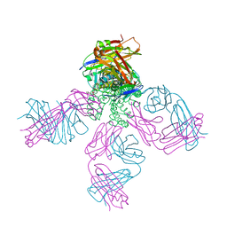
6NWA
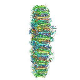 | | The structure of the photosystem I IsiA super-complex | | 分子名称: | 1,2-DIPALMITOYL-PHOSPHATIDYL-GLYCEROLE, 1,2-DISTEAROYL-MONOGALACTOSYL-DIGLYCERIDE, BETA-CAROTENE, ... | | 著者 | Toporik, H, Li, J, Williams, D, Chiu, P.L, Mazor, Y. | | 登録日 | 2019-02-06 | | 公開日 | 2019-05-29 | | 最終更新日 | 2024-03-20 | | 実験手法 | ELECTRON MICROSCOPY (3.48 Å) | | 主引用文献 | The structure of the stress-induced photosystem I-IsiA antenna supercomplex.
Nat.Struct.Mol.Biol., 26, 2019
|
|


