8Y9A
 
 | |
8XX1
 | |
8WD2
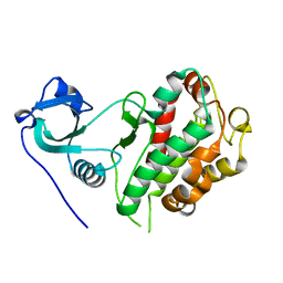
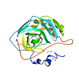
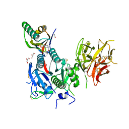
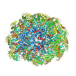
 | | The Crystal Structure of p53 from Biortus. | | 分子名称: | 1,2-ETHANEDIOL, Cellular tumor antigen p53, PHOSPHATE ION, ... | | 著者 | Wang, F, Cheng, W, Yuan, Z, Qi, J, Lu, Y. | | 登録日 | 2023-09-14 | | 公開日 | 2023-10-04 | | 実験手法 | X-RAY DIFFRACTION (1.85 Å) | | 主引用文献 | The Crystal Structure of p53 from Biortus.
To Be Published
|
|
8WF7
 | | The Crystal Structure of integrase from Biortus | | 分子名称: | ACETATE ION, Integrase, SULFATE ION | | 著者 | Wang, F, Cheng, W, Yuan, Z, Qi, J, Li, J. | | 登録日 | 2023-09-19 | | 公開日 | 2023-10-04 | | 実験手法 | X-RAY DIFFRACTION (1.55 Å) | | 主引用文献 | The Crystal Structure of integrase from Biortus
To Be Published
|
|
8WFG
 | |
8WF4
 | | The Crystal Structure of RSK1 from Biortus. | | 分子名称: | 1,2-ETHANEDIOL, Ribosomal protein S6 kinase alpha-1 | | 著者 | Wang, F, Cheng, W, Lv, Z, Qi, J, Li, J. | | 登録日 | 2023-09-19 | | 公開日 | 2023-11-22 | | 実験手法 | X-RAY DIFFRACTION (2.65 Å) | | 主引用文献 | The Crystal Structure of RSK1 from Biortus.
To Be Published
|
|
8WFF
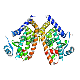
 | | The Crystal Structure of LYN from Biortus. | | 分子名称: | CHLORIDE ION, CITRATE ANION, Tyrosine-protein kinase Lyn | | 著者 | Wang, F, Cheng, W, Yuan, Z, Qi, J, Lu, Y. | | 登録日 | 2023-09-19 | | 公開日 | 2023-11-22 | | 実験手法 | X-RAY DIFFRACTION (1.3 Å) | | 主引用文献 | The Crystal Structure of LYN from Biortus.
To Be Published
|
|
8WD3
 | | The Crystal Structure of JMJD2A(M1-L359) from Biortus. | | 分子名称: | Lysine-specific demethylase 4A, NICKEL (II) ION, ZINC ION | | 著者 | Wang, F, Cheng, W, Lv, Z, Ju, C, Bao, C. | | 登録日 | 2023-09-14 | | 公開日 | 2023-11-22 | | 実験手法 | X-RAY DIFFRACTION (3.3 Å) | | 主引用文献 | The Crystal Structure of JMJD2A(M1-L359) from Biortus.
To Be Published
|
|
8WFQ
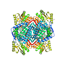 | | The Crystal Structure of RALDH1 from Biortus. | | 分子名称: | 1,2-ETHANEDIOL, 1,4-DIHYDRONICOTINAMIDE ADENINE DINUCLEOTIDE, Aldehyde dehydrogenase 1A1 | | 著者 | Wang, F, Cheng, W, Lv, Z, Qi, J, Shen, Z. | | 登録日 | 2023-09-20 | | 公開日 | 2023-11-22 | | 実験手法 | X-RAY DIFFRACTION (3.5 Å) | | 主引用文献 | The Crystal Structure of RALDH1 from Biortus.
To Be Published
|
|
8ZWV
 | | The Crystal Structure of carbonic anhydrase II from Biortus. | | 分子名称: | 1,2-ETHANEDIOL, Carbonic anhydrase 2, ZINC ION, ... | | 著者 | Wang, F, Cheng, W, Lv, Z, Ju, C, Bao, C. | | 登録日 | 2024-06-13 | | 公開日 | 2024-07-03 | | 実験手法 | X-RAY DIFFRACTION (1.5 Å) | | 主引用文献 | The Crystal Structure of carbonic anhydrase II from Biortus.
To Be Published
|
|
8ZKD
 | | The Crystal Structure of the RON from Biortus. | | 分子名称: | 1,2-ETHANEDIOL, MAGNESIUM ION, Macrophage-stimulating protein receptor beta chain, ... | | 著者 | Wang, F, Cheng, W, Yuan, Z, Qi, J, Pan, W. | | 登録日 | 2024-05-16 | | 公開日 | 2024-06-26 | | 実験手法 | X-RAY DIFFRACTION (2.05 Å) | | 主引用文献 | The Crystal Structure of the RON from Biortus.
To Be Published
|
|
8WFE
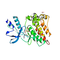
 | | The Crystal Structure of PPARg from Biortus. | | 分子名称: | 1,2-ETHANEDIOL, DI(HYDROXYETHYL)ETHER, Peroxisome proliferator-activated receptor gamma | | 著者 | Wang, F, Cheng, W, Lv, Z, Guo, S, Lin, D. | | 登録日 | 2023-09-19 | | 公開日 | 2023-11-22 | | 実験手法 | X-RAY DIFFRACTION (2.2 Å) | | 主引用文献 | The Crystal Structure of PPARg from Biortus.
To Be Published
|
|
8WFR
 | | The Crystal Structure of PCSK9 from Biortus. | | 分子名称: | 1,2-ETHANEDIOL, 4-(2-HYDROXYETHYL)-1-PIPERAZINE ETHANESULFONIC ACID, GLYCEROL, ... | | 著者 | Wang, F, Cheng, W, Lv, Z, Meng, Q, Lu, Y. | | 登録日 | 2023-09-20 | | 公開日 | 2023-11-22 | | 実験手法 | X-RAY DIFFRACTION (1.95 Å) | | 主引用文献 | The Crystal Structure of PCSK9 from Biortus.
To Be Published
|
|
8UI8
 | |
6NAV
 | | Cryo-EM reconstruction of Sulfolobus islandicus LAL14/1 Pilus | | 分子名称: | M9UD72 | | 著者 | Wang, F, Cvirkaite-Krupovic, V, Prangishvili, D, Krupovic, M, Egelman, E.H. | | 登録日 | 2018-12-06 | | 公開日 | 2019-05-08 | | 最終更新日 | 2024-03-20 | | 実験手法 | ELECTRON MICROSCOPY (4.1 Å) | | 主引用文献 | An extensively glycosylated archaeal pilus survives extreme conditions.
Nat Microbiol, 4, 2019
|
|
4RL1
 | |
6OJ0
 | | Cryo-EM reconstruction of Sulfolobus polyhedral virus 1 (SPV1) | | 分子名称: | Structural protein VP4, Uncharacterized protein | | 著者 | Wang, F, Liu, Y, Conway, J.F, Krupovic, M, Prangishvili, D, Egelman, E.H. | | 登録日 | 2019-04-10 | | 公開日 | 2019-10-02 | | 最終更新日 | 2020-01-08 | | 実験手法 | ELECTRON MICROSCOPY (3.7 Å) | | 主引用文献 | A packing for A-form DNA in an icosahedral virus.
Proc.Natl.Acad.Sci.USA, 116, 2019
|
|
6WL7
 | | Cryo-EM of Form 2 like peptide filament, 29-20-2 | | 分子名称: | peptide 29-20-2 | | 著者 | Wang, F, Gnewou, O.M, Modlin, C, Egelman, E.H, Conticello, V.P. | | 登録日 | 2020-04-18 | | 公開日 | 2020-12-02 | | 最終更新日 | 2024-03-06 | | 実験手法 | ELECTRON MICROSCOPY (3.8 Å) | | 主引用文献 | Structural analysis of cross alpha-helical nanotubes provides insight into the designability of filamentous peptide nanomaterials.
Nat Commun, 12, 2021
|
|
6WL1
 | | Cryo-EM of Form 1 related peptide filament, 36-31-3 | | 分子名称: | peptide 36-31-3 | | 著者 | Wang, F, Gnewou, O.M, Modlin, C, Egelman, E.H, Conticello, V.P. | | 登録日 | 2020-04-17 | | 公開日 | 2020-12-02 | | 最終更新日 | 2024-05-29 | | 実験手法 | ELECTRON MICROSCOPY (4 Å) | | 主引用文献 | Structural analysis of cross alpha-helical nanotubes provides insight into the designability of filamentous peptide nanomaterials.
Nat Commun, 12, 2021
|
|
6WL8
 | | Cryo-EM of Form 2 peptide filament | | 分子名称: | Form 2 peptide | | 著者 | Wang, F, Gnewou, O.M, Xu, C, Su, Z, Egelman, E.H, Conticello, V.P. | | 登録日 | 2020-04-18 | | 公開日 | 2020-12-02 | | 最終更新日 | 2024-03-06 | | 実験手法 | ELECTRON MICROSCOPY (4.1 Å) | | 主引用文献 | Structural analysis of cross alpha-helical nanotubes provides insight into the designability of filamentous peptide nanomaterials.
Nat Commun, 12, 2021
|
|
6WL9
 | | Cryo-EM of Form 2 like peptide filament, Form2a | | 分子名称: | peptide Form2a | | 著者 | Wang, F, Beltran, L.C, Gnewou, O.M, Egelman, E.H, Conticello, V.P. | | 登録日 | 2020-04-18 | | 公開日 | 2020-12-02 | | 最終更新日 | 2024-03-06 | | 実験手法 | ELECTRON MICROSCOPY (4.2 Å) | | 主引用文献 | Structural analysis of cross alpha-helical nanotubes provides insight into the designability of filamentous peptide nanomaterials.
Nat Commun, 12, 2021
|
|
6WL0
 | | Cryo-EM of Form 1 related peptide filament, 36-31-3-RD | | 分子名称: | peptide 36-31-3-RD | | 著者 | Wang, F, Gnewou, O.M, Su, Z, Egelman, E.H, Conticello, V.P. | | 登録日 | 2020-04-17 | | 公開日 | 2020-12-02 | | 最終更新日 | 2024-03-06 | | 実験手法 | ELECTRON MICROSCOPY (4.4 Å) | | 主引用文献 | Structural analysis of cross alpha-helical nanotubes provides insight into the designability of filamentous peptide nanomaterials.
Nat Commun, 12, 2021
|
|
6WKY
 | | Cryo-EM of Form 1 related peptide filament, 29-24-3 | | 分子名称: | peptide 29-24-3 | | 著者 | Wang, F, Gnewou, O.M, Egelman, E.H, Conticello, V.P. | | 登録日 | 2020-04-17 | | 公開日 | 2020-12-02 | | 最終更新日 | 2024-03-06 | | 実験手法 | ELECTRON MICROSCOPY (4.2 Å) | | 主引用文献 | Structural analysis of cross alpha-helical nanotubes provides insight into the designability of filamentous peptide nanomaterials.
Nat Commun, 12, 2021
|
|
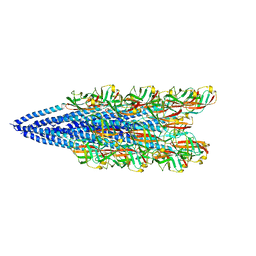
6WQ0
 | | Cryo-EM of the S. solfataricus rod-shaped virus, SSRV1 | | 分子名称: | DNA (301-MER), Structural protein | | 著者 | Wang, F, Baquero, D.P, Beltran, L.C, Prangishvili, D, Krupovic, M, Egelman, E.H. | | 登録日 | 2020-04-28 | | 公開日 | 2020-07-29 | | 最終更新日 | 2024-05-29 | | 実験手法 | ELECTRON MICROSCOPY (2.8 Å) | | 主引用文献 | Structures of filamentous viruses infecting hyperthermophilic archaea explain DNA stabilization in extreme environments.
Proc.Natl.Acad.Sci.USA, 117, 2020
|
|
6WQ2
 | | Cryo-EM of the S. islandicus filamentous virus, SIFV | | 分子名称: | A-DNA, Structural protein MCP1, Structural protein MCP2 | | 著者 | Wang, F, Baquero, D.P, Su, Z, Zheng, W, Prangishvili, D, Krupovic, M, Egelman, E.H. | | 登録日 | 2020-04-28 | | 公開日 | 2020-07-29 | | 最終更新日 | 2024-05-29 | | 実験手法 | ELECTRON MICROSCOPY (4 Å) | | 主引用文献 | Structures of filamentous viruses infecting hyperthermophilic archaea explain DNA stabilization in extreme environments.
Proc.Natl.Acad.Sci.USA, 117, 2020
|
|


